Biochemistry - The Chemical Reactions of Living Cells, Volume 3 - D. Metzler 1980
Metabolism of Nitrogenous Compounds
Metabolism of Aromatic Compounds
Phenylalanine and Tyrosine Metabolism in Animals and Bacteria
Fig. 14-20 illustrates the major Catabolic pathways, along with several biosynthetic reactions, that constitute the METABOLISM of Phenylalanine and Tyrosine in animals. Transamination to phenylpyruvate (reaction a) proceeds quite readily, and the resulting product can undergo oxidative decarboxylation to yield phenylacetate. The latter can be excreted in the form of glutamine conjugates (recall Knoop's classic experiments in which dogs excreted phenylacetate after conjugation with Glycine; Box 9-A). This degradation pathway for phenylalanine is also operative in humans, though apparently to a limited extent; indeed, if excess phenylalanine is not oxidized to tyrosine (reaction b, Fig. 14-20), it exerts toxic effects in humans1).
Considerable attention has been focused on the pteridine-dependent hydroxylation of phenylalanine to tyrosine [Eq. (10-52)], partly because of its relevance to phenylketonuria [118], a severe metabolic disorder in which this reaction fails to occur. Newborn infants with this condition appear initially normal, but soon begin to experience impaired mental development. If dietary phenylalanine intake in such children is restricted to the minimum required for Protein Synthesis alone, The Development of severe mental disability can be prevented. Some children raised on this diet have now reached young adulthood without any cognitive deficits, showing an increased tolerance to phenylalanine as they grow older.
a. Tyrosine Catabolism
The primary pathway for tyrosine breakdown in animals begins with a transamination reaction yielding p-hydroxyphenylpyruvate (Fig. 14-20, reaction c). The enzyme tyrosine aminotransferase has been studied in considerable detail, largely due to the induction of its hepatic synthesis in response to glucocorticoid Hormones (Chap. 11, Sect. E, 7). The synthesis of this enzyme is also regulated at the translational level [119], with the release of newly formed protein from Liver Ribosomes being stimulated by cyclic AMP. In addition, the enzyme undergoes post-translational modification, including phosphorylation [120], and is characterized by an unusually rapid turnover rate [121].
The keto acid p-hydroxyphenylpyruvate is decarboxylated by a dioxygenase, which was discussed in Chap. 10 [Eq. (10-55)]. The reaction product, homogentisic acid, is acted upon by a second dioxygenase (Fig. 14-20), ultimately being converted into fumarate and acetoacetate.
One of the earliest recognized "inborn errors of metabolism" was alkaptonuria, caused by a deficiency of the dioxygenase that cleaves the aromatic ring of homogentisic acid [122]. The disorder is easily identified by a characteristic sign: the urine turns dark brown upon standing (due to The oxidation of homogentisate). Alkaptonuria was correctly characterized by Garrod (Box 1-G) in 1909 as a disorder of tyrosine catabolism.
b. THYROID HORMONES [123, 124].
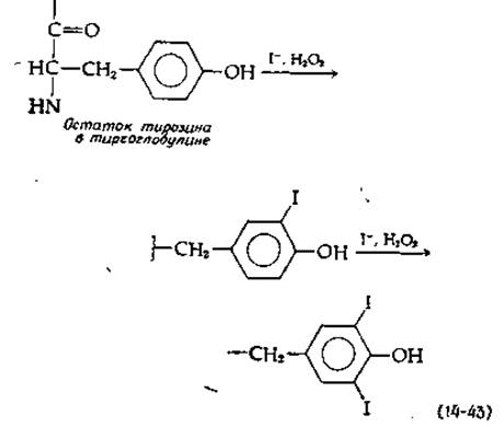
Thyroid hormones, whose principal and most active forms are thyroxine and triiodothyronine, are produced via the metabolic transformation of tyrosine. The Thyroid Gland is rich in iodide ions, which are actively taken up from the plasma and concentrated to levels of ~1 μM free I-. Mediated by a specialized peroxidase [see Eq. (10-7) and accompanying text], iodide ions undergo a reaction that iodinates tyrosine residues within the high-molecular-weight (dimeric) protein thyroglobulin (MW 660,000) [124a]. This iodination of several tyrosine side chains generates mono- and diiodotyrosine residues [Eq. (14-43)].
1) However, the precise mechanism by which excess amino acid damages the Brain remains unknown.
Class="center">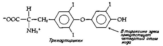
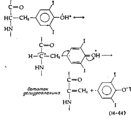
The precise Nature of the ring-coupling reaction, whereby the aromatic group of one mono- or diiodotyrosine residue is linked via an ether bond to a second such residue, is not yet fully understood. It is known, however, that the reaction proceeds readily in the presence of oxygen and peroxidase. One can easily envision the generation of an electron-deficient radical from iodinated tyrosine, which undergoes ξta-elimination to form dehydroalanine and an aromatic radical. The latter then condenses with a second radical to yield triiodothyronine or thyroxine [Eq. (14-44)]. A second possibility involves a Pyridoxal phosphate-dependent ξta-elimination of a radical. A third possibility entails the oxidative attack on keto acids derived from iodotyrosines [125].
Thyroxine and triiodothyronine are released from thyroglobulin through the action of several proteinases. Both proteolytic Cleavage and the subsequent release of thyroid hormones into the bloodstream are stimulated by the pituitary thyrotropic hormone (thyrotropin, TSH). Like Glucagon, TSH likely exerts its effects via a mechanism involving cAMP. Thyroid hormones are distributed throughout the body bound to thyroid-binding globulin, a specialized transport protein, while a fraction of the hormone molecules is carried by other Serum Proteins. Both thyroxine and triiodothyronine exert potent hormonal effects on target Tissues, though the response latency is shorter for triiodothyronine than for thyroxine. Within target Cells, thyroxine apparently first loses an iodine atom to be converted into the more active triiodo form.
The primary function of thyroxine and triiodothyronine is to stimulate Energy Metabolism in peripheral tissues. It has long been established that thyroid hormone deficiency leads to a decreased basal metabolic rate (Chap. 3, Sect. A, 5). Nevertheless, a fully comprehensive chemical theory of their MECHANISM OF ACTION is still lacking. Lardy and his coworkers obtained important evidence indicating that thyroxine uncouples Oxidative Phosphorylation in isolated Cell/35.html">Mitochondria (Chap. 10, Sect. D, 5). When mitochondria isolated from animals treated with excess thyroxine were compared with those from control animals, The rate of electron transfer was found to be higher in the experimental mitochondria than in the controls. However, little or no change in the P/O ratio was observed. Thus, in vivo, the hormone may enhance the rate of electron transfer without compromising the efficiency of ATP synthesis. According to one hypothesis, uncoupling occurs selectively at only one of the phosphorylation steps [126].
Some investigators suggest that the uncoupling of phosphorylation is a secondary effect of thyroid hormones, whereas the primary effect is the induction of mitochondrial Swelling, which can trigger A wide variety of metabolic consequences.
Evidence also points to a direct effect of triiodothyronine on Gene Transcription [127]. The binding of this hormone to a nuclear protein has been observed [127a]; it also associates with other cellular components [127b]. Of particular importance is the ability of triiodothyronine to stimulate the mobilization of Lipids from adipose tissue. It has been suggested that this effect is mediated by the inhibitory Action of Thyroid hormones on membrane-bound cyclic AMP phosphodiesterase [Eq. (7-25)] [128].
A variety of thyroid disorders are known, typically characterized by enlargement of the gland (goiter development). The underlying defects may involve impaired iodine transport into the thyroid or defective formation of iodinated thyroglobulin. Alternatively, they may manifest as a low efficiency in the aromatic ring-coupling reactions required to form iodinated thyronine residues [124].
c. Catecholamines
Decarboxylation coupled with ring hydroxylation of tyrosine leads to The formation of o-dihydroxybenzene (catechol) derivatives, which serve as crucial Neurotransmitters. They are also precursors of melanin, the dark pigment of Skin and Hair. One pathway of catecholamine Biosynthesis proceeds via the decarboxylation of tyrosine to tyramine (Fig. 14-20, reaction d), followed by its oxidation. However, the quantitatively predominant pathway involves hydroxylation catalyzed by tyrosine hydroxylase [129], a reduced pterin-dependent enzyme that yields 3,4-dihydroxyphenylalanine, better known as L-DOPA. This compound has achieved widespread prominence due to its therapeutic efficacy in Parkinson's disease. The profound debilitation accompanying this disorder is attributed to a deficiency of the DOPA decarboxylation product, dopamine (Fig. 14-20), in certain Regions of the brain. Dietary administration of L-DOPA leads to more efficient dopamine production within brain tissue.
Hydroxylation of dopamine by ascorbic acid in the presence of a copper-containing enzyme [Eq. (10-57)] yields norepinephrine (noradrenaline). Subsequent methylation produces the vital hormone epinephrine (adrenaline). Catecholamines are degraded catabolically via two main pathways, illustrated for adrenaline in Fig. 14-20. Monoamine oxidase (MAO) catalyzes oxidative cleavage coupled with deamination. Subsequent oxidative side-chain cleavage combined with methylation yields end products such as vanillylmandelic acid, which is excreted in the urine. The second catabolic route involves direct O-methylation mediated by catechol-O-methyltransferase (COMT), a highly active enzyme present in neural tissues. These metabolites exhibit virtually no significant physiological activity and may be excreted as such or undergo further oxidative breakdown.
d. Melanins [130]
Dihydroxyphenylalanine (L-DOPA) rapidly darkens in the presence of oxygen. This process is greatly accelerated by tyrosinase (Box 10-3), an enzyme that also catalyzes the oxidation of tyrosine to L-DOPA (Fig. 14-20, reaction e). In animals, tyrosinase is localized exclusively within Organelles called melanosomes, found inside specialized melanin-producing cells known as melanocytes. These pigments are formed through a sequential series of enzymatic and nonenzymatic oxidation, decarboxylation, and Condensation reactions. The initial steps are outlined in Fig. 14-21. Following the oxidation of L-DOPA to dopachinone, an intramolecular addition reaction occurs, coupled with tautomerization of the molecule into an indole derivative, leucodopachrome. A second tyrosinase-catalyzed oxidation is followed by decarboxylation and tautomerization to 5,6-dihydroxyindole. The latter can undergo a third oxidation step to yield indole-5,6-quinone. Condensation of these final two products (as shown in Fig. 14-21) produces a dimer, to which further dihydroxyindole units can be added oxidatively, ultimately forming a high-molecular-weight polymer. A related series of red polymers found in red hair and feathers is generated by The addition of Cysteine to dopachinone [131]. Cysteine addition can occur at multiple positions, and the resulting adducts (only one of which is shown) can undergo oxidative cyclization as depicted in the figure.

FIG. 14-21. Proposed biosynthetic pathways for the black melanin pigment and the red pigments of hair and feathers.
d. Catabolism of phenylalanine, tyrosine, and other benzenoid compounds in Bacteria
Bacteria play a crucial role in the biosphere by degrading numerous Aromatic Compounds generated through plant metabolic pathways [132]. The latter include Lignin, the primary component of wood and one of the most abundant plant products, second in quantity only to Cellulose.
In some cases, the degradation of aromatic compounds by bacterial cells begins with elimination reactions. For instance, in certain bacteria, phenol is released from tyrosine via β-elimination. Hydroxylation and the oxidative cleavage of side chains are more commonly observed, leading to the formation of benzoic acid derivatives or various hydroxybenzoic acids [133]. Several Examples are shown in Fig. 14-22. Note that in each case, the Cleavage of the benzene ring requires a specific dioxygenase. These pathways involve intriguing isomerization steps [134], some of which have already been discussed on previous pages.
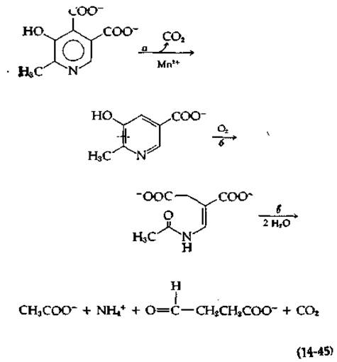
Another example of aromatic compound degradation deserves mention because it involves unusual enzymatic reactions—specifically, the bacterial degradation of Various Forms of Vitamin B6 [136]. In one of the pathways, the initial stages involve the oxidation of the hydroxymethyl group at the 5-position and the substituent group at the 4-position, converting them into carboxylate groups. Subsequently, as shown in equation (14-45), decarboxylation takes place through the action of an unusual dioxygenase.

FIG. 14-22. Several examples of aromatic compound catabolism in bacteria.
This dioxygenase, isolated from a Pseudomonas strain, contains bound FAD, which must be reduced by external NADH. Like a typical dioxygenase, the enzyme incorporates two oxygen atoms into the product. However, it also utilizes reduced FAD to reduce a double-bond system (either before or after attack by molecular oxygen). Another enzyme from the same bacteria is notable for hydrolytically cleaving the oxygenation reaction product into four different compounds without the accumulation of intermediates.
Last update: 06/08/2026
Editorial and Educational Adaptation: This material has been compiled based on the primary/original source text. The project team performed an editorial review, corrected technical inaccuracies, structured sections, and adapted the content for an educational format.
What was processed:
- elimination of formatting defects (OCR errors, structural breaks, corrupted characters);
- editorial organization of content;
- standardization of terminology in accordance with academic sources;
- verification of factual statements against the original source text.
All mentions of the author, publication year, and origin of the primary text have been preserved in accordance with the source.