Chemistry and Biology of Proteins - F. Haurowitz 1953
Electrochemistry of Proteins
Protein Ionization. Combination with Hydrogen Ions
Like Amino Acids, Proteins migrate toward the cathode in acidic solutions and toward the anode in alkaline solutions; similarly, they possess an isoelectric point at which no net migration occurs [9]. It is generally accepted that proteins at their isoelectric point exist as multivalent zwitterions, differing from simple amino acids primarily in the sheer multiplicity of their anionic and cationic groups.
However, this view was not accepted without objection. The point is that proteins exhibit minimal solubility at their isoelectric point [10], whereas it is well established that the solubility of organic acids and bases increases with their ionization. This rule, however, does not hold for ampholytes. The precipitation of proteins at their isoelectric point is driven by electrostatic forces of mutual attraction between positively and negatively charged groups of adjacent zwitterions. This explanation finds considerable support when considering the solubilizing effect of neutral salts added to protein solutions. Due to their electrostatic action on the ionized groups located On the surface of the protein particle, neutral salt ions prevent their mutual aggregation [11].
The amphoteric Nature of the ionized groups in an isoelectric protein is also indicated by the magnitude of the heat of neutralization (by strong acids and bases). The heat of neutralization of proteins can be determined calorimetrically [12] or calculated from the Temperature coefficient of the ionization constant [13]. Both Methods yield a value of approximately +10,000 cal per gram-ion of hydrogen when an acid is added to a weakly alkaline protein solution (pH approximately 8–9). This order of magnitude is comparable to that obtained upon The addition of strong acids (hydrogen ions) to organic bases, but is significantly higher than the heat of ionization [14] for the reaction
Class="center">
From these data, it is evident that the reaction taking place at pH 8–9 does not correspond to the reaction given above, but proceeds According to the equation:

The most compelling proof of the zwitterionic Structure of isoelectric proteins is provided by a comparison of data obtained from electrometric titration in the presence and absence of formaldehyde. Formaldehyde, as we know (see Chapter VII), reacts with the amino groups of proteins, depriving them of their basic properties. When amino acids or proteins are titrated with sodium hydroxide in the presence of formaldehyde, the typical inflection point in The titration curve at pH 9—which corresponds to The conversion of  into NH2—disappears (Figs. 10 and 11). This serves as decisive proof that the buffering action of proteins in this pH range is due to their amino groups rather than The ionization of carboxyl groups [15]. The exact same result is obtained if Amino groups are converted into hydroxyl groups by the action of nitrous acid [16]. The addition of ethanol also alters the course of the electrometric titration curve, noticeably decreasing the dissociation constant of the carboxyl groups, while the dissociation constant of the ammonium groups is only slightly diminished [17].
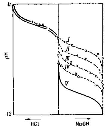
Fig. 10. Titration curve of Glycine in the presence of increasing concentrations of formaldehyde [18].
I — 2% HCHO; II — 80% HCHO; III — 20% HCHO; IV — 0.5% HCHO; V — Water.
Although the curves obtained from the electrometric titration of proteins resemble those of amino acids, certain essential differences remain between these processes. The ionization of monoaminocarboxylic acids is caused by the electrolytic dissociation of the grouping
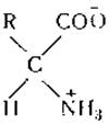
that is, the dissociation of the a-amino group and the adjacent carboxyl group. Because of the close proximity of these two groups, electrostatic interaction is inevitably set up both between the groups themselves and between them and nearby hydrogen and hydroxyl ions. A positively charged hydrogen ion attracted by the negative carboxyl group will be repelled by the neighboring positively charged ammonium group. Conversely, the negative charge of the carboxyl group is effectively dampened by the close proximity of the positive ammonium group. Consequently, the electrostatic interaction between these groups is highly complex and difficult to predict. However, it can be seen from Table 6 that the a-carboxyl groups of monoamino acids (pK approx. 2.2) possess stronger acidic properties than the carboxyl groups of the corresponding Fatty acids (pK approx. 4.8), whereas the amino groups of these acids (pK approx. 9.8) have weaker basic properties than the amino groups of typical organic amines (pK approx. 10.6).
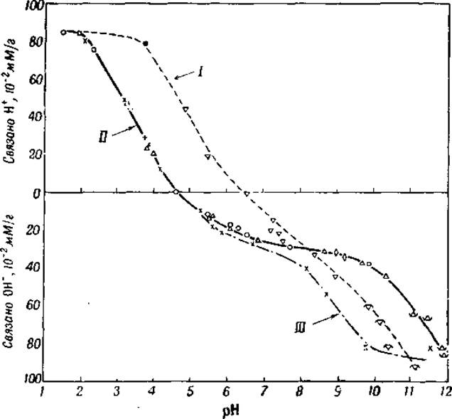
Fig. 11. Electrometric titration of egg albumin in water, 80% ethyl alcohol, and 1% formaldehyde solution [15].
I — ethyl alcohol; II — water; III — formaldehyde.
Proteins possess essentially the same ionic groups as amino acids. However, in a protein molecule, the majority of a-amino groups and a-carboxyl groups are linked together by peptide bonds. The acidic groups of proteins are represented primarily by the free carboxyl groups of aspartic and glutamic acids, the ionization of which corresponds to pK values of 3.87 and 4.28 (Table 7). The basic groups of proteins include the guanidino groups of Arginine (pK 12.48) and the ε-amino groups of Lysine (pK 10.53). The phenolic hydroxyl groups of Tyrosine and The sulfhydryl groups of Cysteine release their protons in approximately the same pH range (pK around 10), whereas the imidazole groups of Histidine are titrated near pH 6 (Table 7).
Due to the buffering action of their carboxyl and amino groups, electrometric titration curves of proteins exhibit distinct inflection points at pH 3–4 and 10–12. However, it is not possible by means of electrometric titration to differentiate the small number of terminal a-carboxyl groups of proteins from the β- and γ-carboxyl groups of aspartic and glutamic acids, nor to distinguish terminal a-amino groups from the ε-amino groups of lysine. Titration data allow for only one Conclusion: the number of terminal a-carboxyl groups cannot be very large (see Chapter VII), otherwise the inflection point would be shifted from pH 3–4 closer to pH 2. The inflection near pH 6–7, which is noticeable on many titration curves, corresponds to the buffering action of the histidine imidazole groups (see Fig. 11).
Electrometric titration is performed by adding strong acids (usually Hydrochloric acid) or strong bases (such as sodium hydroxide) to an isoelectric protein solution. When hydrochloric acid is added to an isoelectric protein solution, a portion of the hydrogen ions (protons) is bound by the COO- groups of the protein, converting them into carboxyl groups (see equation 16); on the other hand, some of the few NH2 groups present in the isoelectric protein bind protons and are converted into positively charged ammonium groups  . The maximum acid binding is determined from the titration curve by the distance between the horizontal line corresponding to the isoelectric point and the horizontal plateau of the titration curve at pH 1–2. Maximum base binding is calculated in a similar manner from the electrometric titration curve of the isoelectric protein with sodium hydroxide. However, accurately determining The amount of bound base is difficult because the endpoint in this case is less sharp than in the titration of acid-binding groups [19, 20]. Furthermore, it should be borne in mind that the hydroxyl ions introduced as a base during electrometric titration are not actually bound by the protein; rather, they combine with protons released either by the positively charged guanidino groups of arginine or ammonium groups of the protein

or by the uncharged hydroxyl groups of tyrosine [21] or sulfhydryl groups of cysteine

Clearly, the process here does not involve actual base binding, but rather The transfer of protons from the acidic groups of the protein to the hydroxyl ions.
For many proteins, electrometric titration results show remarkably good agreement with chemical determinations of Amino Acid Composition. For instance, the total number of anionic and cationic groups determined electrometrically in egg albumin, lactalbumin [19], and serum albumin [22] almost precisely matched the number of acidic and basic amino acids found via corresponding chemical analyses. In other proteins, however, such as Insulin, a significant excess of groups titratable in the alkaline pH range was discovered [22], which is attributed to the presence of A large number of terminal a-amino groups in insulin.
An isoelectric protein contains equal numbers of positively and negatively charged groups; consequently, its net free charge is zero, and no migration toward either pole occurs when an electric field is applied. Nevertheless, an isoelectric protein does not represent a truly homogeneous substance, but rather a mixture of particles possessing a slight excess of either positive or negative charges. For example, in a solution of isoelectric Hemoglobin at pH 6.9, only 22.4% of the hemoglobin molecules are precisely isoelectric (net charge equal to zero), whereas molecules bearing 1, 2, 3, or more positive charges account for 17, 9.4, 3.9, and 1.5%, respectively, and molecules bearing 1, 2, 3, or more negative charges amount to 21.2, 14.2, 3.0, and 2.5%, respectively [23].
The isoelectric point of a protein depends on the number and ionization constants of its ionized groups. Because the dissociation of each ionizable group is influenced by the electrostatic effects of neighboring ionic groups, there is no strictly constant relationship between The ratio of the maximum number of accepted protons to the maximum number of donated protons, on the one hand, and the isoelectric point of the protein, on the other. This is evident from Table 8, where I represents the isoelectric point (the pH of the electrodialyzed protein solution), a is the maximum number of protons accepted by 105 g of protein, and b is the maximum number of protons donated by 105 g of protein (expressed in gram-equivalents).
Table 8 Dependence of the isoionic point of a protein on its degree of ionization in solution
|
Protein |
1 |
а |
b |
c/b |
|
Egg albumin |
4,74 |
110 |
134 |
0,82 |
|
Serum albumin |
5,34 |
148 |
159 |
0,92 |
|
Hemoglobin |
6,97 |
156 |
127 |
1,23 |
The dependence of the isoelectric point on the ionization constants of dissociating groups becomes apparent from determinations of the isoelectric point in the presence of varying concentrations of ethyl alcohol. Because ethyl alcohol primarily decreases the dissociation constant of carboxyl groups (see the previous section), it shifts the isoelectric point toward higher pH values. In gelatin solutions containing 80% ethyl alcohol, the isoelectric point corresponds to pH 6.0, whereas In aqueous solutions it corresponds to pH 4.9 [15].
While interpreting the electrometric titration data of Amino Acids and simple Peptides is relatively straightforward, interpreting protein titration curves presents several difficulties arising from the following circumstances.
1. Proteins are multivalent zwitterions containing a large number of positively and negatively charged groups. Titration curves show that neutralizing 1 g of protein requires approximately 1 mM of acid or base. Since the Molecular Weight of a protein is on the order of 100,000, each protein molecule bears roughly 100 negative and 100 positive groups. These figures agree quite well with analytical data determining the amounts of aminodicarboxylic acids, ammonia, and basic amino acids in the protein molecule. However, accurately establishing the exact number of ionized groups in the molecule is difficult due to some degree of overlap on the titration curve, particularly in the alkaline pH range between 8 and 12; pH 8.5 is conventionally, and somewhat arbitrarily, chosen as the endpoint for neutralizing basic groups [24].
2. Proteins are labile in the presence of acids and bases. Certain proteins undergo Denaturation at pH values below 2 or above 11, making precise determinations of native protein ionization impossible in these pH ranges. The rate of denaturation can be reduced by conducting measurements at low temperatures. If denaturation cannot be avoided, its effect can be partially eliminated by measuring the pH immediately after mixing the protein with the acid or base, and subsequently at defined time intervals. The neutralization of ionized groups occurs instantly, whereas denaturation typically takes longer. This approach allows the two reactions to be distinguished [25].
3. Proteins readily interact with ions other than hydrogen ions, notably calcium, magnesium, phosphate, and bicarbonate ions. The resulting bonds are so strong that these ions cannot be removed by dialysis or Electrophoresis, and only electrodialysis proves successful [26]. The high bond strength between protein molecules and calcium or phosphate ions is due to the strong electrostatic effect of divalent inorganic ions. Monovalent ions, such as potassium, sodium, or chloride, are bound less firmly; hence, sodium hydroxide, potassium hydroxide, and hydrochloric acid are used for electrometric titrations. Recently, however, it has been found that proteins also bind chloride ions [27]. Most likely, the latter are bound to the positively charged groups of the protein molecule.
During the adsorption of counterions, the ionized groups of the protein are neutralized, ultimately altering the electrochemical Properties of the protein [28]. For instance, it has been established that the isoelectric point of a protein depends on The Nature and concentration of ions present in solution [29, 30].
The shift in the isoelectric point is particularly pronounced when multivalent ions, such as phosphate ions, are present in the solution. This complicates The Use of salts with multivalent ions as buffers, which are commonly employed for this purpose within the pH range of 6 to 7. Michaelis [31] therefore proposed using barbiturate as a buffer for this pH range.
As demonstrated above, the isoelectric point of a protein depends on the presence of foreign ions and is therefore not constant. To designate the pH of a pure protein dissolved in water in the absence of salts, the term isoionic point is used [32]. However, determining the isoionic point also encounters many difficulties and is often impossible, as many proteins are insoluble in the absence of salts. Furthermore, the conductivity of salt-free protein solutions is extremely low. The isoionic point of sheep Blood carboxyhemoglobin lies at pH 7.6, whereas the isoelectric point of the same protein in an ammonium phosphate buffer ranges between 6.70 and 7.16 [33].
4. Finally, certain complications in interpreting protein electrometric titration data arise from interactions between the protein zwitterions themselves. Two or more protein molecules may form an aggregate (micelle) through The formation of salt-like linkages between the positively and negatively charged groups of adjacent molecules. As already mentioned, neutral salts inhibit the formation of insoluble protein precipitates. The same holds true for the formation of soluble aggregates. For example, serum globulin molecules form aggregates during electrodialysis when most of the salts have already been removed [35]. Alongside proteins, simple amino acids also exhibit a tendency to bind with one another and form similar salt-like compounds [35].
The difficulties listed above stem from the inherent complexity of Cell/13.html">Protein Structure. In addition to these challenges, any attempt to interpret electrometric titration and electrophoresis results must account for another general complicating factor. In most experiments, hydrogen ion concentrations are determined using standard electrodes. Such measurements do not yield the actual hydrogen ion concentration [H+], but rather their activity in conducting electricity, aH+, which equals [H+]y, where y is the activity coefficient. The results of electrometric titrations and determinations are typically referred to hydrogen ion activity, and the term pH is frequently replaced by pdH+. However, this is also a simplification, as hydrogen ion activity depends on the nature and concentration of other ions present in the solution. The true concentration and activity of hydrogen ions are indeterminable [36]. Nevertheless, the values obtained from standard determinations can serve comparative purposes and be considered close to, though not identical with, the true hydrogen ion activity [37, 38].
Last update: 06/08/2026
Editorial and Educational Adaptation: This material has been compiled based on the primary/original source text. The project team performed an editorial review, corrected technical inaccuracies, structured sections, and adapted the content for an educational format.
What was processed:
- elimination of formatting defects (OCR errors, structural breaks, corrupted characters);
- editorial organization of content;
- standardization of terminology in accordance with academic sources;
- verification of factual statements against the original source text.
All mentions of the author, publication year, and origin of the primary text have been preserved in accordance with the source.