Biochemistry and Molecular Biology - Belyasova, N. A. 2002
Molecular Foundations and Mechanisms of Heredity
Gene Expression
Regulation of Gene Expression at the Transcriptional Level
Every Cell of a whole Organism or population contains the full set of genes characteristic of a given species (strain). However, at any given time, not all genes function (are expressed) in a cell, but only those whose products are needed. This distribution of "duties" among genes is made possible by the existence of Gene Expression regulation mechanisms that operate at various levels. Through these mechanisms, The Cell conserves its resources: at any specific moment, it synthesizes a defined, limited set of substances rather than their entire potential spectrum, and furthermore, coordinates metabolic pathways.
Among the several levels of Introduction/30.html">Regulation of Gene Expression, the most essential and frequently utilized is the Regulation of enzyme synthesis, which takes place at the transcriptional level. The Essence of this type of regulation boils down to the acceleration or deceleration of METABOLISM/31.html">Transcription processes for specific genes, which ultimately affects the rate at which their products are synthesized. Positive and negative Regulation of transcription are distinguished. Negative regulation involves the inhibition of Transcription initiation through the binding of repressor Proteins to the operator region; positive regulation, conversely, encompasses events that "turn on" transcription, which are likewise brought about by the attachment of specific proteins to the operator (in this case termed activators).
The regulation of enzyme synthesis has been best studied in prokaryotes. Their characteristic feature is the Organization of genes involved in a single metabolic pathway into operons. This enables prokaryotes to "turn on" and "turn off" the transcription of a group of genes (comprising the Operon) simultaneously. Several Different types of transcription initiation regulation (operon regulation) will be examined in this topic using two E. coli operons as Examples: the lactose operon, belonging to the catabolic operon group, and the Tryptophan operon, which is anabolic.
The regulation of transcription initiation in eukaryotes is far more complex than in prokaryotes, yet its fundamental regularities still apply.
Regulation of lactose operon expression via induction. The E. coli lactose operon contains a regulatory region (promoter and operator) and three structural genes: lacZ (encoding The Structure of ß-galactosidase), lacY (determining the structure of ß-galactoside permease), and lacA (the structure of ß-galactoside transacetylase) (Fig. 3.5). These Enzymes mediate the uptake of the disaccharide lactose into the cell and its Cleavage into glucose and galactose. Transcription of the structural GENES OF THE lactose operon occurs in a coordinated manner: the lacZ, lacY, and lacA Genes are transcribed into a single polycistronic mRNA, which is translated to yield nearly equal amounts of each enzyme protein. Not far from the lactose operon on the E. coli chromosome lies the I gene, which encodes the STRUCTURE OF THE repressor protein. In its free state, this protein has an affinity for the operator region of the lac operon.
When Escherichia coli Cells are grown on a medium containing glucose as the sole carbon source, they contain very few proteins encoded by the structural genes of the lactose operon: approximately 10 molecules per cell. In the presence of lactose and other ß-galactosides, the concentration of these proteins rises to 10,000 or more molecules per cell. In this case, lactose (ß-galactosides) serves as an inducer for the synthesis of these enzymes, meaning that the corresponding operon is regulated by induction—that is, the enzymes it encodes are synthesized only in the presence of the inducer.
Class="center">
Fig. 3.5. The E. coli lactose operon (lac) and the closely linked lac repressor gene (lac I): P — promoter; O — operator
The Mechanism of induction is as follows. In the absence of the inducer, the free repressor protein (a tetrameric molecule) tightly binds to the operator region of the lac operon (Fig. 3.6). Because the promoter and operator sequences overlap, the binding of the repressor to the operator obstructs the attachment of RNA polymerase to the promoter, resulting in the blockage of structural gene transcription. However, the presence of lactose or another inducer of the lac operon within the cell leads to The formation of an inducer-repressor complex, which loses its affinity for the operator and frees the regulatory region (Fig. 3.6). Transcription of the structural genes proceeds, and the corresponding proteins are synthesized on the newly formed mRNA.
Thus, The phenomenon of induction allows the cell to conserve its resources, as it ensures that inducible genes are transcribed and corresponding enzymes are synthesized not continuously, but only when the inducer is present in the medium.
Mutations have been isolated in E. coli Bacteria that manifest as a decrease or complete loss of the repressor's affinity for the operator. In such mutants, constitutive synthesis of lactose operon enzymes is observed; that is, its expression occurs even in the absence of the inducer.
Regulation of enzyme synthesis by induction belongs to negative control. In addition, the functioning of the lactose operon and many other operons is subject to positive regulation, which can be examined using catabolite repression as an example.
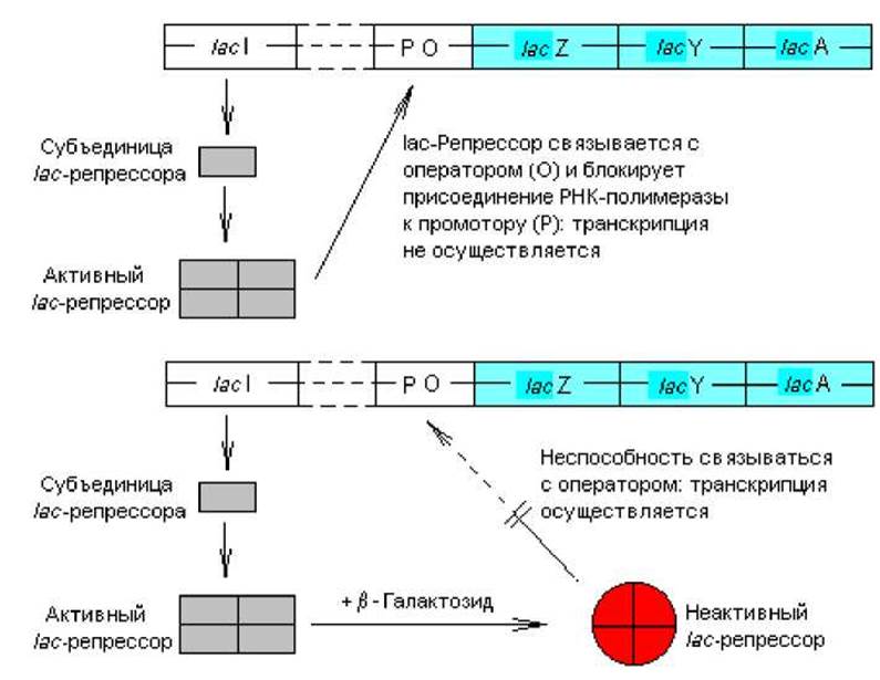
Fig. 3.6. Repression of the lac operon by the homotetrameric lac repressor (top) and Induction of the lac operon following binding of a ß-galactoside inducer to the repressor (bottom)
Regulation of the lactose operon via catabolite repression. Positive regulation of lactose operon transcription involves the binding of an activator complex to a specific sequence located at the very beginning of the lac promoter. This stimulates transcription of the lac operon, resulting in an almost 50-fold increase in The rate of corresponding mRNA synthesis. This phenomenon does not yet have a definitive explanation, but several hypotheses describe The process of transcription stimulation. According to the most widely accepted one, the activator complex binds to the part of the promoter that directly adjoins the RNA polymerase attachment site (Fig. 3.7) and enhances the enzyme's affinity for the promoter. An alternative hypothesis posits that the binding of the activator complex to the promoter prevents RNA polymerase from attaching to a nearby weak promoter, thereby increasing the probability of the enzyme binding to the "correct" promoter site.
In this case, the function of the transcription activator is performed by a complex of cyclic AMP (cAMP) and the catabolite activator protein (CAP). This complex performs analogous Functions in regulating the expression of many other catabolite operons. The free CAP protein is incapable of binding to the specific sequence within the promoter and requires the participation of cAMP.
cAMP is formed from ATP (Fig. 3.8) during enzymatic conversion in response to various cellular events and signals. This messenger molecule participates in numerous processes, mediating the regulation of various aspects of cellular metabolism. The intracellular level of cAMP is controlled by two balancing processes: synthesis involving adenylate cyclase (Fig. 3.8) and degradation mediated by phosphodiesterase. Glucose accelerates The breakdown of cAMP and inhibits its synthesis; consequently, in the presence of glucose, cellular cAMP levels are low, whereas in its absence, they are high.
Thus, the level of cAMP—and, accordingly, of the cAMP-CAP activator complex—depends on the presence of glucose in the cell. In bacteria growing on glucose, the concentration of these substances is very low; therefore, even in the presence of Inducers, transcription of the lactose and similar operons does not occur, and the cell fails to synthesize enzymes involved in the Catabolism of the corresponding sugars (lactose, arabinose, galactose, etc.). This phenomenon is termed "catabolite repression".
Regulation of the tryptophan operon via repression. The E. coli tryptophan operon contains five structural genes (trpE, trpD, trpC, trpB, trpA) that determine the Amino acid sequences of five enzymes involved in The conversion of chorismate to tryptophan. Additionally, the operon includes a leader segment (trpL) and a regulatory region (overlapping promoter and operator sequences) that participate in regulating the transcription of the structural genes (Fig. 3.9). At a separate site on the Escherichia coli nucleoid (at a sufficient distance from the trp operon) lies the trpR gene, which encodes the structure of the repressor protein.
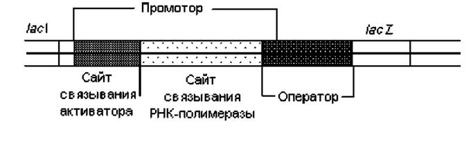
Fig. 3.7. Nucleotide sequences involved in regulating lactose operon expression
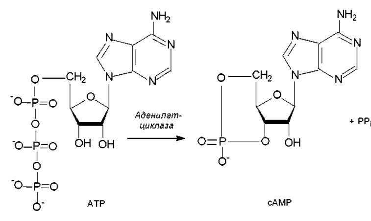
Fig. 3.8 Synthesis of cyclic AMP (cAMP) from ATP
The structural genes of the trp operon are transcribed as a polycistronic mRNA 7,000 NUCLEOTIDES in length. mRNA synthesis is initiated at a promoter whose sequence overlaps with the operator (Fig. 3.9). Transcription is controlled by the interaction of the repressor protein with the operator, utilizing the end product of this biosynthetic pathway—tryptophan—as its effector. When free tryptophan is present in the cell, it binds to the repressor and exerts an allosteric effect on the latter's structure, enabling the repressor to bind tightly to the operator. In this context, tryptophan acts as a corepressor of the tryptophan operon.
The binding of the tryptophan-repressor complex to the operator region prevents the proper interaction of RNA polymerase with the promoter because the operator and promoter sequences overlap. Transcription of the trp operon is blocked, and the enzymes required for tryptophan synthesis are not produced.
In the absence of tryptophan, free repressor protein does not bind to the operator, and operon transcription proceeds unhindered. Under these conditions, there is an intensive Synthesis of the five enzymes that convert chorismate into tryptophan. Due to regulatory mechanisms, the intracellular concentration of these enzymes in E. coli cells can vary up to 700-fold depending on the intracellular tryptophan level.

Fig. 3.9. The E. coli tryptophan operon (trp operon). Products of structural genes: a1 — anthranilate synthase, component I; a2 — anthranilate synthase, component II — phosphoribosylanthranilate transferase; pr — phosphoribosylanthranilate isomerase-indoleglycerol phosphate synthase; t1 — tryptophan synthase ß; t2 — tryptophan synthase a. Intermediates: PR — phosphoribosyl; CdRP — carboxyphenylamino deoxyribulose phosphate; InGP — indoleglycerol phosphate; PRPP — 5-phosphoribosyl-1-pyrophosphate
Repression also regulates the operation of other operons, particularly those involved in The Biosynthesis of Other Amino Acids. Some of these operons feature an additional regulatory mechanism known as attenuation.
Attenuation of tryptophan operon expression. This regulatory mechanism couples two processes: transcription and Translation. It involves a regulatory DNA segment located upstream of the structural gene trpE. This so-called trpL leader segment contains an attenuator sequence approximately 145 nucleotide pairs in length. When free tryptophan is present in the cell, transcription of the attenuator sequence is initiated but prematurely terminated, releasing a short trp leader mRNA (145 nucleotides). In the absence of tryptophan, premature termination does not occur, and full-length tryptophan mRNA is transcribed. Thus, the attenuator sequence contains a signal that regulates the transcription of structural genes.
Sequence Analysis of the attenuator sequence revealed three unusual segments within it: one encodes a short polypeptide consisting of 14 amino acids, including two adjacent tryptophan residues; the other two segments contain inverted repeats capable of forming various hairpin structures through complementary nucleotide pairing. One of these two hairpin structures does not interfere with transcription, whereas the other mediates rho-independent premature termination of transcription. Which of the two hairpin structures forms depends on the cellular tryptophan concentration: in its absence (or at low levels), the short peptide containing the two tandem tryptophan repeats cannot be fully synthesized, which serves as a signal for the Formation of the transcription-permissive hairpin. When tryptophan levels are moderate to high, the short peptide encoded by the attenuator sequence is successfully synthesized; in this case, Ribosomes reach the stop codon in the leader mRNA, triggering the formation of a hairpin structure that halts further transcription. Thus, attenuation of premature transcription termination occurs when tryptophan is scarce or virtually absent in the cell, with the specific position of the ribosome on the leader mRNA serving as the key signal.
Expression of the tryptophan operon, as well as certain other biosynthetic operons (such as Histidine, Threonine, and isoleucine-valine operons), reaches its maximum when repression is absent and transcription termination attenuation is at its minimum.
Last update: 06/08/2026
Editorial and Educational Adaptation: This material has been compiled based on the primary/original source text. The project team performed an editorial review, corrected technical inaccuracies, structured sections, and adapted the content for an educational format.
What was processed:
- elimination of formatting defects (OCR errors, structural breaks, corrupted characters);
- editorial organization of content;
- standardization of terminology in accordance with academic sources;
- verification of factual statements against the original source text.
All mentions of the author, publication year, and origin of the primary text have been preserved in accordance with the source.