INVERTEBRATE ZOOLOGY IN THREE VOLUMES - VOLUME 3 - H.Y. Shcherbak - 1997
PHYLUM MOLLUSCA (MOLLUSCS)
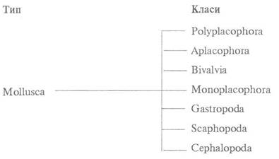
CLASS POLYPLACOPHORA, OR LORICATA (CHITONS)
This Class comprises exclusively marine animals that inhabit primarily the littoral zone, with only a few species descending to greater depths. They are sluggish, predominantly herbivorous animals that attach themselves to rocks and hard substrates using a muscular FOOT. About 1,000 species are known worldwide, 3 of which occur in the Black Sea.
Chitons are mostly small animals. The body length of the smallest species barely reaches 1 cm, while that of the largest ranges from 30 to 40 cm, with a weight of several kilograms. The dorsal surface is typically colored in ochre-yellow or brown tones, adorned with spots of all colors of the rainbow.
The body shape of chitons is usually elongated-oval, dorsoventrally flattened. The body consists of a HEAD, trunk, and foot. Most of the dorsal side is covered by a shell composed of eight separate plates movably articulated with one another (Fig. 11). Along the periphery of the dorsal side, free from the shell, lies the marginal zone of the mantle, known as the perinotum.

Fig. 11. External Anatomy of the chiton Tonicella marmorea:
a, b — dorsal and ventral views; 1 — first shell plate; 2 — marginal zone of the mantle; 3, 4 — fourth and eighth shell plates; 5 — head; 6 — Mouth; 7 — mantle; 8 — mantle furrow; 9, 10, 11 — genital, excretory, and anal openings, respectively; 12 — osphradium; 13 — gills; 14 — foot
The anterior part of the ventral side is occupied by the head bearing the mouth opening, separated from the foot by a transverse furrow. The head lacks the specialized Sense Organs characteristic of other Molluscs. The foot occupies the greater part of the ventral surface and features a large, flat sole. The head and foot are bordered by a wide muscular fold known as the mantle. Between the head/foot and the mantle lies a deep mantle furrow (mantle cavity), which surrounds not only the foot but also the head of the mollusc. The mantle furrow houses the mantle complex of organs: gills (ctenidia), osphradia, the anus, and paired genital and excretory openings.
The chiton shell, as noted previously, consists of eight overlapping plates arranged in a single series such that the posterior edge of each plate overlaps the anterior edge of the next. Each plate is composed of four layers: an outermost, very thin conchiolin periostracum; beneath it, a pigmented layer called the tegmentum; further inward, a thick, unpigmented articulamentum; and the innermost, nacreous hypostracum. The tegmentum is specific to chitons; it consists largely of a Chitin-like substance and represents a thickened continuation of the perinotum cuticle that wraps around the shell margins. It is formed by the deposition of cuticular material On the surface of the articulamentum, which itself is composed primarily of calcium carbonate. The tegmentum does not completely cover the articulamentum. On most plates, the lower layer projects anteriorly and laterally as flat, wing-like processes. The anterior processes are covered by the posterior margins of the preceding plates, while the lateral ones are covered by the folds of the marginal mantle zone.
Numerous pores are scattered across the shell surface, continuing into branching canalicles that pierce the entire thickness of the shell and reach the dorsal epithelium. These canalicles contain aesthetes, which are Sensory Organs unique to chitons (see below).
In some species, the shell may be reduced. For instance, in Cryptoplax, the plates are diminished and spaced apart, giving the animal a worm-like appearance due to its narrowed and elongated body. In other forms, such as Cryptochiton, the mantle completely grows over the shell, making it invisible from the exterior.
The Skin (integument) of chitons consists of an epithelium covered with a cuticle, and a Connective Tissue layer; its Structure varies across different Regions of the body. The foot sole, mantle cavity, gills, and the dorsal body surface beneath the shell are covered by a standard epithelium with a thin cuticle; the epithelial Cells on the gill lamellae are partially ciliated. Among the epithelial cells are glandular mucous cells, which are particularly abundant on the SOLE OF THE foot.
The skin of the marginal mantle zone (the perinotum) has a unique structure. It is covered with a thick layer of cuticle containing numerous calcareous spicules and chitinous needles on the dorsal side, as well as sharp-edged calcareous plates on the ventral side.
The connective tissue of the skin is fibrous in structure and densely penetrated by numerous Muscle fibers.
In addition to cutaneous Muscles, chitons possess a well-developed musculature associated with the shell plates: longitudinal, oblique, and transverse muscles connect adjacent plates. By contracting these shell muscles, a chiton can curl up ventrally when threatened, protecting its soft body parts. The robust musculature of the foot is thoroughly permeated by muscle fibers running in various directions: longitudinal, transverse, oblique, and dorsoventral (Fig. 12). The dorsal ends of the latter attach to the shell plates and, passing on either side of the Internal Organs, fan out across the foot. The foot serves as an organ of locomotion and substrate adhesion. The mantle margins are also penetrated by muscle fibers in all directions, enabling the mantle to fit tightly against the microscopic irregularities of the substrate when the animal adheres. There is also specialized musculature associated with the Oral Cavity, Pharynx, radula, etc.

Fig. 12. Anatomy of the musculature, circulatory, and excretory systems of the chiton Tonicella marmorea (dorsal dissection; most viscera removed):
1 — head; 2 — mouth; 3 — aorta; 4 — oblique muscle; 5 — transverse muscle; 6 — straight muscle; 7 — gills; 8 — mantle; 9 — Pericardium; 10 — external genital opening; 11 — excretory opening; 12 — ventricle; 13 — rectum; 14 — osphradium; 15 — anus; 16 — internal opening of the Kidney into the pericardium; 17 — Ureter; 18, 19 — outer and inner loops of the kidney; 20 — foot
The coelom of chitons consists of two independent structures: the gonadal coelom, located in the middle of the body and forming the cavity of the Gonads, and the pericardial coelom, located closer to the posterior end of the body, which houses The Heart (Fig. 13). Each coelomic compartment has its own pair of coelomoducts; the coelomoducts of the gonadal coelom function as genital ducts, whereas those of the posterior (pericardial) coelom function as Kidneys. The large body cavity housing the viscera is a primary body cavity, or schizocoel. Parenchyma is prominently developed in the ventral body wall of the head, the marginal zone of the mantle, and particularly within the foot.

Fig. 13. Diagram of the coelom and Reproductive System structure in Polyplacophora:
1 — mouth; 2 — gonad; 3 — kidney; 4 — genital duct; 5 — genital opening; 6 — internal opening of the kidney into the pericardium; 7 — excretory opening; 8 — pericardium; 9 — gills
The Digestive System begins with the mouth on the underside of the head and consists of the oral cavity, pharynx with accessory glands, Esophagus, Stomach with Liver, and the Midgut and Hindgut (Fig. 14). The pharynx possesses well-developed muscles. Rising from the floor of the pharyngeal cavity is a longitudinal muscular ridge, the Tongue. Its surface is covered by a thick cuticular layer bearing transverse rows of Teeth, known as the radula. The anterior end of the tongue with the radula can protrude from the pharynx, while its posterior end is embedded in a long radular sheath. At the bottom of this sheath lies a cluster of epithelial cells (odontoblasts) that secrete the horny radular teeth. The radula grows much like a fingernail: its anterior teeth gradually wear down, while new ones are continuously formed at the Base of the radular sheath and pushed forward onto The surface of the tongue. The radular teeth are extremely durable, containing iron in the form of magnetite. The tongue and radula are operated by a well-developed musculature, moving back and forth like a rasp to scrape Algae off rocks and stones.
Opening into the pharynx are a pair of small Salivary Glands and a pair of large, so-called sugar glands, the secretions of which help convert dietary starches into sugars. The pharynx leads into a narrow esophagus, which continues into the midgut. The anterior part of the midgut forms a sac-like expansion, The Stomach, into which the ducts of the large, bilobed liver open. The remainder of the midgut (the Small Intestine) is very long, forming several loops before transitioning into the ectodermal hindgut, which lies beneath the pericardium and terminates in the anus at the posterior end of the mantle groove.
The excretory system consists of a pair of relatively large kidneys (coelomoducts) located on either side of the body (see Figs. 12, 13). Each kidney is shaped like a U-shaped, highly branched tube with both ends directed posteriorly. One end terminates in a ciliated funnel (nephrostome) opening into the pericardium (forming the reno-pericardial aperture), while the other opens via an excretory pore on the floor of the mantle groove just in front of the anus.
The Circulatory system in chitons is open; it comprises the heart, Blood Vessels, lacunae, and sinuses (see Figs. 12, 14). The heart is situated on the DORSAL SIDE OF the body within the pericardium and consists of a single ventricle flanked by two auricles. The ventricle is an elongated muscular sac; the auricles also have muscular walls, though thinner than those of the ventricle. The auricles communicate with the ventricle through valved openings that prevent the backflow of blood. Posteriorly, the two auricles are connected to each other by a narrow channel. The posterior end of the ventricle is blindly closed, whereas its anterior end extends into an aorta that runs along the midline of the body toward the head, opening broadly into the cephalic sinus. The aorta gives rise to numerous branches (Arteries) that penetrate the gonads.

Fig. 14. Internal anatomy of a female Tonicella marmorea (dorsal dissection):
1 — pharynx; 2 — salivary gland; 3 — Diaphragm; 4 — pharyngeal retractor muscles; 5 — aorta; 6 — liver; 7 — gonadal arteries; 8 — Ovary; 9 — intestine; 10 — sectioned renal caeca; 11 — oviduct; 12 — auriculo-ventricular aperture; 13 — interauricular channel; 14 — ventricle; 15 — auricle; 16 — pericardium; 17 — body wall; 18 — stomach; 19 — sugar gland;
20 — esophagus
The rest of the circulatory system consists of narrow clefts in the connective tissue known as lacunae, alongside large, well-defined cavities called sinuses. From the cephalic sinus, blood flows to the intestine and liver, and then enters a system of lacunae that coalesce into three longitudinal pedal sinuses (Fig. 15). 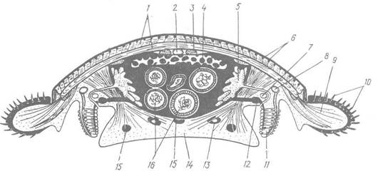
Fig. 15. Diagrammatic cross-section of a chiton:
1 — shell valve; 2 — aorta; 3 — longitudinal muscle; 4 — ovary; 5 — kidney; 6 — aesthete; 7 — pleurovisceral cord; 8 — afferent branchial vessel; 9 — perineotium muscles; 10 — cuticular spines; 11 — gills; 12 — efferent branchial vessel; 13 — pedal cord; 14 — foot; 15 — blood lacuna; 16 — intestine
From these sinuses, venous blood travels to the afferent branchial sinuses and, after being oxygenated in the gills, is carried via efferent sinuses to the auricles, which pump it back into the ventricle.
The respiratory organs of chitons are ctenidia (gills) located in a single row on each side of the mantle groove floor (see Fig. 11). Unlike most Mollusks, their number ranges from 4 to 80 pairs, with the single pair situated just posterior to the excretory organs being larger than the rest. Each ctenidium is bipectinate, consisting of a tapered central axis with gill filaments branching off on both sides. The surface of the filaments is covered by ciliated epithelium whose beating circulates Water around the gills, facilitating gas exchange.
The Nervous System of chitons comprises a nerve ring surrounding the anterior gut and two pairs of longitudinal nerve cords connected to it: the pedal cords and the pleurovisceral cords (Fig. 16). The supra-esophageal portion of the nerve ring is termed the cerebral arch (or Brain) and appears as a thick, ganglion-free connective. The sub-esophageal portion (sub-cerebral commissure) is significantly thinner and is associated with small paired buccal and sub-radular ganglia. The pedal cords run through the musculature of the foot, which they innervate, while the pleurovisceral cords extend through the trunk above the mantle
groove, fusing at the posterior end of the body just above the anus. The pedal cords are interconnected with each other and with the pleurovisceral cords by numerous irregularly arranged transverse commissures. The circum-esophageal ring and its associated ganglia innervate the head, oral cavity, and gut; the pleurovisceral cords supply the gills, mantle, body musculature, kidneys, and heart; and the pedal cords innervate the foot.
Sense organs in chitons are poorly developed, reflecting their sluggish, sedentary lifestyle. They lack true eyes, cephalic tentacles, and statocysts. Sensory capabilities are largely limited to Touch and chemoreception. The organs of chemical sense (smell) include a strip of sensory epithelium located on the floor of the mantle groove near the base of the gills, as well as small osphradia positioned on either side of the anus (see Fig. 11). The latter appear as groups of tall, pigmented sensory cells. The ORGAN OF TASTE is the so-called sub-radular organ, an invagination of the oral cavity lined with sensory cells.

Fig. 16. Central Nervous System of the chiton Acanthochiton discrepans:
1 — cerebral arch; 2 — buccal ganglion; 3 — nerves to Oral Cavity and pharynx; 4 — sub-radular ganglion; 5 — pedal cord; 6 — pedal commissures; 7 — pleurovisceral cord; 8 — commissure of pleurovisceral cords
The sense of touch is mediated by structures called aesthetes, which enable the animal to perceive water currents and the pressure of the water flowing over its body. Aesthetes are situated on the dorsal surface of the shell plates within canals of the tegmentum. There are two types of aesthetes: large megalaesthetes and numerous tiny micraesthetes that branch off from the megalaesthetes (Fig. 17). Each megalaesthete is surrounded by more than 20 micraesthetes. A cuticular cap forms over each aesthete, shielding the sensory and glandular cells beneath. The inner ends of the sensory cells continue as nerve fibers that run through the shell canals and connect directly to the pleurovisceral cords.

Fig. 17. Sectional detail through a chiton shell valve showing aesthetes:
1 — periostracum; 2 — micraesthete cap; 3 — megalaesthete cap; 4 — thread-like cells of megalaesthete; 5 — glandular Cell; 6 — processes of thread-like cells; 7 — nerve fibers; 8 — articulamentum; 9 — tegmentum; 10 — micraesthete cell
Chitons are dioecious animals, though females and males cannot be distinguished by external features alone. The gonad (ovary or Testis) is unpaired and lies along the body midline above the gut; paired ducts (oviducts or sperm ducts) lead from it and open via a pair of genital pores into the mantle groove. Copulatory organs are absent.
Chitons spawn directly into the water, releasing eggs singly or in gelatinous strings, while some species brood their eggs within the mantle cavity, which Functions as a brood chamber.
Cleavage is spiral, and Gastrulation occurs by invagination. In chitons, paired mesodermal bands are formed, but they never become segmented; eventually, they break down into individual cells that disperse throughout the primary body cavity. On either side of the gut, they form two clusters in which cavities appear; these clusters give rise to the pericardium and kidneys. The musculature and connective tissue (parenchyma) also develop from the mesoderm. The gonad separates later from two outgrowths of the pericardium, which subsequently fuse into a single unpaired gonad.
The egg hatches into a typical trochophore, similar to that of annelid worms. It possesses an apical plate with a tuft of cilia and a prototroch (Fig. 18). Subsequently, it acquires certain features characteristic of chitons: the primordia of eight shell plates appear on the prospective dorsal side—one developing anterior to the prototroch and seven posterior to it. On the ventral side, the foot primordium appears as a ciliated outgrowth; one or two pairs of ocelli form below the prototroch. The larva swims in the water Column for a short time, grows uniformly, and then settles to the bottom. The larval organs (prototroch, ocelli, etc.) degenerate; a longitudinal mantle furrow forms along the sides of the body, within which the gills develop. The body gradually acquires the characteristic shape of a chiton.
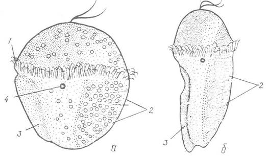
Fig. 18. Development of the chiton Ischnochiton magdalensis:
a — early metamorphosis trochophore stage; b — later stage; 1 — prototroch; 2 — shell plate primordium; 3 — foot primordium; 4 — ocellus
Most chitons inhabit shallow waters, and many species populate the intertidal zone. They adhere firmly to stones or rocks, with the foot and the ventral side of the mantle acting as a powerful sucker. If a wave does dislodge the mollusk, it rolls up into a ball, thereby protecting its soft ventral surface. Chitons use this same defensive mechanism against predators; there are only isolated records of them being found in the stomachs of fish and starfish.
Some chitons are used for food, for example, Chiton tuberculatus, Cryptochiton stelleri, and others; the muscular foot and, in some cases, the "roe" of these mollusks are consumed.
In the northern seas of Eastern Europe, the marbled chiton Tonicella marmorea, the white chiton Trachydermon albus, and Lepidopleurus arcticus are common. In the Far Eastern seas, the large Steller's sea cucumber (Cryptochiton stelleri), reaching up to 18 cm in length, is found; in this species, the mantle covers the entire dorsal surface, rendering the shell plates invisible.
Polyplacophorans cannot tolerate significant freshening and therefore mostly inhabit seas with oceanic salinity. Only three species are known in the Black Sea: the most common is Middendorffia caprearum, which lives at shallow depths under coastal stones and shell debris, along with Acanthochitona fascicularis and Lepidochitona cinerea, which settle on the surface of submerged stones and rocks.
Last update: 13/08/2026
Editorial and Educational Adaptation: This material has been compiled based on the primary/original source text. The project team performed an editorial review, corrected technical inaccuracies, structured sections, and adapted the content for an educational format.
What was processed:
- elimination of formatting defects (OCR errors, structural breaks, corrupted characters);
- editorial organization of content;
- standardization of terminology in accordance with academic sources;
- verification of factual statements against the original source text.
All mentions of the author, publication year, and origin of the primary text have been preserved in accordance with the source.