INVERTEBRATE ZOOLOGY IN THREE BOOKS - BOOK 3 - H.I. Shcherbak - 1997
PHYLUM MOLLUSCA
CLASS APLACOPHORA (OR SOLENOGASTRES)
This Class includes exclusively marine animals distributed across all the world's oceans from the Arctic to the Antarctic. They are benthic organisms living among hydrozoan and coral polyps, with some species burrowing into silt. They are found at various depths—from the intertidal zone to the greatest depths of the World Ocean (about 9,000 m). A total of about 150 species are known.
In appearance, aplacophorans differ significantly from other Molluscs. Consequently, for a long time, these animals were classified either as worms or as Echinoderms, and their molluscan affinities were established only through The Study of their internal anatomy.
Aplacophorans are predominantly small, worm-like organisms, ranging from 0.3 to 3 cm in length, with the largest reaching up to 30 cm. Their bilaterally symmetrical body is cylindrical, greatly elongated, and lacks the characteristic molluscan division into HEAD, trunk, and FOOT. In some species, the body is laterally compressed and features a small keel on the dorsal side (Fig. 19). On the ventral side, in place of the foot, part of the aplacophorans (subclass Solenogastres) have a longitudinal groove lined with ciliated epithelium; it begins at the anterior end of the body, just behind the Mouth in a pit, and extends to the posterior end (hence their name, Solenogastres, meaning furrow-bellied). Within the groove lies a narrow keel, also covered with ciliated epithelium, which represents a modified foot. In others (subclass Caudofoveata), the groove is shortened, restricted to the posterior end of the body, or entirely absent.

Fig. 19. Aplacophora:
a — Chaetoderma nitidulum; b — Neomenia carinata; c, d — Rhopalomenia aglaopheniae and Myzomenia sp., respectively, on hydrozoan colonies; 1 — gills; 2 — mouth; 3 — ventral groove
Externally, the body of these molluscs is covered by a cuticle containing numerous calcareous spines and scales (Fig. 20). Some are sharp, resembling an awl or a knife blade, while others are blunt, shaped like a spatula or a spoon. They are typically directed backward, imparting a silky gloss to the body surface. Structurally, the Skin of aplacophorans resembles the girdle (perinotum) of chitons.
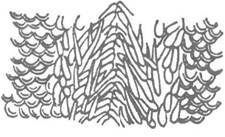
Fig. 20. Calcareous spines and scales of the aplacophoran cuticle
The musculature is represented by a well-developed cutaneous-muscular sac consisting of circular, oblique, and longitudinal Muscles, In addition to specialized Muscles of the Pharynx and cloaca. Aplacophorans are more active than chitons; they crawl among the branches of cnidarian colonies, or less frequently burrow into silt, relying on their epidermal spines for traction.
The coelom in aplacophorans is a single cavity consisting of two parts: an anterior reproductive part and a posterior pericardial part (Fig. 21). Gametes develop within the walls of the coelomic reproductive section (paired or unpaired), which thus Functions as a gonad. The posterior (unpaired) part of the coelom surrounds the hindgut and The Heart; a pair of coelomoducts extends from the coelom, which do not serve an excretory function but act solely as gonoducts for gamete release.
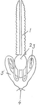
Fig. 21. Diagram of the coelomic Structure of Rhopalomenia acuminata:
1 — reproductive section of the coelom; 2 — Pericardium; 3 — seminal receptacle; 4 — cloacal opening; 5 — coelomoduct
Aplacophorans feed primarily on animal matter: species inhabiting hydrozoan and alcyonacean colonies graze on the polyps and coenenchyme; benthic-dwelling species (detritivores and microphages) feed on foraminiferans, radiolarians, and small crustaceans. Their gut is short, appearing as a straight tube with a poorly developed radula that directly overlies the pharyngeal cuticle rather than resting on a Tongue as in other molluscs. In some species, the radula is absent. The digestive gland (Liver) is also weakly differentiated and mostly appears as numerous lateral, symmetrically arranged pockets—outgrowths of the midgut lined with secretory epithelium. The hindgut opens into the cloaca, which also contains one or two genital openings.
Excretory Organs are absent in aplacophorans; their function is performed by the intestine.
The Circulatory system OF aplacophorans is open and has a very simple structure. True Blood Vessels are absent, and blood circulates through sinuses and lacunae. There are two longitudinal sinuses: dorsal and ventral. A heart forms from the posterior part of the dorsal sinus (Fig. 22); auricles are absent. Due to this rudimentary circulatory architecture, Blood Circulation is very weak.
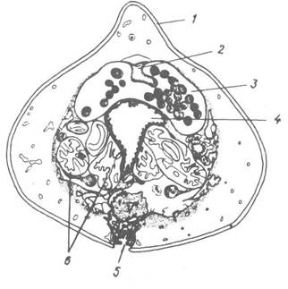
Fig. 22. Cross-section through the posterior region of Neomenia:
1 — cuticle; 2 — heart; 3 — pericardium with ova; 4 — intestine; 5 — ventral groove; 6 — loops of the oviduct
Respiration in most aplacophorans occurs through the body wall, primarily via the ventral surface. In some species, skin folds form within the cloaca and function as gills, while Representatives of the family Chaetodermatidae even possess a pair of small ctenidia.
The Nervous system of aplacophorans shares a structural type similar to that of chitons, but unlike the latter, solenogastres possess well-developed cerebral ganglia. The remainder of the Central Nervous System consists of two pairs of nerves
trunks: pedal and pleurovisceral, interconnected by metamerically arranged commissures (Fig. 23). Nerve Cells along the trunks are not evenly distributed; instead, they concentrate near the origins of the commissures, forming ganglion-like structures, while the intervening sections of the trunks contain only scattered nerve cells. However, this does not yet represent true complete ganglionization.
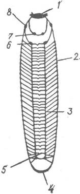
Fig. 23. Central nervous system of solenogasters:
1 — cerebral ganglion; 2 — pleurovisceral trunk; 3 — pedal trunk; 4 — posterior commissure of the pedal and pleurovisceral trunks; 5 — pedal commissures; 6 — anterior pedal ganglion; 7 — sublingual ganglion; 8 — lateral ganglion
Specialized Sense Organs are absent, though sensory papillae are scattered across the body, alongside isolated sensory cells within the cutaneous epithelium. Recently, a balancing organ—the statocyst—has been discovered in certain species.
Most aplacophorans are hermaphroditic, although gonochoric (separate-sex) species also occur, such as those belonging to the genus Chaetoderma. The Gonads may be paired or single and unpaired; the efferent ducts (oviducts or sperm ducts) are invariably paired, though their terminal sections may fuse in some species. These ducts open into a cloaca at the posterior end of the body. Copulatory organs are absent. Fertilization is external.
Eggs are spawned into the Water; in some forms (such as the genus Halomenia), they are brooded within the cloacal cavity. Cleavage is typically spiral. The egg hatches into a trochophore larva featuring an apical tuft, a prototroch, and a telotroch. A ventral furrow forms on the larval ventral side, within which a narrow keel (the foot) subsequently develops. The larva grows uniformly, elongates, and gradually acquires a worm-like shape. In most studied species of the genera Nematomenia and Neomenia, metamorphosis is accompanied by the degeneration of the larval skin epithelium and The formation of a new one. In certain species, following the reduction of the ciliated larval coat, seven calcareous plates develop on the DORSAL SIDE OF the larva. These plates overlap like roof tiles and bear a striking resemblance to the shell Valves of chitons (Fig. 24). Later, these plates are shed and replaced by the small spines and scales characteristic of adult animals.

Fig. 24. Metamorphosis in aplacophorans:
a, b, c — trochophore and late larval stages of Epimenia verrucosa; d — larva of Myzomenia; 1 — prototroch; 2 — mouth; 3 — ventral furrow; 4 — telotroch; 5 — remnants of the episphere; 6 — calcareous plates
The class Aplacophora is divided into two subclasses: the more species-rich Caudofoveata and the solenogasters proper (Solenogastres). In Solenogastres, the ventral furrow is well-developed, whereas in Caudofoveata it is either absent or small and restricted to the posterior end of the body. There are also certain internal Anatomical Features that distinguish these two subclasses.
Aplacophorans are marine inhabitants adapted to full oceanic salinity and cannot tolerate significant freshening, which explains their absence in the Black and Azov Seas.
In northern seas at depths of 30–80 m, the species Chaetoderma nitidulum (see Fig. 19, a) is widely distributed, reaching up to 8 cm in length; deeper-water species, such as Proneomenia sluiteri (growing up to 14 cm long) and P. thulensis (2 cm long), are found at depths ranging from 100 to 150 m.
Last update: 13/08/2026
Editorial and Educational Adaptation: This material has been compiled based on the primary/original source text. The project team performed an editorial review, corrected technical inaccuracies, structured sections, and adapted the content for an educational format.
What was processed:
- elimination of formatting defects (OCR errors, structural breaks, corrupted characters);
- editorial organization of content;
- standardization of terminology in accordance with academic sources;
- verification of factual statements against the original source text.
All mentions of the author, publication year, and origin of the primary text have been preserved in accordance with the source.