INVERTEBRATE ZOOLOGY - H. I. Shcherbak - 2008
KINGDOM MULTICELLULAR ANIMALS (METAZOA)
SUBKINGDOM TRUE MULTICELLULAR ANIMALS (EUMETAZOA)
SECTION TRIPLOBLASTIC (TRIPLOBLASTICA) OR BILATERAL (BILATERIA) ANIMALS
SUBSECTION SPIRALIA (SPIRALIA)
PHYLUM FLATWORMS (PLATHELMINTHES)
SUBPHYLUM TURBELLARIANS (TURBELLARIA) OR ARCHIDERMATA
This subphylum comprises about 3,000 predominantly free-living species distributed in marine environments, freshwater habitats, and occasionally in damp soil. Some species form symbiotic relationships with Echinoderms, crustaceans, Annelids, etc.; a small number of species are parasitic. Over 200 species inhabit the fresh and saline waters of Ukraine.
Marine turbellarians are often brightly colored, ranging in length from a few millimeters to 5–6 cm, while certain tropical terrestrial species can reach up to 60 cm (for instance, Representatives of the genus Bipalium).
Morphology. The body shape can be leaf-like, ribbon-like, etc. The Skin-muscular sac is characterized by the presence of a ciliated epithelium, the beating of cilia enabling turbellarians to swim. In most turbellarians, the epithelial Cells are cylindrical, with their basal ends resting on a structureless basal membrane (Fig. 109).
Class="center">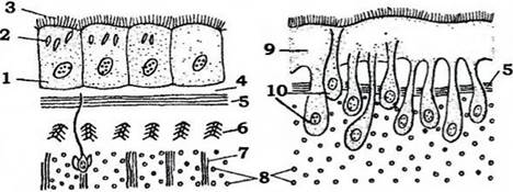
Fig. 109. Diagram of the skin-muscular sac Structure in turbellarians (adapted from Ivanov):
a, b - with typical ciliated and sunken epithelium, respectively:
1 - epithelial cells; 2 - rhabdites; 3 - cilia; 4 - basal membrane; 5 - circular Muscles; 6 - diagonal muscles; 7 - dorsoventral muscles;
8 - longitudinal muscles; 9 - cytoplasmic plate; 10 - sunken cytoplasmic regions containing nuclei
The tegument contains numerous mucous glands; mucus lubricates the body, facilitating free locomotion across the substrate or suspension beneath the surface film of Water. Shiny rod-like structures—rhabdites—are located within the epidermal cells. Upon irritation, they discharge, swell in water, and transform into a sticky, toxic mass. Rhabdites function in defense and serve as a tool for capturing prey. In some species, the epithelium bears long spines or poison glands that perform a similar function.
The Muscles of the skin-muscular sac are typically arranged in three layers: circular, oblique (diagonal), and longitudinal. Their contraction allows turbellarians to crawl along the substrate surface or swim. Bundles of dorsoventral muscles extend between the dorsal and ventral body walls, ensuring body flattening.
The parenchyma, which fills the spaces between the skin-muscular sac and Internal Organs, consists of loosely arranged cells of various shapes (Fig. 107) with large intercellular spaces filled with fluid. It performs supportive, transport, and storage Functions. It houses mobile phagocytic cells—some acting defensively by engulfing Bacteria, while others (athrocytes) accumulate excretory products (excretory function).
Additionally, the parenchyma contains specialized cells known as neoblasts, which drive regeneration processes (replacing old cells with new ones or restoring lost body parts).
The structure of the Digestive System varies significantly: ranging from entirely unformed (in acoeloms, class Acoelomorpha) to highly complex, featuring a branched gut (Fig. 110). In most turbellarians, it consists of an ectodermal muscular Pharynx and an endodermal blind-ending midgut, which can be straight or branched.

Fig. 110. STRUCTURE OF THE digestive system in representatives of various turbellarian classes (adapted from Beklemishev, with modifications):
a - Neorhabdocoela; b, d - Seriata, c - Polycladida:
1 - oral opening; 2 - pharynx; 3 - midgut
The degree of gut branching depends to some extent on the body size of the turbellarian because the intestine, In addition to Digestion, transports nutrients to distant PARTS OF THE body; consequently, the larger the body, the more extensively branched the midgut becomes. Digestion is primarily extracellular, though phagocytosis of small food particles by midgut epithelial cells also plays an important role.
Indigested food remnants are expelled through the Mouth. Turbellarians are predominantly carnivorous, preying on hydras, various worms, crustaceans, insect larvae, and the like. Some species consume diatoms. Such species swallow small prey whole. Predators that hunt relatively large animals exhibit extracellular digestion.
The excretory system is protonephridial. There are one or two main canals opening to the exterior via excretory pores (Fig. 108). The excretory function is also performed by the aforementioned specialized parenchyma cells known as athrocytes, which accumulate Metabolic waste products. These cells either remain in the parenchyma or exit to the exterior through the canals of the protonephridial system.
The Nervous system is most primitive in Acoela turbellarians: a diffuse plexus of nerve fibers resembling an irregular net is located beneath the integumentary epithelium and connects with the cerebral ganglion—a cluster of Nerve Cells at the anterior end of the body surrounding the statocyst, an Organ of Equilibrium.
In most turbellarians, nerve cells are concentrated into several pairs of longitudinal cords connected by numerous transverse bridges, or commissures, and linked to the cerebral ganglion located near the anterior end of the body. This forms a more or less regular lattice known as an orthogon (Fig. 111). Further Evolution of the nervous system involves a reduction in the number of nerve cords (concentration) and their immersion into the parenchyma.
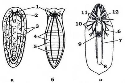
Fig. 111. Structure of the turbellarian nervous system (after Beklemishev, modified):
a - diffuse plexus with nerve cords in acoel turbellarians; b - orthogon in triclads; c - orthogon in rhabdocoels: 1 - cerebral ganglion; 2 - statocyst; 3 - longitudinal cords;
4 - transverse nerves; 5 - nerve strands of the diffuse plexus; 6 - dorsal cord; 7 - ventral cord;
8 - intestine; 9 - circumpharyngeal nerve plexus; 10 - Branches of the dorsal cord;
11 - sensory nerves extending from the Brain; 12 - eyes
Sense Organs are diverse. Visual organs—eyes—belong to the everted or inverted type (Fig. 112). Such an eye consists of a pigmented cup whose concave side faces the body surface. The cavity of the cup contains the expanded photosensitive parts of visual cells, whose Cell bodies lie in front of the cup's opening. Light rays must pass through The Cell bodies before reaching the photosensitive parts.
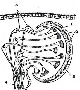
Fig. 112. Diagram of the turbellarian eye structure (after Dogel):
1 - photosensitive part of receptor cells; 2 - nuclei of pigment cup cells; 3 - pigment cup;
4 - nerve processes of receptor cells forming the Optic nerve; 5 - nuclei of receptor cells
In most other animals, eyes are non-inverted, meaning the photosensitive ends of the visual cells are directed toward the light source. Typically, turbellarians have a single pair of eyes situated above the cerebral ganglion, with which the processes of the visual cells connect. In some large species, there may be numerous eyes—up to several hundred—located on the anterior margin or the dorsal surface of the body.
The functions of mechano- and chemoreception are performed by single or clustered sensilla. Chemoreceptors are concentrated in auricular organs, which are pits at the anterior end of the body containing ordinary ciliated cells alongside chemoreceptive sensilla.
Many turbellarians possess specialized organs of equilibrium known as statocysts. They resemble a small vesicle containing one or more statoliths inside.
The Reproductive System is hermaphroditic, though structural details vary considerably across the subtypic group. Acoel turbellarians feature a diffuse hermaphroditic gland scattered throughout the parenchyma, which produces both ova and spermatozoa; distinct genital ducts are absent. Most turbellarians possess separate Ovaries and Testes (Fig. 113).
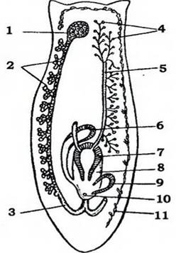
Fig. 113. Diagram of the turbellarian reproductive system (after Ioffe):
1 - Ovary; 2 - vitellaria; 3 - oviduct; 4 - testes;
5 - vas deferens; 6 - copulatory bursa; 7 - copulatory organ; 8 - genital cloaca;
9 - glandular organ; 10 - genital pore; 11 - excretory system
The Male Reproductive System typically consists of numerous testes scattered throughout the parenchyma; vasa efferentia extend from them and unite into two vasa deferentia. The latter merge to form a single ejaculatory duct that penetrates the copulatory organ and opens into the genital cloaca.
In most turbellarians, the FEMALE REPRODUCTIVE SYSTEM consists of a pair of ovaries with oviducts extending from them. Many groups feature vitellaria (yolk glands), whose ducts empty into the oviducts. The oviducts merge to form a Vagina that opens into the genital atrium. In some species, a copulatory bursa containing a seminal receptacle also empties here, receiving the partner's sperm after mating.
Reproduction. Fertilization is internal and typically cross-fertilization. Cleavage of the egg is spiral. Development is either direct or involves metamorphosis. In many marine polyclads (class Polycladida), a ciliated Müllerian larva hatches from the egg (Fig. 114). In the middle of its sac-like body are eight lobes with long cilia that help it swim in the water Column. The larva possesses a tuft of sensory cilia at its apical pole, a pair of eyes, and a mouth leading to a sac-like gut.

Fig. 114. Müllerian larva (adapted from Joffe):
1 - cerebral ganglion; 2 - sac-like gut; 3 - mouth; 4 - perioral lobes; 5 — eyes
The larvae lead a planktonic lifestyle, and after some time, they settle to the bottom and metamorphose into juvenile worms.
Turbellarians possess a high capacity for regeneration. This phenomenon underlies asexual reproduction, which occurs via transverse fission (for example, in many freshwater macropharyngids).
The subphylum includes eight classes grouped into two superclasses. Let us examine some of them.
Last update: 13/08/2026
Editorial and Educational Adaptation: This material has been compiled based on the primary/original source text. The project team performed an editorial review, corrected technical inaccuracies, structured sections, and adapted the content for an educational format.
What was processed:
- elimination of formatting defects (OCR errors, structural breaks, corrupted characters);
- editorial organization of content;
- standardization of terminology in accordance with academic sources;
- verification of factual statements against the original source text.
All mentions of the author, publication year, and origin of the primary text have been preserved in accordance with the source.