Practical Course in Zoology: A Study Guide - T. A. Dauda 2014
Multicellular Animals
Molluscs
Class Bivalvia, or Lamellibranchia
Swan mussel (Anodonta cygnea).
Materials and equipment. Various species of the genus Anodonta are common in freshwater bodies with muddy bottoms and slow-flowing or standing Water. The swan mussel is a typical representative of the Class Bivalvia and the primary object for studying this class.
Before examining the external Morphology or dissecting the animals, they are euthanized with a 1% chloral hydrate solution in which they should remain for about a day, or placed in water heated to 50°C. Specimens collected during summer field practice and fixed in 5-10% formalin are also suitable for study.
Required tools and materials: Microscope; hand lens; microscope slides and coverslips; dissection instruments (tray, forceps, scissors, scalpel, needles, pins); beakers of water, pipettes; freshly killed or formalin-fixed swan mussels; separate shell Valves; wet mount preparation of a swan mussel (internal anatomy).
Assignment. Perform an external examination and draw the shell. Open the shell and body of the swan mussel, examine, and draw the internal anatomy. Examine and draw the larvae of the swan mussel — glochidia.
Study of external anatomy. Take the swan mussel (Fig. 70) and examine The Structure of the bivalve shell enclosing its body. Hold the mussel over a dissection tray, as water may leak from the shell. Locate the ligament, which joins the two shell valves together along the dorsal side. The opposite side of the shell is the ventral side. Additionally, distinguish the anterior end, which is broader and more rounded, and the posterior end, which is narrower and pointed.
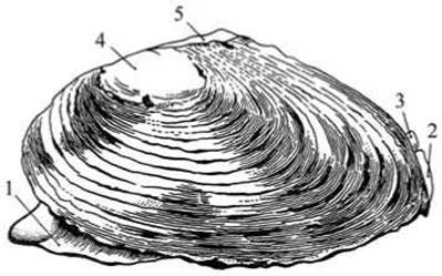
Fig. 70 External Anatomy of the swan mussel (Anodonta cygnea):
1 — FOOT; 2 — inhalient (branchial) siphon; 3 — exhalient (cloacal) siphon; 4 — umbo (the conchiolin layer is eroded, revealing the inner porcelain-like layer); 5 — ligament.

Fig. 71 Cross-section of the shell and mantle of the swan mussel (Anodonta cygnea):
1 — conchiolin layer of the shell; 2 — porcelain layer of the shell; 3 — nacreous layer; 4 — outer epithelium of the mantle; 5 — middle Connective Tissue layer of the mantle; 6 — inner epithelium of the mantle.
Examine the STRUCTURE OF THE swan mussel valve (Fig. 71). The outer layer has a brownish-greenish color and is formed by the organic substance conchiolin. Scraping it away reveals the middle, porcelain-white layer.
Examine the inner surface of the empty shell valves, which is covered with a nacreous (mother-of-pearl) layer. The porcelain and nacreous layers consist of calcium carbonate crystals. Note the curved lines on the shell surface running parallel to its anterior margin — the annual growth lines. The age of the mollusk can be determined by counting these lines.
Draw the shell of the swan mussel.

Fig. 72 Inner view of the swan mussel shell valve:
1 — cardinal hinge Teeth; 2 — lateral plate-like tooth; 3 — Muscle scars.
Opening the shell. Slightly pry open the shell valves using the handle of a scalpel. Look through the resulting gap to observe the inner surface of the valves, which is lined with a fold of Skin known as the mantle.
Insert the blunt end of the scalpel between the left valve and the mantle, and carefully separate the mantle from the shell. You will encounter resistance from two adductor Muscles located at the anterior and posterior ends of the shell, closer to the dorsal side. Cut through the adductor muscles with the scalpel blade. To avoid damaging the mantle, keep the scalpel pressed firmly against the inner surface of the valves throughout the process. With the adductor muscles severed, the shell springs open automatically due to the elasticity of the ligament. The ligament and adductor muscles act as antagonists.
Examine the left shell valve, freed from the mantle: its inner surface is covered with the nacreous layer, showing the attachment scars of the severed muscles (Fig. 72). The impression of the large anterior adductor muscle is clearly visible at the anterior end of the valve.
Next to it, smaller impressions of the foot muscles are visible: the anterior foot retractor and the foot protractor. At the posterior end of the valve, the attachment sites for the posterior adductor muscle and the posterior retractor can be seen.
Place the swan mussel in the tray resting on its right valve, fold back the left valve, cover the mollusk with water, and begin your examination.
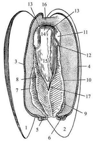
Fig. 73 Swan mussel (shell opened):
1, 2 — right and left valves of the shell; 3, 4 — edges of the right and left mantle lobes; 5, 6 — edges of the respiratory (branchial, or inhalant) siphon; 7-10 — outer and inner gill lamellae of the right and left gills; 11 — outer and 12 — inner lamellae of the "velums," or "labial palps"; 13 — edges of the oral opening; 14 — oral opening; 15 — foot; 16 — anterior shell adductor muscle; 17 — edge of the gill cleft (water exit to the cloacal siphon).
Examine the swan mussel without removing the valves of the open shell. It is clearly visible that the body is covered by the mantle. On the dorsal side, the mantle forms a continuous covering. The free edges of the mantle (anterior, ventral, and posterior) are thickened. Locate the severed muscles near the anterior and posterior edges. Note that the posterior edges of the mantle, folded together, form the siphons (Fig. 73).
The siphon located closer to the ventral side is the inhalant, or branchial, siphon; through it, water enters the gills. The dorsal siphon is called the exhalant, or cloacal, siphon—through it, water flows out, carrying away the mollusk's Metabolic waste products. The walls of the siphons are darker than the mantle, and the branchial siphon has a fringed edge. Note that the foot protrudes from beneath the ventral edge of the mantle. By lifting the free left edge of the mantle upward toward the dorsal side, the mantle cavity is exposed: here lies the body of the mollusk—the foot and two pairs of plate-like gills.
Locate the oral opening at the anterior upper end of the body, flanked on each side by a pair of triangular oral or labial lobes ("velums"). The swan mussel lacks a distinct HEAD. Locate the anus, which is situated above the posterior adductor muscle and opens into the cloacal siphon.
Examine the pair of outer gill lamellae adjacent to the mantle and the pair of inner gill lamellae located medially to the outer ones. The right and left inner lamellae are fused behind the body by their dorsal edges, while at the anterior end they attach to the Base of the foot. The gill leaflets are formed by a dense network of fine rods resembling a lattice. The surface of the gills is lined with ciliated epithelium. The inner surface of the mantle and the labial palps are covered with the same type of epithelium. The beating of the cilia of the epithelial Cells generates a water current. Water enters the mantle cavity through the branchial siphon, washes over the gills, and supplies oxygen. Along with the water, Algae, Ciliates, small crustaceans, rotifers, and other tiny planktonic organisms that serve as food for the swan mussel enter the mantle cavity. The beating of the cilia drives the food into the Mouth; thus, feeding is passive. The water current passing through the mantle cavity ensures gas exchange and Nutrition for the mollusk, collects metabolic waste products and excrement, and expels them outward through the cloacal siphon.
Examine the foot—it is yellowish or yellowish-orange in color, covered by the gill lamellae. The foot is keel-shaped, with a sharp end pointing forward. Its coloration indicates that it is divided into two parts. The lower, muscular, firm part of the foot serves for locomotion, while the upper, soft part represents the body proper, where Internal Organs (the gonad and intestinal loops) are embedded in the parenchyma.
Dissection of the swan mussel's body (simultaneously examine a preserved wet specimen of a dissected swan mussel). Cut the ligament of the swan mussel (the same one used for opening the shell) and remove the left valve.
Trim the adductor muscles on the right side and free the swan mussel's body from the right valve. Place the animal in a dissecting pan, pull the right and left edges of the mantle apart, and pin them down. Examine the body from the dorsal side. The internal organs are visible through the integument. Posterior to the anterior adductor muscle, the Liver is visible as a grayish-green patch, and behind it lies the brownish-red organ of Keber. Closer to the posterior adductor muscle, two dark stripes of the Kidneys (Bojanus' organs) can be seen on the sides of the body.
The secondary body cavity (coelom) in the swan mussel is reduced and filled with parenchyma; only the pericardial cavity (Pericardium) and the cavity housing the Gonads remain.
Lift the dorsal edge of the mantle upward with forceps, carefully cut it with scissors, and open the underlying pericardial cavity. Inside, The Heart ventricle is visible as a pear-shaped sac with thin, transparent walls. The intestinal tube passes right through the ventricle. Carefully widen the incision with forceps and locate the two auricles on the sides of the ventricle; they appear as transparent, triangular sacs (Fig. 74). They are so thin and delicate that they easily tear away from the ventricle during dissection.
Sketch the pericardial cavity and the heart within it; indicate the intestine passing through the heart with a dashed line.

Fig. 74 Swan mussel from the dorsal side, pericardial cavity opened:
1 — foot; 2 — mantle; 3 — openings of the exhalant siphon; 4 — anterior shell adductor muscle (severed); 5 — posterior shell adductor muscle (severed); 6 — opened pericardial cavity; 7 — heart (ventricle); 8 — auricle; 9 — hindgut; 10 — organ of Bojanus (Kidney); 11 — organ of Keber.
Examine the excretory Organs of the swan mussel. Clusters of glandular tissue of an orange-red or brownish-red color lie on the inner anterior wall of the pericardium. This is the paired pericardial gland, or organ of Keber, which performs an excretory function. The kidneys (Bojanus' organs) also belong to the excretory system. If the heart is pushed aside, the kidneys become clearly visible. The kidneys of the swan mussel are of the metanephridial type. Each consists of a bent tube with overgrown walls. One end of the tube (the nephrostome/funnel) communicates with the pericardium, while the other opens into the mantle cavity. The waste products of Keber's organ enter the pericardium and are subsequently excreted from it by the kidneys into the mantle cavity. From the mantle cavity, the excretory products are flushed out by the water current through the cloacal siphon.

Fig. 75 Internal Structure of the swan mussel:
1 — mantle; 2 — foot; 3 — branchial siphon; 4 — cloacal siphon; 5 — adductor muscles; 6 — labial palps; 7 — oral opening; 8 — Stomach; 9 — liver; 10 — intestine; 11 — anus; 12 — heart; 13 — pericardial sac; 14 — kidney; 15 — organ of Keber; 16 — cerebral ganglion; 17 — pedal ganglion; 18 — visceral ganglion.
Detach the swan mussel from the wax bottom of the dissecting pan. Examine the structure of The Nervous system. The nervous System of the swan mussel is of the scattered ganglion type. It consists of three pairs of ganglia: cerebral ganglia, located in the pharyngeal region; pedal ganglia, situated within the foot; and visceral, or trunk, ganglia, located beneath the hindgut. The cerebral ganglia are connected by paired nerve cords to the pedal and visceral ganglia. Fine branches extending from the nerve ganglia innervate various organs (Fig. 75).
Locate the cerebral ganglia by carefully scraping away the integument in the anterior part of the body, in the space between the foot retractor and protractor muscles. Cut the seam between the inner gill lamellae with scissors and locate the visceral ganglia. They should be sought in the posterior part of the incision; they are easily distinguishable.

Fig. 76 Swan mussel larva — glochidium:
1 — larval shell; 2 — adductor muscle; 3 — larval gland; 4 — bundles of sensory bristles; 5 — teeth with denticles.
Cut away the mantle and gills with scissors. Make a longitudinal incision with a scalpel through the muscular part of the foot down to the base (try to ensure the cut runs right down the middle).
Fold back one of the cut halves and locate the intestine on the other (it lies within the parenchyma, so it cannot be fully dissected out). The oral opening is easily identified by the perioral palps, followed by a short Esophagus, and then a voluminous sac-like stomach. The liver is clearly visible—it is located on the sides of The Stomach and consists of A large number of lobules. The intestine extends from the stomach. It descends, forms loops within the foot parenchyma, turns upward, passes through the pericardium, penetrates the heart ventricle, and opens via the anus above the posterior adductor muscle, near the cloacal siphon.
Make a sketch of the dissected swan mussel.
Among the dissected swan mussels, find a specimen with swollen outer gills filled with glochidia, the larvae of the swan mussel (Fig. 76).
Take a small amount of the contents from the swollen gill, place it on a Glass slide, cover with a coverslip, and examine the glochidium under a microscope. Note that the larva is equipped with a bivalve shell.
Identify the large teeth, covered with small denticles, along the free edges of the valves. A strong adductor muscle is stretched between the valves. Most of the glochidium's organs are underdeveloped. Occasionally, a thread of adhesive substance—the byssus—can be seen hanging from the body of the glochidium.
Maturation of the reproductive products in the swan mussel occurs during the summer. Eggs are laid between the gill lamellae. Spermatozoa released from the Testes are carried by water currents into the female's mantle cavity via the branchial siphon. Glochidia develop from the fertilized eggs. When a fish swims past a female carrying glochidia, the mollusk expels a batch of larvae into the water through the excurrent siphon. They attach to the fish's gills or fins using the byssus thread and by snapping shut their spiky shell valves. The fish's epithelium grows over the larva, enclosing it within a small cyst. Here the larva grows, feeding osmotically on the Body Fluids of the fish. The young swan mussel eventually detaches from the fish and drops to the bottom, where it develops into an adult, free-living mollusk.
Last update: 13/08/2026
Editorial and Educational Adaptation: This material has been compiled based on the primary/original source text. The project team performed an editorial review, corrected technical inaccuracies, structured sections, and adapted the content for an educational format.
What was processed:
- elimination of formatting defects (OCR errors, structural breaks, corrupted characters);
- editorial organization of content;
- standardization of terminology in accordance with academic sources;
- verification of factual statements against the original source text.
All mentions of the author, publication year, and origin of the primary text have been preserved in accordance with the source.