Practical Guide to Zoology: Study Guide - T. A. Dauda 2014
Protozoa
Apicomplexa
Class Coccidiomorpha
Unlike gregarines, coccidian-like Protozoa are intracellular parasites for the major part of their life cycle. Among the orders belonging to this Class, we will examine two: Haemosporidia and Coccidiida.
Order Haemosporidia
This order includes several dozen species, part of whose life cycle takes place within the erythrocytes of vertebrates, such as mammals, birds, and reptiles. Unlike coccidia, sporogony in Blood-dwelling Sporozoans never occurs in the external environment, but rather inside the bodies of blood-sucking insects (most commonly mosquitoes), which act as the vectors of these parasites. The malaria parasite, represented by four species, also belongs to the haemosporidians.
The malaria parasite (Plasmodium vivax) (Fig. 22) is a blood parasite. During one stage of its life, its host is a human; during another, it is a malaria mosquito. When the plasmodium enters The Human Body, the individual contracts malaria. Similar diseases caused by other haemosporidians are also observed in farm animals. These conditions are known as piroplasmoses.

Fig. 22 Life Cycle of the malaria parasite:
1, 2 — sporozoites penetrating the Cells of human Liver Blood Vessels; 3, 4 — schizogony (formation of merozoites); 5 — penetration of a merozoite into an erythrocyte; 6-10 — growth of the merozoite and schizogony; 10 — release of merozoites from the erythrocyte; 11, 12 — Development of the macrogametocyte (11a, 12a — development of the microgametocyte); 13 — macrogamete; 14 — microgametocyte; 15 — formation of microgametes; 16 — gamete copulation; 17 — zygote; 18 — ookinete; 19-23 — development of the ookinete in the wall of the mosquito's Stomach; 24 — release of sporozoites; 25 — penetration of sporozoites into the mosquito's Salivary Glands.
Materials required: diagram of the malaria parasite's life cycle; Microscope with an immersion objective; immersion oil; permanent slides of blood smears from malaria patients.
Assignment. Study The life cycle of the malaria parasite. Examine and sketch the various stages of its life cycle.
Studying the life cycle of the malaria parasite using the diagram (see Fig. 22). When a mosquito of the genus Anopheles infected with the malaria parasite bites a human, it introduces parasites at the sporozoite stage into the blood along with its saliva. These spindle-shaped parasites invade the cells of various Internal Organs, specifically the endothelial cells of blood vessels, the reticuloendothelial Cells of the liver, and others. Approximately 5 days after human infection, the sporozoites transform into oval-shaped schizonts measuring up to 40-60 µm. The schizonts of the extraerythrocytic developmental stage are non-pigmented, as they contain no pigment, unlike the schizonts of the erythrocytic stage. The nuclei of the non-pigmented schizonts divide, producing numerous other nuclei around which Cytoplasm condenses, resulting in The formation of new stages within the schizont—the merozoites.
When The Cell infected by the plasmodium ruptures, the merozoites are released into the Blood Plasma. This marks The process of schizogony—multiple fission, which results in the accumulation of merozoites. The stay of free merozoites in the plasma is short-lived. They rapidly invade new tissue cells, repeating the asexual reproduction cycle there, or penetrate erythrocytes, where the erythrocytic stage of development begins.
Upon entering an erythrocyte, a vacuole appears within the merozoite, and it assumes a ring-like shape as its plasma and Nucleus are displaced to the periphery—transforming into a trophozoite. The duration of this stage does not exceed 8-10 hours. The plasmodium then adopts an amoeboid shape and enters the proper schizont stage. This is the longest stage, lasting 32-34 hours in Pl. vivax. During this time, the schizont grows, feeding on the Hemoglobin of THE RED BLOOD cell.
Waste products of its METABOLISM—small clumps of melanin pigment (a breakdown product of erythrocyte hemoglobin)—are deposited in the schizont's plasma. Next, the process of schizogony takes place. The schizont's nucleus divides repeatedly, cytoplasm condenses around each nucleus, and the schizont breaks apart into merozoites. The erythrocyte membrane ruptures, and the merozoites are released into the blood plasma to invade other red Blood Cells.
Following a series of schizogonies, a large quantity of parasite metabolic products accumulates in the human blood, released into the bloodstream when the merozoites exit the erythrocytes. These Metabolic waste products of the parasite are toxic to the host.
The human body's response to this intoxication manifests as a malaria attack. The onset of the attack, characterized by chills in the patient, coincides with The breakdown of erythrocytes and the release of merozoites and plasmodial metabolic products into the blood plasma. Malaria attacks recur at regular intervals depending on the duration of asexual reproduction of the plasmodia within the erythrocytes. This is a species-specific biological characteristic of plasmodia: for Pl. vivax (the CAUSATIVE AGENT OF tertian malaria), the interval between attacks is 46-48 hours; for Pl. malariae (the agent of quartan malaria), it is 72 hours; and for Pl. falciparum (the agent of the most severe form of the disease—malignant tertian malaria), the interval is 40-48 hours, occasionally shortening to 24 hours. The incubation (latent) period of malaria lasts from two weeks to several months, after which malaria attacks begin, alternating with resting periods lasting 48 or 72 hours.
After a series of asexual generations, gametocytes (gamonts)—immature sexual forms of the parasite—appear among the merozoites. Gametocytes are differentiated into male (microgametocytes) and female (macrogametocytes) forms. Gametocytes, much like merozoites, invade erythrocytes.
Macro- and microgametocytes can mature and fuse (copulate) only if they enter the definitive host—a mosquito of the genus Anopheles. In The Stomach of a female mosquito that has fed on the blood of a malaria patient, the merozoites and erythrocytes are digested, gametocytes are liberated, and under favorable temperatures, the sexual reproductive cycle of the plasmodia begins. Upon maturing, the macrogametocyte transforms into a mature macrogamete. Upon maturing, the microgametocyte divides into 12-20 small, worm-like microgametes. Micro- and macrogametes fuse in pairs to form zygotes.
During the first 24 hours at optimal Temperature, the zygote transforms into a worm-like, motile body known as an ookinete. On the second day, the ookinete migrates between the epithelial cells of the gut wall and reaches the outer surface of the intestine. Here, the ookinete rounds off, becomes covered with a thick membrane, and transforms into an oocyst. The Contents of the oocyst divide repeatedly to produce slender, mononuclear, highly motile sporozoites. Several thousand spindle-shaped sporozoites are formed within each oocyst. The oocyst enlarges, its membrane ruptures, and the sporozoites enter the body cavity and are distributed via the hemolymph throughout the mosquito's body, with some reaching the salivary glands. When biting, the mosquito injects sporozoites into human blood along with its saliva, thereby transmitting malaria.
Thus, the asexual stage of plasmodium development takes place in the blood of a human or animal, while the sexual stage occurs in the mosquito's gut. The Anopheles mosquito serves not only as the definitive host of the plasmodium but also as its vector.
Since the transmission of plasmodia occurs directly from human to mosquito and from mosquito to human without the plasmodium ever entering the external environment, the plasmodium is classified as a sporozoan that lacks a spore stage with protective membranes.
Examination of the slide. Examine a stained prepared slide—a blood smear from a patient with tertian malaria (causative agent: Pl. vivax)—using an immersion objective. To do this, apply a drop of oil directly onto The surface of the smear and lower the immersion objective into it. Focus the image, adjust the illumination by lowering the condenser or slightly closing the Diaphragm.
Scanning the blood smear, locate erythrocytes infected with the plasmodium; they are slightly enlarged in size. Pay attention to the coloration of the erythrocytes, leukocytes, and plasmodia. The erythrocyte cytoplasm is pale pink, the nuclei of leukocytes and lymphocytes are dark purple, the cytoplasm of the plasmodia is bluish, and their nuclei are bright red.
Find the merozoites that have penetrated the erythrocytes, which initially appear amoeboid, and the trophozoites (an early developmental stage of plasmodia within the erythrocyte)—they look like rings (Fig. 23). A vacuole is visible in their center, the cytoplasm forms a bluish rim, and the red-stained nucleus lies at the periphery of the ring. Observe that in later developmental stages, the regular ring-like shape of the schizont (a parasite increasing in size and preparing for division) is lost, though the vacuole is typically retained.
Moving the slide, locate later Developmental Stages of the schizont—amoeboid schizonts with a single nucleus and numerous cytoplasmic projections. Observe grains of dark brown pigment, melanin, within the cytoplasm of such schizonts. Melanin is a breakdown product of hemoglobin utilized by the parasite. Young schizonts contain very few melanin granules.

Fig. 23. Various Forms of the malaria parasite (Plasmodium vivax) from human blood:
1, 2 — ring stages; 3, 4 — amoeboid schizonts; 5, 6 — schizogony and merozoite formation; 7, 8 — gametocytes.
Sketch the Stages of the trophozoite and young schizonts, then proceed to examine schizonts preparing for division. Such a schizont is oval or round in shape, lacking a vacuole and pseudopodia. It occupies the greater part of the infected erythrocyte. Its large nucleus is typically located at the periphery, and the melanin granules are gathered in clusters.
Locate schizonts with multiple nuclei (the process of schizogony) as well as erythrocytes containing 12 to 20 merozoites. The cytoplasm of these merozoites is blue, and The Nucleus is cherry-red. You can also observe compact clusters of melanin lying between the merozoites.
Continuing the work, find the sexual forms of the parasite in the slide: unlike mature schizonts, gametocytes are mononuclear. The microgametocyte is distinguished by pale blue cytoplasm containing coarse melanin granules, and a loose, large nucleus. The central part of the nucleus is stained cherry-red, while the peripheral part has a pinkish hue. The macrogametocyte is larger in size than the microgametocyte and the adjacent uninfected erythrocytes. Its nucleus is compact, larger than that of a schizont, but smaller than that of a microgametocyte, and is stained red; the cytoplasm is darker.
Sketch the multinucleated schizonts, schizonts with merozoites, and gametocytes.
Order Coccidiida (Coccidia)
Coccidia are parasites living in the intestine and various organs (such as the liver) of certain fish, rabbits, cattle, chickens, turkeys, ducks, and geese. They are sometimes found in humans as well. The disease caused by coccidia is called coccidiosis.
Coccidia are characterized by two modes of reproduction—sexual and asexual—which alternate regularly. Most species have a single host, and sporogony takes place in the external environment. Let us examine the life cycle of the monoxenous coccidian Eimeria magna, which parasitizes the rabbit intestine.
Materials and Equipment. Several species of the genus Eimeria are found in the rabbit intestine. The most common are E. magna, E. perforans, and E. media. In addition, E. stiedae infects the liver. The presence of the parasite in the intestine is determined by examining feces under a microscope for the presence of oocysts. Rabbits completely free of coccidia are almost never found. Young rabbits aged 1–2 months turn out to be heavily infected.
A richer and more uniform material can be obtained by experimental infection of young animals aged 18–22 days. For this purpose, a drop of a saturated NaCl solution containing oocysts of a particular Eimeria species is introduced with a pipette into the Oral Cavity of the rabbit to be infected. Knowing the timing of the individual stages of the Eimeria life cycle (see the monograph: Kheysin E. M. Coccidia of the Rabbit Intestine, p. 51), researchers choose the timing for dissecting the infected rabbits to fix the material. E. magna localizes in the lower part of the Small Intestine over a length of about 60 cm from its junction with the Large Intestine. In case of heavy infection, intestinal damage is clearly visible to the naked eye, manifested by Swelling of the mucosa and a reddish tint of its inner wall. A smear is made from the inner surface of the intestine on a coverslip, fixed, and stained with iron hematoxylin.
Required for work: microscope; diagram of the coccidian life cycle; fixed slide of a rabbit intestine section showing various stages of coccidian development.
Assignment. Familiarize yourself with the life cycle of coccidia. Examine and sketch the various stages of the life cycle.
Studying the coccidian development cycle using the diagram. Animals and humans contract coccidiosis by ingesting coccidian oocysts along with food, which contain spores with germs—sporozoites (Fig. 24). In the host's intestine, the oocyst wall dissolves, the spore walls burst, and the sporozoites actively penetrate the epithelial cells of the intestine or liver, where they begin to reproduce via schizogony. When the schizont divides into merozoites (from 8 to 60), the epithelial cells of the intestine or liver are destroyed, and the merozoites emerge from them and penetrate new epithelial cells. The process of schizogony repeats many times. The intensity of infection also depends on the number of spores that have entered the host.
The asexual process is succeeded by the sexual process, which takes place within the same host. During the sexual process, merozoites in the epithelial cells begin to transform into immature male and female sex cells (macro- and microgametocytes). Upon maturation, each macrogametocyte transforms into a macrogamete, while each microgametocyte divides into microgametes. Gamete copulation then takes place, forming a zygote. It becomes covered with membranes and transforms into an oocyst.
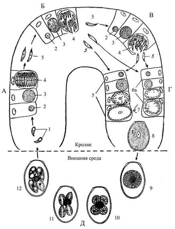
Fig. 24. Life cycle of the coccidian (Eimeria magna) parasitizing the rabbit:
A — first generation of schizogony; Б — second generation of schizogony; В — third generation of schizogony; Г — gametogony; Д — sporogony; 1 — sporozoites; 2 — young schizont; 3 — growing multinucleated schizont; 4 — schizont broken down into merozoites; 5 — merozoites; 6 — development of the macrogamete; 6а — development of microgametes; 7 — microgamete; 8 — oocyst; 9 — oocyst initiating sporogony; 10 — sporocyst with four sporoblasts and residual body; 11 — development of sporoblasts; 12 — mature oocysts with four spores, each containing two sporozoites.
Oocysts pass into the intestinal lumen and are expelled into the external environment along with the host's excrement. For the further development of oocysts, specific temperature, humidity, and free atmospheric oxygen are required. During division inside the oocyst, sporoblasts are formed; they turn into spores containing sporozoites.
Compare the life cycle of coccidia with that of the malaria parasite. The complex life cycle of coccidia and hemosporidians—associated with their transition into the external environment in coccidia, and with the transfer to another host in blood sporozoans—presents numerous obstacles to their spread and results in a high mortality rate among the parasites.
Schizogony is an adaptation for enhanced reproduction that guarantees the survival and propagation of both coccidia and blood sporozoans.
Examination of the Slide. Examine various Selection/3.html">Stages of development of the coccidian E. magna in the rabbit intestine under high magnification of the microscope (Fig. 25). A more detailed study is carried out using immersion. Moving the slide and adjusting the illumination, locate the epithelial cells covering the intestinal villus and schizonts in the early stage of development. Schizonts at an early stage of development have a regular spherical shape and are mononuclear.
One can find schizonts whose nuclei have divided into 2, 4, and 8, as well as schizonts with numerous nuclei (at the schizogony stage). The stage of merozoite formation is easily detected. Schizonts with formed merozoites attract attention because the merozoites lie close to one another like orange segments. Sometimes, sexual forms of coccidia can also be found and observed in the slide.
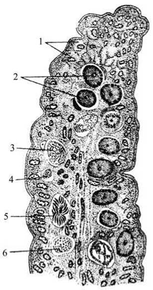
Fig. 25. Section of a rabbit intestinal villus showing various developmental stages of Eimeria magna:
1 — epithelial cells; 2 — macrogametes; 3 — microgametocyte with microgametes; 4 — schizont at the beginning of growth; 5 — schizont divided into merozoites; 6 — schizont at the end of growth with numerous nuclei.
Macrogametes at Cytology/cytology/16.html">Early stages of development are difficult to distinguish from young schizonts. At later stages of development, they differ from schizonts in that the nucleus does not divide and has a vesicular shape.
Macrogametes are initially spherical in shape and then become oval. The microgametocyte is characterized by containing numerous elongated microgametes. The oocyst has two membranes.
Draw the individual stages of coccidian development: a young schizont at the division stage, a schizont with multiple nuclei, a schizont with merozoites, a macrogamete, a microgametocyte with microgametes, and oocysts.
Last update: 13/08/2026
Editorial and Educational Adaptation: This material has been compiled based on the primary/original source text. The project team performed an editorial review, corrected technical inaccuracies, structured sections, and adapted the content for an educational format.
What was processed:
- elimination of formatting defects (OCR errors, structural breaks, corrupted characters);
- editorial organization of content;
- standardization of terminology in accordance with academic sources;
- verification of factual statements against the original source text.
All mentions of the author, publication year, and origin of the primary text have been preserved in accordance with the source.