Workshop in Zoology: A Study Guide - T. A. Dauda 2014
Protozoa
Sporozoans
Class Gregarines
The phylum Apicomplexa comprises Protozoans characterized by an exclusively parasitic lifestyle. Representatives of this phylum exhibit a highly complex life cycle featuring a distinct alternation of asexual reproduction (schizogony), sexual process (gametogony), and sporogony. During sporogony, sporozoites—the dispersive life-cycle stages responsible for spreading the species—develop inside an oocyst (a zygote enclosed within a thick protective wall).
The phylum is divided into two classes: Gregarinina (gregarines) and Coccidiomorpha, each of which comprises several orders.
The material presented below is selected to introduce one representative from each order, with a life cycle studied in as much detail as possible. Among gregarines, the cycle of Gregarina blatarum is examined, while Eimeria magna serves as the representative for coccidians. From the Blood-dwelling sporozoites (order Haemosporidia), we will examine the portion of the malaria parasite's life cycle (Plasmodium sp.) that takes place in the blood of a human patient with malaria.
Gregarines are the largest Representatives of the phylum Apicomplexa. They parasitize the intestine and, less commonly, the body cavity of various invertebrates, primarily worms and Arthropods (insects and their larvae). Infection occurs via spores.
Materials and Equipment. To obtain gregarines, larvae of the mealworm beetle (incorrectly referred to as "mealworms") can be used. To do this, use scissors to cut off the anterior and posterior ends of the larva's body as shown in Figure 19. By applying gentle pressure with your fingers, squeeze out the intestine—a thin, brown tube—from the larva's body. Grasp it with forceps and place it on a watch Glass. Thoroughly mince the intestine with dissecting needles and add physiological saline using a pipette. Place a drop of this turbid fluid onto a glass slide and cover it with a coverslip.
Class="center">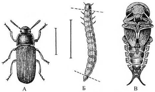
Fig. 19 Mealworm beetle (Tenebrio molitor):
A — adult beetle; B — its larva, the "mealworm" (dashed lines indicate where to cut the HEAD and posterior ends of the larva's body to extract the intestine for obtaining gregarines); C — pupa.
To obtain gregarines from the intestine of the oriental cockroach, euthanize the cockroach with ether and dissect it. Use scissors to cut along the lateral sides of its body from the anterior to the posterior end. Place the cockroach ventral side down in a dissecting tray, pinning down the head and the posterior end of the body. Remove the dorsal body wall (tergum) using forceps and a dissecting needle. Wash away the fat body, which surrounds the intestine, with Water from a pipette. Locate the midgut (situated between the blind caeca and Malpighian tubules), excise it, transfer it to a watch glass, and macerate it with dissecting needles. Add physiological saline. Prepare the wet mount.
Required equipment: a Microscope; dissecting instruments (tray, forceps, scissors, needles, pins); watch glasses, glass slides, and coverslips; physiological saline in a small beaker; pipettes; mealworm beetle larvae (Tenebrio molitor) or oriental cockroaches; gregarine slide preparations.

Fig. 20 Various species of gregarines:
A — from the cockroach intestine: 1 — protomerite; 2 — deutomerite; 3 — cuticle; 4 — Nucleus; 5 — ectoplasm; 6 — endoplasm with paraglycogen granules; B, C, and D — from the mealworm.
Assignment. Extract the intestine from a mealworm beetle larva or dissect a cockroach to isolate its intestine. Examine the intestinal contents and locate single and paired gregarines. Make a drawing of a paired gregarine, labeling the body regions of each individual, the pellicle, ectoplasm, endoplasm with paraglycogen granules, and The Nucleus.
Examination of the Preparation. Examine the intestinal Contents of the oriental cockroach and the mealworm beetle larva under low magnification of the microscope to locate gregarines. Large Gregarina blatarum specimens are found in the intestine of the oriental cockroach. Smaller forms, such as Gr. polymorpha, Gr. steini, and Gr. cuneata, occur in the intestine of the mealworm larva (Fig. 20).
Observe the body of a gregarine under low magnification. It consists of two distinct regions: the posterior region, which contains the nucleus (the deutomerite), and the anterior region (the protomerite). Locate both solitary gregarines and those joined in pairs (syzygy) under low power. Examine a paired gregarine, sketch the body regions of each individual, and locate the transparent nucleus within the deutomerite.
Examine the Cytoplasm, which is differentiated into a fluid, granular endoplasm and a denser, transparent ectoplasm.
The ectoplasm forms septa that divide the gregarine's body into distinct compartments. The endoplasm appears opaque due to numerous inclusions of paraglycogen carbohydrate granules, which serve as a reserve nutrient and structural material. Microscopic examination reveals that the outer layer of the ectoplasm is condensed into a cuticle—a tough pellicle covering the body of the gregarine and maintaining its constant shape. Located in the ectoplasm beneath the cuticle are thin, dense fibrils that provide structural support (skeletal fibrils), as well as longitudinal and transverse contractile fibers known as myonemes. These structures are typically not visible in standard preparations. Gregarines lack specialized feeding or respiratory Organelles, and contractile vacuoles are likewise absent. Nutrition, Respiration, and excretion take place osmotically across the cuticle over the entire surface of the body. Gregarines exhibit various modes of locomotion. In some species, movement is achieved through the contraction of myonemes; in others, it relies on a jet-propulsion mechanism: tiny streams of fluid are expelled backward from the body through numerous Pores in the cuticle, creating a reactive counter-thrust that slowly and smoothly propels the parasite's anterior end forward. Examine the ULTRASTRUCTURE OF THE gregarine under high magnification and add the structural details to your drawing.
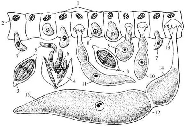
Fig. 21 Diagram of The life cycle of intestinal gregarines:
1 — intestinal epithelial Cells with nuclei; 2 — nucleus; 3 — spore; 4 — opened spore; 5 — sporozoites; 6 — residual body; 7 — penetration of sporozoite into epithelium; 8 — sporozoite within an epithelial Cell; 9–12 — growth of the sporozoite and its transformation into a gregarine; 13 — epimerite; 14 — protomerite; 15 — deutomerite.
Study the Life Cycle of the gregarine using the diagram (Fig. 21). Gregarines reproduce sexually. Occasionally, gregarines can be found attached in pairs, with one individual attached to the posterior end of another; this represents preparation for sexual reproduction. The anterior individual is typically female, while the posterior one is male. Through repeated karyokinetic Divisions of the protoplasm and nucleus of each gregarine, numerous sporoblasts are formed. Those originating from the female individual function as female Gametes, whereas those derived from the male correspond to male gametes. Male and female gametes fuse in pairs (copulate) to form a zygote (a fertilized egg cell), which becomes enclosed within a thick wall and develops into a spore. Inside each spore, eight worm-like sporozoites are formed. As a result of the sexual process, each pair of gregarines transforms into a cyst packed with A large number of spore-filled sporozoites. Cysts are expelled with the host's feces into the external environment, serving to infect other animals of the same species. Upon accidental ingestion of the cysts (e.g., with food), the sporozoites are released from the spores within the new host's intestine and invade the intestinal epithelial cells. The sporozoite grows, transforming into a gregarine that attaches to the intestinal wall exclusively via its epimerite. Upon reaching maturity, the epimerite is shed, and the parasite lives freely within the intestinal lumen.
Gregarines have no practical economic or medical significance. In zoology practical courses, they are studied as large and accessible model organisms for introducing the phylum Apicomplexa.
Last update: 13/08/2026
Editorial and Educational Adaptation: This material has been compiled based on the primary/original source text. The project team performed an editorial review, corrected technical inaccuracies, structured sections, and adapted the content for an educational format.
What was processed:
- elimination of formatting defects (OCR errors, structural breaks, corrupted characters);
- editorial organization of content;
- standardization of terminology in accordance with academic sources;
- verification of factual statements against the original source text.
All mentions of the author, publication year, and origin of the primary text have been preserved in accordance with the source.