IMMUNOLOGY - Roitt I. - Mir 2000
Chapter 27. Transplantation and Rejection
PREVENTION OF TRANSPLANT REJECTION
Graft rejection can be mitigated by careful donor-recipient matching.
The ideal transplant combination is an isogeneic donor and recipient, such as identical twins. However, such matches are rarely available, and in most cases, there are disparities between the donor and recipient regarding the MHC and/or minor histocompatibility loci. In clinical practice, it is generally sufficient to match for major Histocompatibility Antigens (MHC, known as HLA in humans). Compatibility can be assessed using serological typing (Fig. 27.19), a technique that takes only a few hours and can therefore be performed while the donor organ is being preserved on ice. Recently, a sensitive and precise typing method utilizing the Polymerase Chain Reaction (PCR; see Ch. 29) has been developed to identify the HLA genes of both donor and recipient.
Class="center">
Fig. 27.19. Serological tissue typing is performed using the microlymphocytotoxicity test. Typing sera (e.g., anti-HLA-B8), Complement, and trypan blue dye are added to the test Cells. Cell death, indicated by dye uptake, demonstrates that the test cells carry the specific antigen in question (in this case, HLA-B8). The microphotograph on the right shows dead cells stained with trypan blue (dark).
While it is virtually impossible to ensure compatibility for all known HLA antigens, excellent results can be achieved when the donor and recipient share the same class II MHC antigens, particularly HLA-DR (Fig. 27.20), as these directly activate the recipient's Th cells.
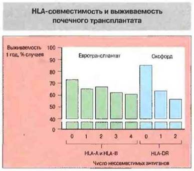
Fig. 27.20. Results from two independent studies showing 1-year graft survival rates for cadaveric Kidney transplants. In the first study (Eurotransplant), Donors were typed for HLA-A and HLA-B antigens (class I). In the second study (Oxford), donors were typed for HLA-DR antigens (class II).
The number of currently known class I (HLA-A, HLA-B, and HLA-C) and class II (HLA-DP, HLA-DQ, and HLA-DR) HLA antigens is quite large (Fig. 27.21), making complete compatibility between two randomly chosen individuals highly unlikely.
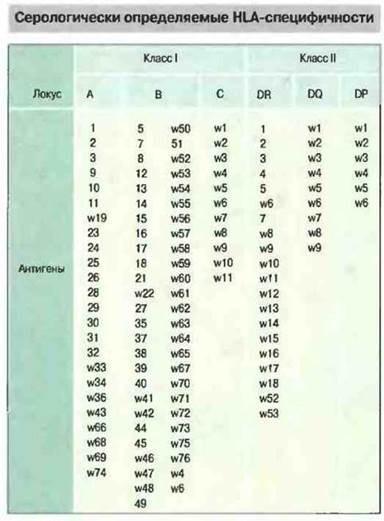
Fig. 27.21. Approximately 80 different class I molecules (HLA-A, HLA-B, and HLA-C) and over 35 different class II molecules (HLA-DP, HLA-DQ, and HLA-DR) have been identified in humans. Molecular Genetic Methods reveal an even greater number of variants, although not all of these new variants can be distinguished serologically.
The mixed lymphocyte reaction (MLR) can also be used to assess the reactivity of recipient lymphocytes against antigens expressed on donor cells (Fig. 27.22). A low response in a mixture of donor and recipient cells correlates with excellent graft survival. However, performing an MLR takes 4–5 days, which poses a major clinical obstacle, as Organs retrieved from cadavers or patients pronounced dead by Brain criteria cannot be preserved for more than 24–48 hours. The MLR test is therefore reserved for living-related donor transplants. The results of this assay are particularly critical in Bone Marrow transplantation, as they determine whether the donor's bone marrow cells are capable of reacting against recipient antigens and causing GVHD.
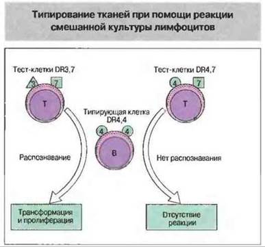
Fig. 27.22. In the mixed lymphocyte reaction, test cells are incubated with 'typing' cells of known HLA Specificity (in this case, DR4,4). DR3,7 cells recognize the typing cells as foreign, which is detected by the transformation and proliferation of the typing cells (the typing cells are pre-treated so that they cannot divide themselves when interacting with test cells). In contrast, DR4,7 cells, sharing the same specificity as the typing cells (DR4), do not recognize or react against them.
Graft rejection can be prevented by nonspecific immunosuppression
There are two forms of immunosuppressive therapy: antigen-nonspecific and antigen-specific. Nonspecific immunosuppression suppresses or dampens The activity of The Immune System against all antigens, but it leaves the transplant recipient more vulnerable to infections. For instance, high-dose X-irradiation prevents rejection but simultaneously causes severe adverse effects, including the suppression of anti-microbial Immunity. Most nonspecific immunosuppressive agents currently in use are drugs that exert a selective, or semi-selective, effect on the immune system when administered according to specific regimens. In the future, this approach will be refined to eliminate only those lymphocyte clones specific to donor antigens while leaving other clones intact. This will preserve defense mechanisms against infections and avoid other side effects. Such highly specific immunosuppression remains something of a Holy Grail in transplantation immunobiology (see below).
Currently, the three most widely used nonspecific immunosuppressive agents in clinical practice are Steroids, cyclosporine, and azathioprine (Fig. 27.23).

Fig. 27.23. Widely used clinical agents—steroids, cyclosporine, and azathioprine—suppress rejection through different pathways. Steroids possess anti-inflammatory properties, inhibiting macrophage and APC Functions while also reducing MHC antigen expression. Cyclosporine blocks cytokine production, whereas azathioprine suppresses the proliferation of activated cells.
Steroids possess anti-inflammatory properties, suppress activated macrophages, inhibit APC functions, and decrease the expression of MHC antigens. This therapeutic action stems from their ability to reverse many of the effects of IFNγ on macrophages and transplanted Tissues.
Cyclosporine is a cyclic polypeptide antibiotic produced by soil Fungi, notable for its high immunosuppressive potency. Its primary MECHANISM OF ACTION involves the inhibition of lymphokine synthesis (via effects on lymphokine Gene activation) and the direct or indirect downregulation of IL-2 receptor expression on lymphocytes that have received an activation signal. Other fungal-derived cyclic Polypeptide Antibiotics, such as FK506 (tacrolimus) and rapamycin, also exhibit immunosuppressive properties. FK506 inhibits lymphokine production by Th cells through a mechanism similar to that of cyclosporine. Rapamycin blocks the Intracellular Signaling pathways originating from the IL-2 receptor, thereby inhibiting IL-2-dependent lymphocyte activation. The structures of cyclosporine, FK506, and rapamycin are illustrated in Fig. 27.24.
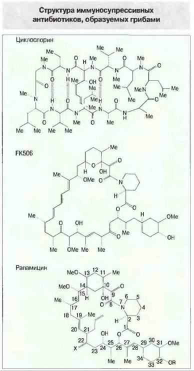
Fig. 27.24. Immunosuppressive cyclic polypeptide antibiotics produced by fungi differ in Structure and exert distinct effects on lymphocytes: cyclosporine and FK506 inhibit lymphokine production, whereas rapamycin blocks signal Transduction mediated by the IL-2 receptor (IL-2R).
Transplant rejection is driven by the rapid division and differentiation—proliferation—of lymphocytes. This process can be targeted using the antiproliferative agent azathioprine. Its metabolic product is incorporated into the DNA of dividing cells, preventing their further proliferation. Currently, novel antiproliferative drugs are under investigation, particularly mycophenolic acid derivatives.
All of these immunosuppressive agents can be effective as monotherapies; however, achieving the desired clinical outcome requires high doses, which increases the likelihood of adverse toxic effects. When used in combination, immunosuppressants produce a synergistic effect because they target different Stages of the same immune process. Consequently, the doses of individual components can be reduced, thereby minimizing side effects. The Introduction of cyclosporine significantly improved clinical transplantation outcomes (with 1-year survival rates for kidney, Heart, and Liver grafts reaching 85–90%). Nevertheless, the half-life of transplanted Kidneys remains 7–8 years, as cyclosporine failed to resolve the issue of chronic rejection, and its long-term use is still associated with adverse effects. Further progress is anticipated from the Introduction of new pharmaceutical agents into clinical practice.
Research is currently underway on novel agents that also exhibit non-specific immunosuppressive properties but act with greater selectivity (Fig. 27.25). Monoclonal Antibodies directed against cell-surface antigens—specifically CD3, CD4, CD8, and the IL-2 receptor—can be used to eliminate cells or block their function. To enhance the efficacy of these antibodies, they can be conjugated with cytotoxic agents. Another similar approach involves coupling a toxin with IL-2: cells expressing the IL-2 receptor, which are activated in response to transplant antigens, bind the IL-2-toxin conjugate and are selectively inactivated by the toxin.
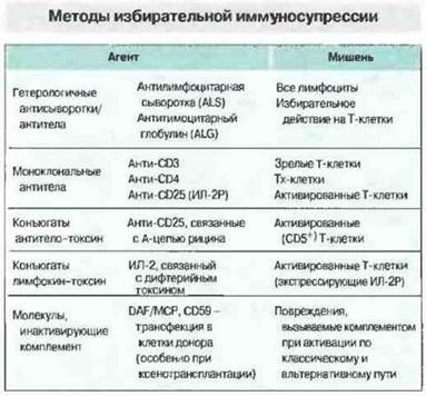
Fig. 27.25. Antibodies and lymphokines can specifically target certain Cells of the immune system. In contrast, standard medications may exert undesirable effects on non-lymphoid tissues, such as nephrotoxicity and hepatotoxicity. The efficacy of biological agents is enhanced by conjugating them with chemical agents or toxins. (DAF — decay-accelerating factor; MCP — membrane cofactor protein.)
Specific immunosuppression dampens the Immune Response to a graft without increasing susceptibility to infections
The intensity, type, and specificity of immunological reactions are regulated through various feedback mechanisms (see Ch. 13). Experimentally, transplant rejection can be prevented by manipulating these regulatory pathways using three classical approaches: the induction of neonatal tolerance, active enhancement, and passive enhancement of tolerance.
Administering donor antigens to newborn animals can induce unresponsiveness to a graft. Unlike humans, mature T cells in rodents begin leaving the Thymus during the neonatal period (the corresponding developmental stage in humans occurs at 16–20 weeks of gestation). If newborn mice are provided with a continuous source of antigen (such as proliferating living cells) or are repeatedly injected with it, The Development of mature T cells reactive to that antigen is suppressed. In a classical experimental setup, this is performed as follows. Bone marrow cells from (A x B)F1 mice are injected into newborn line B mice. (The Use of F1 hybrid cells prevents the anti-B graft-versus-host disease that would occur if line A cells were injected into line B mice instead of (A x B)F1 cells.) The transplanted bone marrow serves as a continuous source of donor antigens. When line B mice reach adulthood, they are found to be unresponsive to the antigens of strain A to which they were exposed postnatally. These animals exhibit tolerance to strain A antigens present in Skin grafts and other tissues from line A or (A x B)F1 donors. The Mechanism of neonatal tolerance induction is illustrated in Fig. 27.8 and described in detail in Ch. 12.
An antigen can selectively activate certain lymphocyte subpopulations. According to current models, T helper cells are divided into two main populations—Th1 and Th2 (see Ch. II). Mice with neonatally induced tolerance may exhibit a deficiency in donor-specific Th1 cells and an elevated number of donor-specific Th2 lymphocytes. As shown in Fig. 27.23, Th1 cells produce IFN-γ and IL-2 and participate in graft rejection. Conversely, Th2 cells produce other lymphokines, including IL-10 and a factor that inhibits lymphokine synthesis by Th1 cells. The presence of few Th1 cells and A large number of donor-specific Th2 cells in such mice signifies a shift in the balance between rejection and engraftment processes, leading to the development of tolerance. Strictly speaking, this form of tolerance represents not unresponsiveness per se, but rather immune deviation. Interestingly, cyclosporine may act preferentially on Th1 cells while leaving Th2 cells intact.
Finally, an antigen can activate suppressor T cells (Ts cells). Their exact nature remains elusive. The primary observation supporting the activity of Ts cells is that The transfer of T lymphocytes from a donor tolerant to a strain A skin graft prevents the rejection of strain A-bearing grafts in a recipient. This demonstrates the adoptive transfer of suppression, with the mediating cells likely being Th or Ts lymphocytes. Experimental data clearly indicate that T cells possessing suppressive functions do exist, yet opinions regarding The Nature of Ts cells and their Mechanisms of action remain highly controversial. These cells are resistant to cyclosporine and may contribute to its effects by mediating tolerance through active immunosuppression.
Specific immunosuppression in humans. A state equivalent to neonatally induced tolerance in animals cannot be achieved in humans. However, a somewhat similar situation arises when specific techniques are used to manipulate the human immune system. Total lymphoid irradiation (TLI) leads to a drastic depletion of lymphoid tissue, while bone marrow shielding preserves normal hematopoiesis. As a result, humans develop a state resembling neonatally induced tolerance in rodents. Indeed, TLI followed by antigen administration induces profound tolerance. Nevertheless, applying TLI in routine clinical practice is quite hazardous. In heart transplantation, antithymocyte globulin—obtained from animals immunized with human lymphocytes—is widely used. The Effect of such serum is the elimination of circulating recipient T cells. Monoclonal antibodies directed against mature T-cell antigens provide a safer yet equally effective means of T-cell depletion, and anti-CD3 antibodies have found established clinical utility.
Unresponsiveness to a graft can be induced in humans via Blood transfusions. In some cases, graft survival can be prolonged, occasionally indefinitely, by prior administration of donor antigens (Fig. 27.26). This is the exact opposite of what one would expect from immunizing a recipient with donor antigens—namely, accelerated or hyperacute graft rejection. This phenomenon is known as active enhancement of graft survival. The route of antigen administration is critical, apparently due to the involvement of different compartments of the lymphoid tissue. For instance, in rat kidney transplantation experiments, intravenous administration of donor blood to the recipient one week prior to transplantation ensured long-term survival of the grafted organ, whereas subcutaneous injection of the same amount of donor blood triggered accelerated rejection. This effect is immunologically specific, which is why the blood donor and the kidney donor must share at least some common antigens.
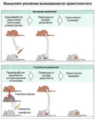
Fig. 27.26. Prior intravenous administration of donor antigens to recipients can enhance the survival of a subsequently transplanted allograft. This phenomenon is termed active immune enhancement of tolerance, as it is driven by the recipient's active immune response. (Note that administering blood from the same donor via a different route can lead to rapid graft rejection.) In contrast, administering donor-specific antibodies to the recipient at the time of transplantation can induce passive enhancement of tolerance. Both active and passive enhancement are immunologically specific, as they suppress responses exclusively against the given donor's antigens; survival is not prolonged (enhanced) for a "third-party" graft taken from an unrelated, outsider donor.
The method of active enhancement of tolerance has found clinical application through donor-specific blood transfusion (DSBT). For example, prior to a living-related kidney transplantation from a parent to a child, the child receives a transfusion of that parent's blood. Unfortunately, approximately 20% of patients undergoing DSBT develop antidonor antibodies, making the planned kidney transplant impossible due to the risk of hyperacute rejection. However, in the remaining 80% of patients, transplantation is successful in 95–100% of cases.
A beneficial effect of pretransplantation blood transfusion (the blood transfusion effect) has also been observed in recipients who received blood from randomly matched donors (i.e., when the blood donor is not the donor of the transplanted organ). This effect is likely due to a chance sharing of antigens between the blood donors and the graft (Fig. 27.27). This explanation is supported by evidence that the transfusion effect increases with the number of transfusions received from different donors. At one time, most transplant centers adopted a strategy of pretransfusing prospective recipients with blood from arbitrary donors. However, this always carried the risk of patient sensitization and transmission of HIV; the subsequent availability of potent immunosuppressive agents has made this Procedure largely unnecessary.
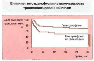
Fig. 27.27. Transplanted kidney survival in patients ($n = 102$) who received pretransplant donor blood transfusions is significantly higher than in patients ($n = 71$) who received no blood transfusions.
For active enhancement to occur, the patient must mount an active immune response against the administered donor antigens. Potential mechanisms include the induction of anergy, selective activation of Th2 cells, or activation of Tc cells driven by blood antigens, similar to the processes seen in neonatally induced tolerance. In other instances, "enhancing antibodies" may be produced which, by blocking the recognition of specific donor antigens, suppress graft rejection or destroy highly immunogenic "passenger" leukocytes contained within the graft. Alternatively, these enhancing antibodies may interact with antigen receptors on cells reactive against donor antigens, leading to their elimination or altering antigen presentation in a way that selectively activates specific subsets, such as Th2 and Tc lymphocytes, post-transplantation.
In the transplant recipient, antibodies can exert regulation via feedback mechanisms. The administration of antidonor antibodies (passive enhancement) to rats undergoing kidney transplantation can ensure long-term survival of the grafted organ (Fig. 27.26).
Questions for Thought
■ How do responses induced by the direct activation of recipient T cells by the donor antigenic peptide–donor MHC molecule complex differ from those developing via indirect activation by the donor peptide–recipient MHC molecule complex?
■ Pretransplant blood transfusion can prolong graft survival by inducing immunological unresponsiveness. However, some patients develop antibodies against foreign donor Blood Cells, making the success of subsequent organ transplantation uncertain. How can The production of antidonor antibodies be prevented?
■ The effector mechanisms of chronic and acute rejection differ. What modifications must be made to the Treatment regimen to prevent chronic rejection?
■ The primary barrier in xenotransplantation is posed by pre-existing antibodies in the recipient directed against donor antigens. Suppose hyperacute rejection could be avoided, for instance, when transplanting a porcine organ into a human. Nevertheless, the development of a cellular response remains probable. How will the recognition processes of foreign antigens differ between xenotransplantation and allotransplantation?
■ What are the mechanisms contributing to graft rejection under Direct and Indirect pathways of immunization?
■ Which combination of drugs, antibodies, and antigens would, in your opinion, be most effective for transplanting a histoincompatible graft?
Alexander G.P.J., Latime D., Gianello P. et al. 1991. Preformed cytotoxic antibodies and ABO-incompatible grafts. Clin. Transplant. 5: 583.
Bach F.H. 1991. Xenotransplantation: problems for consideration. Clin. Transplant. 5: 595.
Bjorkman P.J., Saper M.A., Samaouri B. et al. 1987. The foreign antigen binding site and T Cell Recognition regions of class I histocompatibility antigens. Nature 329: 512.
Burdick J.F. 1991. Chronic rejection Clin. Transplant. 5: 489.
Concar D. 1994. The organ factory of the future? New Scientist 1930: 24-29.
Dallman M.J., Clark G.J. 1991. CYTOKINES AND THEIR receptors in transplantation. Curr. Opin. Immunol. 3. 729.
Graff R.J., Bailey D.W. 1973. The non-H-2 histocompatibility loci and their antigens. Transplant. Rev. 15: 26-49.
Hall B.M., Dorsch S., Roser B. 1978. The Cellular Basis of allograft rejection in vivo. I. The cellular requirements for first set rejection of heart grafts. J. Exp. Med. 148: 878.
Halloran P.F. Broski A.P., Batiuk T.D. et al. 1993. The molecular immunology of acute rejection: an Overview. Transplant. Immunol. 1: 3-27.
Hunt S., Billingham М. 1991. Long-term results of cardiac transplantation. Ann. Rev. Med. 42: 437.
Hutchinson I.V. 1991. Cellular mechanisms of allograft rejection. Cure Opin. Immunol. 3: 722.
Lechler R.I., Lombardi G., Batchelor J.R. et al. 1990. The Molecular Basis of alloreactivity. Immunol. Today 11:83.
Mason D.W., Morris P.J. 1986. Effector mechanisms in allograft rejection. Annu. Rev. Immunol. 4: 119.
Masoor S., Schroeder T.J., Michler R.E. et al. 1986. Monoclonal antibodies in organ transplantation: an overview. Transplant. Immunol. 4: 176-89.
Opelz G. 1989. Effect of HLA matching in heart transplantation. Transplant. Proc. 21: 794.
Piatt J.L., Bach F.H. 1991. The barrier to xenotransplantation. Transplantation 52: 937.
Sablinski T., Hancock W.W., Tilney N.L. et al. 1991. CD4 monoclonal antibodies in organ transplantation. A review of progress. Transplantation 52: 579.
Sachs D.H., Bach F.H. 1990. Immunology of xenograft rejection. Human Immunol. 28: 245.
Stemmuller D. 1985. Which T cells mediate allograft rejection? Transplantion 40: 229.
Thomson A.W. 1994. Immunosuppressive drugs and the induction of transplantation tolerance. Transplant. Immunol. 2: 263-70.
Waldman H., Cobbold S. 1993. The use of monoclonal antibodies to achieve immunological tolerance. Immunol. Today 14: 247-51.
Waldmann H. 1989. Manipulation of T-cell responses with monoclonal antibodies. Annu. Rev. Immunol. 7: 407.
Wood K.J. 1991. Transplantation tolerance. Curr. Opin. Immunol. 3: 710.
Last update: 13/08/2026
Editorial and Educational Adaptation: This material has been compiled based on the primary/original source text. The project team performed an editorial review, corrected technical inaccuracies, structured sections, and adapted the content for an educational format.
What was processed:
- elimination of formatting defects (OCR errors, structural breaks, corrupted characters);
- editorial organization of content;
- standardization of terminology in accordance with academic sources;
- verification of factual statements against the original source text.
All mentions of the author, publication year, and origin of the primary text have been preserved in accordance with the source.