IMMUNOLOGY - Roitt I. - Mir 2000
Chapter 2. Cells Executing the Immune Response
POLYMORPHONUCLEAR GRANULOCYTES, MAST CELLS, AND PLATELETS
Polymorphonuclear granulocytes (often referred to simply as granulocytes) are predominantly neutrophils (PMNs), which are released from the Bone Marrow at a rate of approximately 7 million/min. In contrast to monocytes and macrophages, which can persist for months or years, granulocytes are short-lived Cells (only 2–3 days). They account for 60–70% of total Blood Leukocytes and are also found in Tissues. Like monocytes, PMNs can adhere to the endothelial cells lining Blood Vessels ("margination") and leave the bloodstream by squeezing between endothelial cells (see Fig. 1.17). This process is known as diapedesis. PMN adhesion is induced by chemoattractants (chemokines) such as IL-8 (see Ch. 5) and is mediated by granulocytic receptors that interact with ligands on endothelial cells.
Granulocytes do not possess any "innate" antigenic Specificity, but they play a crucial role (usually alongside Antibodies and Complement) in the acute protective inflammatory response to infection. The primary function of these cells is phagocytosis. Their importance is underscored by patients with reduced blood granulocyte counts or rare inherited immunodeficiencies where PMNs fail to migrate from vessels in response to a chemotactic stimulus: both situations are characterized by an increased susceptibility to infection.
Neutrophils
These leukocytes comprise over 95% of circulating granulocytes and are characterized by a multi-lobed (segmented) Nucleus and a diameter of 10–20 µm (Figs. 2.32 and 2.33).
Class="center">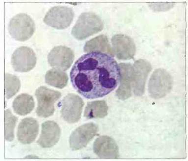
Fig. 2.32. Morphology of a neutrophil. A mature neutrophil with a lobed (segmented) nucleus in a blood smear. The clear identification in one such Cell of condensed Chromatin in the form of a "drumstick", representing the inactivated X chromosome, indicates that the blood belongs to a female. Giemsa stain, x 1500. After Zucker-Franklin D., Greaves M.F., Grossi C.E. et al. 1988. Atlas of Blood Cells: Function and Pathology Vol. II. 2nd edn. Milan: E.E. Ermes, Philadelphia: Lea and Febiger.

Fig. 2.33. Ultrastructure of a neutrophil. The Cytoplasm contains primary (P) and secondary (S) granules. Primary (more electron-dense) granules are far fewer in number. The Nucleus (N) is segmented; arrows indicate Pores in the nuclear envelope, x 17,500. After Zucker-Franklin D., Greaves M.F., Grossi C.E. et al. 1988. Atlas of Blood Cells: Function and Pathology. Vol. II. 2nd edn. Milan: E.E. Ermes, Philadelphia: Lea and Febiger.
Neutrophil chemotaxis is induced by protein fragments generated through complement activation (e.g., C5a), factors of the fibrinolytic and kinin systems, as well as products of leukocyte, platelet, and bacterial origin. Under METABOLISM/18.html">The Influence of chemotactic stimuli, margination (adhesion to endothelial cells) and diapedesis of neutrophils occur. This process is described in detail in Ch. 5.
Neutrophils possess a wide array of antimicrobial Proteins stored in Two Types of granules (Fig. 2.33). Primary (azurophilic) granules are Lysosomes containing acid Hydrolases, myeloperoxidase, and muramidase (Lysozyme). In secondary (specific) granules, lactoferrin is found In addition to lysozyme. Besides Enzymes and lactoferrin, these granules contain high concentrations of antimicrobial proteins—defensins, serprocidins, cathelicidins, and bacterial permeability-increasing protein. Microbes phagocytosed by neutrophils reside within vacuoles (called phagosomes) that fuse with lysosomes to form phagolysosomes (Fig. 2.34).
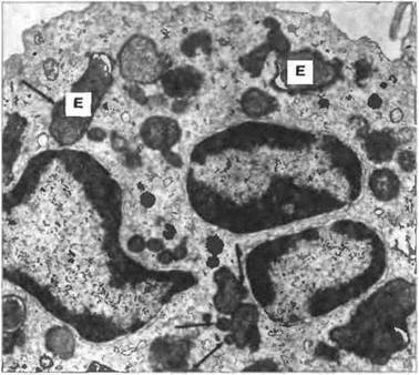
Fig. 2.34. A neutrophil that has phagocytosed Escherichia coli (E) cells. Numerous cells of this bacterium are visible within the phagolysosomes. The neutrophil is almost completely degranulated, with only a few lysosomes (indicated by arrows) still in The process of fusing with phagosomes, x 15,000. After Zucker-Franklin D., Greaves M.F., Grossi C.E. et al. 1988. Atlas of Blood Cells: Function and Pathology. Vol. II. 2nd edn. Milan: E.E. Ermes, Philadelphia: Lea and Febiger.
Upon activation by immune complexes via Fcγ receptors, neutrophils are also capable of releasing granule contents and cytotoxic compounds into the extracellular space. This mechanism likely underlies the Pathogenesis of immune complex diseases (type III hypersensitivity; see Ch. 25).
Eosinophils
Human blood eosinophils typically contain a bilobed nucleus and numerous cytoplasmic granules that stain with acidic Dyes such as eosin (Fig. 2.35). Among the leukocytes of a healthy, non-allergic individual, eosinophils account for 2–5%. They appear capable of phagocytosing and destroying ingested microbial cells, although this is not among their primary functions. The granules of mature eosinophils are membrane-bound cellular Organelles with a "crystalline" core, distinguishable against the surrounding matrix by their high electron density (Fig. 2.36).
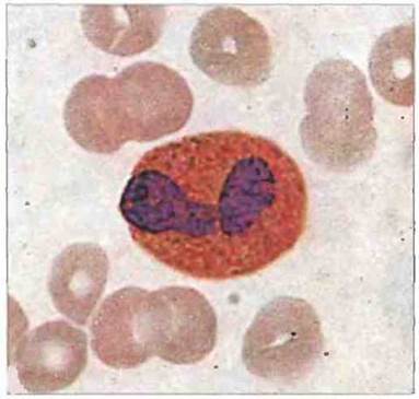
Fig. 2.35. Eosinophil morphology. The bilobed nucleus and eosinophilic cytoplasmic granules are visible. Giemsa stain, x 1000. After Zucker-Franklin D., Greaves M.F., Grossi C.E. et al. 1988. Atlas of Blood Cells: Function and Pathology. Vol. II. 2nd edn. Milan: E.E. Ermes, Philadelphia: Lea and Febiger.
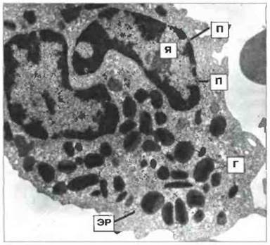
Fig. 2.36. Ultrastructure of a human eosinophil. A mature eosinophil contains granules (G) with a crystalline core in its cytoplasm. N - nucleus; ER - Endoplasmic reticulum; P - nuclear pores, x 17,500. After Zucker-Franklin D., Greaves M.F., Grossi C.E. et al. 1988. Atlas of Blood Cells: Function and Pathology. Vol. II. 2nd edn. Milan: E.E. Ermes, Philadelphia: Lea and Febiger.
Certain stimuli induce eosinophil degranulation, i.e., the fusion of granules with The Plasma Membrane and the release of their contents into the extracellular environment. The degranulation response is one of the mechanisms by which eosinophils use the toxic contents of their granules to destroy large targets (such as schistosomula) that resist phagocytosis (Fig. 2.37). Another mechanism involves the generation of toxic reactive oxygen metabolites. Both mechanisms likely form The basis of anti-helminth Immunity, in which eosinophils are hypothesized to play a specialized role (see Ch. 18).
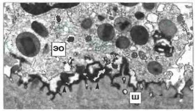
Fig. 2.37. Destruction of a schistosomulum by an eosinophil. An eosinophil (EO) has attached to an IgG-opsonized juvenile Schistosoma mansoni (S). Electron-dense material (indicated by arrows) containing major basic protein and eosinophil cationic protein can be observed at the contact sites, x 19,000. After Zucker-Franklin D., Greaves M.F., Grossi C.E. et al. 1988. Atlas of Blood Cells: Function and Pathology. Vol. II. 2nd edn. Milan: E.E. Ermes, Philadelphia: Lea and Febiger.
The recruitment of eosinophils to the site of parasite invasion is triggered by the release of specific products from T cells, mast cells, and basophils, such as eosinophil chemotactic factor of anaphylaxis (ECFA). In our example, these cells bind to The surface of schistosomula opsonized with specific IgG or IgE antibodies and undergo degranulation to release a toxin called major basic protein. This toxin is contained within the crystalline core of the eosinophil granule, whereas the granule matrix contains another toxic substance, eosinophil cationic protein. Eosinophils also release histaminase and arylsulfatase, which inactivate mast cell products such as histamine and the slow-reacting substance of anaphylaxis (SRS-A). Thus, the products released by eosinophils suppress the inflammatory response and, specifically, the migration of granulocytes to the site of invasion.
Basophils and Mast Cells
Basophils circulate in the blood in very low numbers, accounting for less than 0.2% of total leukocytes (Fig. 2.38). Mast cells are never found in Circulation and differ from basophils in several properties.

Fig. 2.38. Morphology of a basophil. A Wright-stained blood smear showing a typical basophilic granulocyte with dark purple granules, x 1000.
Two types of mast cells are known: mucosal mast cells and Connective Tissue mast cells. Unlike the latter, the proliferation of mucosal mast cells appears to depend on T cells. Both types of mast cells can be visualized by light Microscopy in preparations stained with basic dyes (Fig. 2.39). The cytoplasm of mature blood basophils contains irregularly distributed, membrane-bound granules (Fig. 2.40). The granules of basophils and mast cells contain heparin, SRS-A, histamine, and ECF-A.

Fig. 2.39. Human connective tissue mast cell in a histological section. Dark blue cytoplasm with purplish granules is visible. Alcian blue and safranin staining, x 600. (Photo courtesy of Dr. T.S. Orr.)
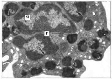
Fig. 2.40. Ultrastructure of a basophil. Showing the segmented nucleus (N) and large cytoplasmic granules (G), x 11,000. From Zucker-Franklin D., Greaves M.F., Grossi C.E. et al. 1988. Atlas of Blood Cells: Function and Pathology. Vol. II. 2nd edn. Milan: E.E. Ermes, Philadelphia: Lea and Febiger.
Often, a specific allergen (an antigen that triggers an allergic reaction) serves as a stimulus for the degranulation of mast cells or basophils. To achieve this, it must cross-link adjacent IgE molecules bound to high-affinity IgE receptors (FcεRI) on the plasma membrane of the mast cell or basophil. Degranulation results in the immediate release of the entire granule contents. First, the granules fuse with one another within the cytoplasm, and then their contents are expelled from The Cell (Fig. 2.41). Mediators secreted As a result of degranulation, such as histamine, cause pathological manifestations of allergy, but on the other hand, they play a positive role in antiparasitic immunity by enhancing the inflammatory response. Functional markers of granulocytes and mast cells are summarized in Fig. 2.42.

Fig. 2.41. Mast cell degranulation. Before the granule contents are expelled, the granules fuse with one another within the cytoplasm (indicated by arrows), x 5000. From Zucker-Franklin D., Greaves M.F., Grossi C.E. et al. 1988. Atlas of Blood Cells: Function and Pathology. Vol. II. 2nd edn. Milan: E.E. Ermes, Philadelphia: Lea and Febiger.
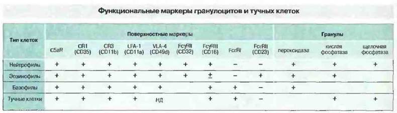
Fig. 2.42. Neutrophils, eosinophils, basophils, and mast cells respond chemotactically to C5a and therefore possess receptors for it. All of these cells also possess C3 receptors and express Cell Adhesion molecules - LFA-1 (CD11a) and VLA-4 (CD49d). In addition, they express FcγRII (CD32) and FcγRIII (CD16). Of these cells, only basophils and mast cells bear the high-affinity IgE receptor (FcεRI). Certain other Glycoproteins are detected on individual granulocytes, including CD13 and CD14 (expressed weakly). Furthermore, all these cells carry glycolipid molecules such as the Lewis X blood group antigen hapten (CD15) and lactosylceramide (CD17). The granules of different cell types vary in enzyme composition. ND - no data.
Platelets (Thrombocytes)
In addition to blood clotting, platelets also participate in the Immune Response, particularly in inflammatory reactions. They are produced from bone marrow megakaryocytes and contain granules (Fig. 2.43). In an adult human, 1011 new platelets are produced daily; on average, 30% of these cells are sequestered in the Spleen. Platelets express MHC class I proteins, IgG receptors (FcγRII; CD32), and low-affinity IgE receptors (FcεRII; CD23). In addition, megakaryocytes and platelets bear receptors for blood clotting factor VIII and other functionally important molecules, such as the GpIIb/IIIa complex (CD41) and the GpIb/GpIx complex (CD42). The former is a cytoadhesin responsible for binding to fibrinogen, Fibronectin, and vitronectin. Both of these complexes also serve as receptors for von Willebrand factor. There is also an additional vitronectin receptor, CD51. Both receptors and adhesion molecules are essential for platelet activation. Upon endothelial injury, they adhere to the subepithelial surface of the damaged vascular wall, forming aggregates. Simultaneously, the contents of two types of platelet granules, including serotonin and fibrinogen, are released, leading to increased capillary permeability, complement activation, and consequently, the recruitment of leukocytes.
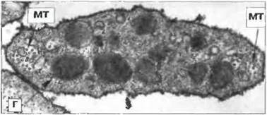
Fig. 2.43. Platelet ultrastructure. A cross-section of a blood platelet showing two types of granules (G) and bundles of microtubules (MT) at the poles, x 42,000. From Zucker-Franklin D., Greaves M.F., Grossi C.E. et al. 1988. Atlas of Blood Cells: Function and Pathology. Vol. II. 2nd edn. Milan: E.E. Ermes, Philadelphia: Lea and Febiger.
Questions for Review
■ What functional subpopulations of T lymphocytes exist, and how do they differ?
■ Why are so many different cells required to participate in the immune response?
■ What molecules are used by lymphocytes (a) as antigen-binding receptors and (b) for interactions among themselves and with other cells? What are the names of the various families of these molecules?
SUGGESTED READING
Levy O. Antibiotic proteins of polymorphonuclear leukocytes. Eur. J. Haematol. 56: 263-77, 1996.
Moller G. (ed.). Accessory molecules in the immune response. Immunol. Rev. 153: 1996.
Peters J.H., Gieseler R., Thiele B., Steinbach F. Dendritic cells: from ontogenetic orphans to myelomonocytic descendants. Immunol. Today 17: 273-8, 1996.
Playfair J.H.L. Immunology at a glance, 6th edn. Oxford: Blackwell Scientific Publications 1996.
Razin E., Pecht I., Rivera J. Signal Transduction in the activation of mast cells and basophils. Immunology Today 16: 370-3, 1995.
Reth M. The B-cell antigen receptor complex and coreceptors. Immunol. Today 16: 310-13, 1995.
Roitt I.M. Essential Immunology, 9th edn. Oxford: Blackwell Scientific Publications, 1997.
Romagnani S. Lymphokine production by human T cells in disease states. Annu. Rev. Immunol. 12: 227-57, 1994.
Wardlaw A.J., Moqbel R., Kay A.B. Eosinophils: biology and role in disease. Adv. Immunol. 60: 151-266, 1995.
Last update: 13/08/2026
Editorial and Educational Adaptation: This material has been compiled based on the primary/original source text. The project team performed an editorial review, corrected technical inaccuracies, structured sections, and adapted the content for an educational format.
What was processed:
- elimination of formatting defects (OCR errors, structural breaks, corrupted characters);
- editorial organization of content;
- standardization of terminology in accordance with academic sources;
- verification of factual statements against the original source text.
All mentions of the author, publication year, and origin of the primary text have been preserved in accordance with the source.