Molecular Biology: Protein Structure and Functions - Stepanov V.M. 2005
Post-translational protein modification
Protein glycosylation. Glycoproteins
Glycoproteins are Proteins that contain a covalently bound carbohydrate component. Based on the Structural Role of this carbohydrate part, glycoproteins can be divided into two classes. One Class includes so-called Proteoglycans, whose peptide chains are so densely studded with oligosaccharide chains that the molecule as a whole behaves like a branched polysaccharide, completely concealing its polypeptide backbone. Proteoglycans play a major role in forming the Extracellular matrix in animal Tissues. The second class comprises glycoproteins in which the polypeptide chain forms a Spatial Structure According to the usual rules characteristic of proteins, while the carbohydrate components are attached to the protein globule. The Discussion that follows will focus specifically on these glycoproteins.
There are two primary ways in which oligosaccharide chains are covalently attached to proteins.
11.4.1. N-Glycoproteins
In these glycoproteins, the carbohydrate component is attached via an N-glycosidic bond (hence the name) to the amide nitrogen of an asparagine residue embedded in the peptide chain. The first monosaccharide unit forming the N-glycosidic bond with asparagine in this group of glycoproteins is always N-acetylglucosamine, so the attachment "junction" has the following structure:

The N-glycosidic bond is readily cleaved in an acidic environment, making N-glycoproteins acid-labile. In this type of glycoprotein, an oligosaccharide is attached to the asparagine residue, consisting of a "junctional" N-acetylglucosamine residue linked via a 1-4 bond to another N-acetylglucosamine residue, followed by several mannose residues. Other monosaccharide units such as N-acetylglucosamine, galactose, neuraminic acid, and fucose may also be present.
The Biosynthesis of N-glycoproteins takes place in the rough Endoplasmic reticulum. A specialized enzyme transfers a highly complex oligosaccharide structure, Glc3Man9GlcNAc2, in a single step to the amide group of asparagine (where Glc is a glucose residue, Man is mannose, and GlcNAc is N-acetylglucosamine). This structure then undergoes more or less complex Processing—the Cleavage of terminal monosaccharide units, specifically the complete removal of glucose residues by specific glucosidases and partial removal of mannose by mannosidases. This leaves a so-called "core" to which other Monosaccharides are subsequently attached:
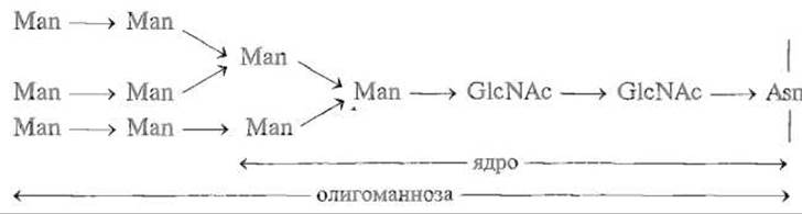
This is followed by the sequential incorporation of individual monosaccharide units, attaching trisaccharide structures such as NeuNAc → Gal → GlcNAc → to the "mannose core" (where NeuNAc is acetylneuraminic acid, Gal is galactose, and sometimes other monosaccharides like fucose — Fuc are also included). These reactions are specific, resulting in a completed oligosaccharide structure that varies among different proteins. Below is a typical branched STRUCTURE OF THE carbohydrate components in certain N-glycoproteins:
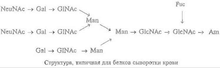
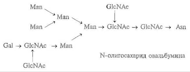
Only a small fraction of the asparagine residues in proteins undergo N-glycosylation. For example, in pancreatic Ribonuclease, out of 34 asparagine residues, only a single one located in the Asn-Leu-Thx sequence is glycosylated, leading to The formation of so-called ribonuclease B. As is often the case with post-translational modifications, the reaction goes to completion in only a fraction of the molecules, leaving a significant portion of the enzyme unglycosylated (ribonuclease A).
Studies of numerous N-glycoproteins have shown that the presence of a hydroxy amino acid—Threonine or Serine—separated by one residue from asparagine serves as a signal for a specific enzyme, oligosaccharyltransferase, which glycosylates the asparagine within the sequence Asn-Xaa-Ser(Thr), where Xaa is any amino acid. This makes it possible to predict potential N-glycosylation sites based on the Primary Cell/13.html">Protein Structure. The enzyme catalyzing N-glycosylation, oligosaccharyltransferase, "recognizes" the amide group of asparagine separated by a single amino acid residue from serine or threonine, provided that the polypeptide chain is unfolded. Apparently, the folding of the respective region into a spatial structure prevents glycosylation. This explains why one of the two asparagine residues in Ovalbumin is not glycosylated in vivo, whereas in vitro Oligosaccharides can be successfully attached to both residues within the characteristic N-glycosylation sequence mentioned above.
The antibiotic tunicamycin blocks protein N-glycosylation. The enzyme glycopeptidase F selectively cleaves oligosaccharides from N-glycoproteins.
N-glycosylation is characteristic of secretory proteins entering The endoplasmic reticulum, although it is not an absolute prerequisite for secretion.
11.4.2. O-Glycoproteins
In this type of glycoprotein, the carbohydrate component is attached via an O-glycosidic bond to the hydroxyl group of a serine or threonine residue, with an N-acetylgalactosamine residue always occupying the "junctional" position:

Unlike N-glycosylation, the biosynthesis of O-glycoproteins proceeds in a stepwise manner. Following the attachment of the "junctional" N-acetylgalactosamine, sequentially acting Glycosyltransferases add further monosaccharide units, forming, for example, the following structure:
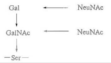
No unambiguous "signal" in the Introduction/19.html">Primary Structure of O-glycoproteins has been found that determines which serine or threonine residue is to be modified. However, there is evidence that glycosylation targets the hydroxyl group of a serine or threonine residue that follows a Proline residue and is part of a β-turn. This indicates that glycosylation occurs after the Spatial structure of the protein has already formed.
The O-glycosidic bond is cleaved in an alkaline environment, with the carbohydrate component separating from the protein moiety along with the hydrogen atom belonging to the α-carbon atom of serine, leaving behind a dehydroalanine residue in the polypeptide chain:

O-glycosylation is less common in Globular proteins and is more typical of mucins (substances that determine Blood Groups)—glycoproteins belonging to the proteoglycan class. However, IMMUNOGLOBULINS, for instance, feature both types of glycosylation simultaneously.
The functional role of carbohydrate components in glycoproteins is a highly complex question. In a few instances, researchers have successfully determined the spatial structure of glycoproteins. It turned out that oligosaccharide chains do not form extensive contacts with the protein globule, apart from a few Hydrogen Bonds near the attachment site to the protein; instead, they project away from the globule and are immersed in the Water surrounding the glycoprotein. For only one of the immunoglobulins G has it been shown that the carbohydrate component, containing six monosaccharide units, lies across the interface between two CH2 domains and apparently acts as a kind of hydrophilic spacer with an area of about 500 Å2, preventing the hydrophobic surfaces of these domains from sticking together (Fig. 11.1). In other cases studied, it is difficult to speak of a structure-forming role for the carbohydrate components.
The carbohydrate component almost never participates in The primary function of a protein; that is, it is not part of the catalytic center or enzyme-binding domain, nor is it found in the active sites of regulatory proteins, and so forth. The activity measured for pairs of glycosylated and unglycosylated Enzymes invariably turns out to be identical. An exception must be made, however, for cases where the function of a protein is signal Transduction—in such proteins, such as certain Hormones, the carbohydrate component is often critically important for activity.
As already mentioned, glycosylation accompanies protein secretion in animals, yet secretion can also occur without glycosylation; consequently, the incorporation of carbohydrate components is a result, rather than a prerequisite, of secretion. This is well illustrated by data on the proportion of glycoproteins among the proteins contained in pancreatic secretions (4.7%), cow's milk (58%), and Serum proteins synthesized in the Liver (59.7%). However, in a chicken egg, approximately 97% of the total proteins are glycosylated.
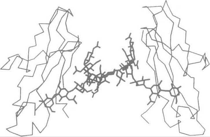
Fig. 11.1. Arrangement of carbohydrate chains attached to the asparagine residues Asn-297 (highlighted in bold lines) between the CH2 domains of rabbit immunoglobulin G.
Carbohydrate structures form a hydrophilic spacer between the domain surfaces
Apparently, glycosylation is important not for the structure or function of the protein as such, but rather for its interactions with Other components of The Cell or Organism, and for its "behavior" within these extraordinarily complex systems. Indeed, it is known that the phosphorylation of one of the mannose residues in the oligosaccharide structures of cytoplasmic enzymes—for example, the intracellular aspartyl proteinase cathepsin D—"targets" them to Lysosomes. The presence of another monosaccharide, sialic acid, in blood glycoproteins helps retain them in the Circulation; proteins that have undergone cleavage of their terminal sialic acid residues are rapidly cleared by the liver. For instance, ceruloplasmin, which circulates in the blood for an average of about 50 hours, is cleared within a few minutes after the removal of its terminal sialic acid.
The Physiological Role of carbohydrate components in membrane glycoproteins is undeniable. They are invariably located in the extracellular portion of the molecule, yet are almost completely absent from the cytoplasmic domains of these proteins. Carbohydrate components are involved in intercellular recognition, cellular differentiation and transformation, phagocytosis, Fertilization processes, cell-virus interactions, and the binding of hormones to receptor proteins.
Of particular interest are the glycoproteins of Antarctic fish, which lower the freezing point of Body Fluids—their functional role is quite obvious. The polypeptide chain of one such protein contains numerous Ala—Ala—Thr repeats, with disaccharide units of galactosyl-N-acetylgalactosamine attached to the hydroxyl groups of threonine.
Last update: 13/08/2026
Editorial and Educational Adaptation: This material has been compiled based on the primary/original source text. The project team performed an editorial review, corrected technical inaccuracies, structured sections, and adapted the content for an educational format.
What was processed:
- elimination of formatting defects (OCR errors, structural breaks, corrupted characters);
- editorial organization of content;
- standardization of terminology in accordance with academic sources;
- verification of factual statements against the original source text.
All mentions of the author, publication year, and origin of the primary text have been preserved in accordance with the source.