Tuberculosis Study Guide - M.M. Savula 2002
Pathogenesis and Pathological Anatomy
Pathogenesis. Following the initial entry of mycobacteria into The Human Body (which most commonly occurs during childhood or adolescence), active disease does not always develop. Humans possess a natural resistance to tuberculosis, driven by a complex of innate and acquired defense mechanisms. In children, these mechanisms are still immature, making infection particularly dangerous for them. The likelihood of developing the disease depends on the dose of MTB and the frequency of exposure. In the respiratory tract, MTB encounter a series of natural barriers. These primarily include the ciliated epithelium of the Bronchi and mucus containing active neutralizing substances. When these systems function normally and the number of pathogens is small, they are cleared from the respiratory tract without causing harm to the body.
However, if mycobacteria penetrate the respiratory mucosa into the Tissues or alveoli, they are engulfed by macrophages, initiating the interaction between the pathogen and the host. This process is accompanied by a complex cascade of immune responses, The ultimate outcome of which depends on many factors: age (children are more susceptible to tuberculous infection, and resistance also declines in old age), sex (men are more frequently affected by tuberculosis), individual genetically determined resistance or susceptibility to tuberculosis, comorbidities that lower resistance, as well as environmental and social factors.
The immunological reactions involving macrophages, T- and B-lymphocytes, and mediators of the Immune Response (cytokines) culminate in The Development of two main phenomena: 1) "acquired cellular resistance," which results in macrophage activation, enabling them to inhibit the Replication of mycobacteria and even destroy the intracellular pathogen; 2) "delayed-type hypersensitivity reactions," which lead to The formation of a cellular wall around the mycobacteria, isolating the pathogen (forming a granuloma). In the center of the granuloma, Cells die, resulting in caseous necrosis. Thus, this protective reaction (isolation of MTB) also carries a tissue-damaging effect. As a result of these processes, the number of mycobacteria decreases, although a certain population persists in the body as viable but metabolically inactive microorganisms. The process stabilizes and becomes contained, preventing the disease from developing. However, if the body is incapable of such a response, the process progresses. Macrophages fail to overcome the mycobacteria. A large number of macrophages, as well as polymorphonuclear leukocytes, disintegrate, releasing Proteolytic Enzymes that destroy surrounding tissues, while MTB multiply rapidly. Uncontrolled geometric progression of mycobacterial replication can lead to an enormous number of microorganisms—over 540 million within 20 days.
Thus, depending on the interplay of these complex processes, the infection may either be contained, leaving the individual healthy, or progress to active disease. In both cases, the delayed-type hypersensitivity to the tuberculosis pathogen and its breakdown products, which develops as a result of these processes and indicates MTB infection, can be detected using the Mantoux tuberculin Skin test (the first-time appearance of a positive reaction or its intensification in a BCG-vaccinated child). This change in sensitivity is commonly referred to as tuberculin conversion.
Morphological reactions developing in response to the penetration of mycobacteria into tissues begin with the formation of a tuberculous tubercle (Fig. 2 - see Appendix) (granuloma) measuring 0.4-0.6 mm. At its center, surrounding the mycobacteria, is an area of caseous necrosis (a manifestation of the alterative phase of inflammation). It is surrounded by epithelioid cells formed through the transformation of monocytes-macrophages, plasma cells, and histiocytes. Among them are occasional Langhans giant cells, surrounded by lymphoid cells. All of this represents the proliferative phase of inflammation. In a favorable course, the tubercle partially resolves, undergoes scarring, and the process subsides.
With the progression of tuberculosis (Fig. 3), multiple tubercles form and coalesce, creating foci (2) (tuberculous inflammatory foci up to 1 cm in size), which, in turn, can merge, enlarge, and form infiltrates (4). In the center of the infiltrate, there is an area of caseous necrosis that liquefies under METABOLISM/18.html">The Influence of proteolytic enzymes from leukocytes. The bronchial wall becomes involved and its integrity is compromised, allowing the liquefied caseous masses to break through into the bronchial lumen. Following the expulsion of these caseous masses, a cavity forms in the center of the infiltrate (cavitating infiltrate) (7), and once its walls are established, it becomes a tuberculous cavity (8), which serves as a source for further spread of the infection. Mycobacteria, along with the cavity contents and sputum, are disseminated throughout the bronchial tree, and fresh tuberculous foci of bronchogenic dissemination develop where they settle. This is a schematic representation of The Mechanism of Pulmonary Tuberculosis progression. Healing is possible at any of these stages. However, if extensive scarring (fibrous) changes (9) develop in the cavity wall and surrounding lung tissue, healing is impeded, and the process becomes irreversible.
Class="center">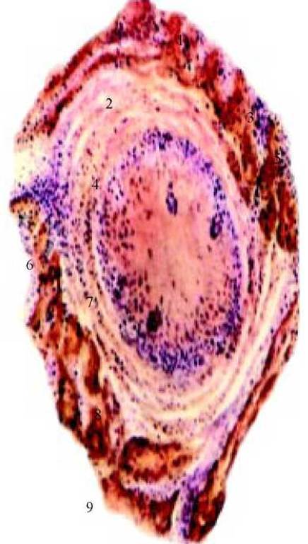
Fig. 3. Schematic diagram of tuberculosis progression: 1 - tubercle (granuloma); 2 - focus; 3 - scar; 4 - infiltrate; 5 - foci surrounded by fibrosis; 6 - Tuberculoma; 7 - cavitating infiltrate; 8 - cavity; 9 - old, fibrotic, deformed cavity surrounded by fibrosis.
Another route of infection spread is through the lymphatic and Blood Vessels, where mycobacteria travel from the hilar Lymph Nodes into the Thoracic duct, the SUPERIOR VENA CAVA system, the right side of The Heart, and from there into the Branches of the pulmonary artery. This is how hematogenous dissemination develops. The entry of the infection into the systemic Circulation lays the groundwork for the development of various Extrapulmonary tuberculosis sites, such as in the bones, Kidneys, Meninges, etc.
If the tuberculous process progresses immediately after the initial infection with mycobacteria, primary forms of tuberculosis develop. These are characterized by mandatory involvement of the lymph nodes (predominantly intrathoracic lymph nodes with massive caseation), a tendency to spread via lymphatic and blood vessels, and recent tuberculin conversion. These forms primarily develop in children and adolescents.
More commonly, the tuberculous tubercles formed during the primary infection heal, and the individual remains healthy, but "dormant" (ultrasmall, L-form) yet viable Mycobacterium tuberculosis persist within these residual lesions for a long time. When host resistance declines, sometimes many years later, these mycobacteria begin to multiply rapidly, leading to the development of so-called secondary tuberculosis. It develops against the Background of a certain acquired Immunity and therefore typically presents as localized organ damage. It is diagnosed predominantly in adults and rarely in children or adolescents.
Another possible pathway for the development of secondary tuberculosis is reinfection with virulent mycobacteria from a patient with active pulmonary tuberculosis.
Questions
1. What are the main reasons for the increase in tuberculosis cases worldwide and in Ukraine today?
2. What is the CAUSATIVE AGENT OF tuberculosis, and which of its species cause disease in humans?
3. How long can the tuberculosis pathogen survive in the environment, and what agents effectively destroy it?
4. What are the sources of human tuberculosis infection, and what is the most common route of transmission?
5. What factors reduce the body's resistance to tuberculosis?
6. Describe The Structure of a tuberculous tubercle, focus, and infiltrate.
7. WHAT IS A tuberculous cavity, and how does it form?
8. By what routes does tuberculous infection spread within the human body?
9. What are Primary tuberculosis and secondary tuberculosis?
Last update: 10/08/2026
Editorial and Educational Adaptation: This material has been compiled based on the primary/original source text. The project team performed an editorial review, corrected technical inaccuracies, structured sections, and adapted the content for an educational format.
What was processed:
- elimination of formatting defects (OCR errors, structural breaks, corrupted characters);
- editorial organization of content;
- standardization of terminology in accordance with academic sources;
- verification of factual statements against the original source text.
All mentions of the author, publication year, and origin of the primary text have been preserved in accordance with the source.