Review of Medical Physiology - William F. Ganong 2002
Functions of the Nervous System
Neural Basis of Instinctive Behavior and Emotions
Neurochemistry and Behavior
Pharmacological agents that modify human behavior include hallucinogenic substances (agents that induce hallucinations and other manifestations of psychosis); tranquilizers (agents that suppress anxiety and other psychiatric symptoms); and antidepressants (agents that elevate mood, enhance interest, and stimulate core drives). These and many other agents modify neurotransmission at synaptic contacts within the Brain. The Neurochemistry of known and putative synaptic Transmitters in both the peripheral and central nervous systems is examined in Chapter 4. This section focuses on The Role of these substances in instinctive behavior and emotions.
Aminergic Systems of the Brain
Four major aminergic systems are distinguished, whose neuronal Cell bodies are located in relatively sparse Regions of the brain; however, the axons of these Neurons are extensively branched and form connections with nearly all parts of The Nervous system (Figure 15-6): the serotonergic, noradrenergic, adrenergic, and histaminergic systems. The Cell bodies of dopaminergic neurons are located in multiple regions of the brain, yet their axons likewise project to diverse areas.
Serotonin
The cell bodies of serotonin-containing neurons are located in the median raphe nuclei of the Brainstem, and their axons project to the Hypothalamus, limbic system, neocortex, and Spinal Cord (see Figure 15-6).
The hallucinogenic agent lysergic acid diethylamide (LSD) is a serotonin agonist that exerts its effects by activating 5-HT2 receptors (see Chapter 4) in the brain. The transient hallucinations and other psychiatric disturbances triggered by this substance were first discovered by a chemist who accidentally inhaled it during synthesis. This discovery drew researchers' attention to the relationship between behavior and fluctuations in brain serotonin levels. Psilocybin, a compound found in certain mushrooms, and N,N-dimethyltryptamine (DMT) also produce hallucinogenic effects and, like serotonin, are tryptamine derivatives. The compound 2,5-dimethoxy-4-methylamphetamine (DOM), mescaline, and related compounds—which are likewise classical hallucinogens—are phenethylamines rather than indolamines. Nevertheless, the action of all these hallucinogens is evidently mediated by binding to 5-HT2 receptors. The compound 3,4-methylenedioxymethamphetamine (MDMA), or ecstasy, is also addictive. It induces euphoria, but is subsequently followed by difficulty concentrating, depression, and (in monkeys) insomnia. This agent causes the release and subsequent depletion of serotonin; thus, euphoria may result from the initial excessive release of serotonin, while the ensuing symptoms stem from its depletion.
As noted in Chapter 11, evidence indicates that serotonin agonists act as Sleep suppressors. Firing rates in serotonergic neurons are rapid during wakefulness, slow during drowsiness, even slower during sleep onset, and completely absent during rapid eye movement (REM) sleep (see Chapter 11).
Serotonin is believed to perform many other Functions in the brain. It may play a stimulatory role in regulating prolactin secretion (see Chapter 23). Descending serotonergic pathways likely inhibit pain transmission in the dorsal horns of the spinal cord. Furthermore, there is significant serotonergic input to the suprachiasmatic nuclei of the hypothalamus, and serotonin may be involved in The regulation of circadian rhythms (see Chapter 14). From a pathophysiological perspective, action potentials firing in the dorsal raphe serotonergic neurons likely trigger migraines, whereas antimigraine medications act precisely by slowing the propagation of such impulses.
Patients with depression exhibit a decreased concentration of the primary serotonin metabolite 5-HIAA in their CEREBROSPINAL FLUID (CSF) (see Figure 4-23). This fact has been used to challenge the hypothesis that depression is caused by low extracellular norepinephrine concentrations in the brain (see below), given that drugs inhibiting norepinephrine reuptake are crucial for treating depression. However, these agents also inhibit serotonin reuptake, and substances such as fluoxetine (Prozac), which inhibit serotonin reuptake without blocking norepinephrine reuptake, have proven to be equally effective antidepressants. Consequently, the focus in clinical depression has shifted from norepinephrine to serotonin. Interestingly, all these compounds require a delayed onset of administration—meaning their antidepressant efficacy manifests only after four to six weeks. This suggests that their therapeutic effect is secondary rather than a direct consequence of the initial reuptake inhibition that improves patient status.
Class="center">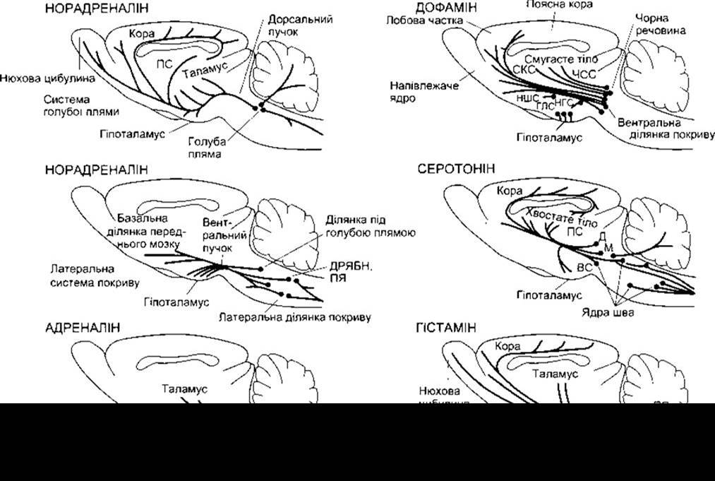
Figure 15-6. Aminergic pathways in the rat brain. Similar pathways exist in the human brain. The two primary noradrenergic systems (locus coeruleus and lateral tegmental area) are depicted separately. StSt = stria terminalis; DMNX = dorsal motor Nucleus of the Vagus nerve; NTS = nucleus tractus solitarius; PAG = periaqueductal gray; Nigi = nigrostriatal system; Mesoc = mesocortical system; Periv = periventricular system; Hypoth = hypothalamus-undetermined system; Tub = tuberoinfundibular system; D, M, and SC = dorsal, medial, and superior central reticular raphe nuclei.
As noted in Chapter 4, the blockade of NK-1 receptors, which mediate the effects of substance P, also alleviates depression through a mechanism that remains to be fully elucidated.
Mice with a knockout of the MAO type A Gene (see Chapter 4) and humans with mutant MAO A genes exhibit heightened aggressive behavior. In such mice, brain serotonin levels are markedly elevated. Animals lacking the 5-HT1B autoreceptor similarly display increased aggression.
Norepinephrine
The cell bodies of norepinephrine-containing neurons in the brain are located in the locus coeruleus and other nuclei of the Pons and Medulla Oblongata. The axons originating from the locus coeruleus form the locus coeruleus system. They descend into the spinal cord, project to the Cerebellum, and ascend to the paraventricular, supraoptic, and periventricular nuclei of the hypothalamus, the thalamus, the Base of the Telencephalon, and throughout the neocortex (see Figure 15-6). Axons of noradrenergic neurons arising from the dorsal motor nucleus of the vagus nerve, The Nucleus tractus solitarius, and the dorsal and lateral tegmental areas of the Midbrain form the lateral tegmental system, which transmits impulses to the spinal cord, brainstem, all regions of the hypothalamus, and the base of the telencephalon. Ascending fibers originating from the locus coeruleus constitute the dorsal noradrenergic bundle, whereas ascending fibers of the lateral tegmental system form the ventral noradrenergic bundle (see Figure 15-6).
Agents that increase brain norepinephrine concentrations improve mood, whereas those that decrease them induce depression. However, as noted above, current theories regarding the Pathogenesis of depression have largely shifted their focus from norepinephrine to serotonin. Furthermore, individuals with congenital DBH deficiency do not exhibit depression. Naturally, the role of monoamines in brain function is complex, as high extracellular neurotransmitter concentrations can trigger secondary effects, notably on receptors.
The function of the locus coeruleus system remains unclear, although it is known that its electrical activity increases in response to sudden sensory stimuli, which may be related
to alertness and wakefulness. The ventral noradrenergic tegmental system influences the secretion of hypophysiotropic Hormones that regulate anterior pituitary hormone production (see Chapter 14). Both norepinephrine and serotonin are apparently involved in body Temperature regulation.
Epinephrine
The medulla oblongata contains a system of neurons that express phenylethanolamine N-methyltransferase and project to the hypothalamus. These neurons synthesize epinephrine, though their exact function is not fully understood. Epinephrine-producing neurons maintain connections with the thalamus, periaqueductal gray, and spinal cord. Significant amounts of tyramine have also been detected in the Central Nervous System, though its precise function remains undefined.
Dopamine
The brain contains numerous dopaminergic systems. Based on the length of their axons, these systems are conventionally classified as ultrashort, intermediate, and long. Ultrashort dopaminergic neurons include Cells located between the inner nuclear and inner plexiform layers of the retina (see Chapter 8) and periglomerular Cells of the olfactory bulb (see Chapter 10). Dopaminergic neurons with intermediate-length axons comprise the following systems: the tuberoinfundibular system (see Figures 15-6 and 15-7), where dopamine is released into hypophysial portal vessels to inhibit prolactin secretion; the incertohypothalamic system, connecting the hypothalamus to the lateral septal nuclei; and a periventricular group of neurons in the medulla oblongata, whose cells are scattered along the walls of the Fourth ventricle. Long-axon dopaminergic systems (see Figure 15-6) include the nigrostriatal system, whose neuronal axons extend from the substantia nigra to the striatum and participate in motor control (see Chapter 12); and the mesocortical system, whose axons project from the midbrain tegmentum to the limbic cortex, olfactory tubercle, nucleus accumbens, and associated subcortical limbic structures. Modern studies utilizing PET scanning in healthy human subjects (see Chapter 32) have demonstrated that the number of dopamine receptors in the Basal Ganglia declines significantly with age. This reduction is more pronounced in males than in females.
The relationship between the mesocortical system—specifically its ventral portion extending from the tegmentum to the nucleus accumbens—and addiction was discussed earlier. Dysfunction of the mesocortical system is believed to account for at least a portion of schizophrenic symptoms. Initially, researchers focused on the hyperstimulation of D2 dopamine receptors in the limbic system. Amphetamine, which (like norepinephrine) stimulates dopamine release in the brain, induces a Schizophrenia-like psychosis. Given that the density of brain D2 receptors is elevated in schizophrenia, There is a logical positive correlation between the antipsychotic potency of many pharmacological agents and their ability to block D2 receptors. However, many modern antipsychotic drugs block D4 rather than D2 receptors, prompting ongoing research into whether these receptors are altered in individuals with schizophrenia.

Figure 15-7. Tuberoinfundibular dopaminergic system. Transverse section through the rat hypothalamus (Fluorescence Microscopy). Note the dopaminergic cell bodies of the arcuate nucleus on either side of the Third ventricle and the floor of the ventricle, as well as the dopaminergic terminals on the Vessels of the hypophysial portal system in the outer layer of the median eminence (reproduced with permission from Hökfelt T, Fuxe K: On the Morphology and the neuroendocrine role of the hypothalamic catecholamine neurons. In: Brain-Endocrine Interaction. Knigge K, Scott D, Weindl A [editors]. Karger, 1972).
Histamine
As noted in Chapter 4, the cell bodies of histaminergic neurons are located in the tuberomammillary nucleus of the ventral posterior hypothalamus. The axons of these neurons project to all PARTS OF THE brain (see Fig. 15-6). The exact function of these diffuse histaminergic systems remains unclear, but it is evident that brain histamine influences arousal, Sexual Behavior, Blood pressure, Water intake, pain threshold, and the regulation of anterior pituitary hormone secretion.
Acetylcholine
Acetylcholine is widely distributed throughout the central nervous system (CNS). High concentrations are found in the Cerebral Cortex, thalamus, and various nuclei of the basal Forebrain. The distribution of Choline acetyltransferase and acetylcholinesterase parallels that of acetylcholine. While the majority of acetylcholinesterase is localized in neurons, it has also been detected in glial cells. Pseudocholinesterase is found in many regions of the CNS. As discussed in Chapter 4, the brain contains various types of nicotinic and muscarinic receptors. Multiple nicotinic cholinergic subunits, along with the genes and Proteins encoding them, assemble into pentameric heterodimers with diverse configurations. Both postsynaptic and presynaptic nicotinic cholinergic receptors are present in the brain, exhibiting region-specific distributions.
The application of Antibodies specific to choline acetyltransferase, combined with immunocytochemical Methods, has made it possible to map cholinergic pathways in the brain. The distribution of cholinergic neurons resembles that of the monoaminergic system in that a portion of them project diffusely to many brain regions; however, it differs in the presence of cholinergic interneurons and short cholinergic systems throughout the CNS. Numerous projections from the nucleus basalis of Meynert and adjacent nuclei to the amygdala and the entire neocortex have also been documented, and these pathways are essential for motivation, sensory awareness, and cognitive function. A significant loss of neurons in this system is observed in Alzheimer's disease (see Chapter 16). The PGO-wave system, responsible for REM sleep, is likewise cholinergic. The relationship between nicotine and dependence was discussed above. In high doses, muscarinic blocking agents such as atropine can induce hallucinations, whereas scopolamine exerts a sedative effect.
As noted in Chapters 12 and 13, somatic motor, preganglionic, and some postganglionic neurons of the Autonomic nervous system are cholinergic. Cortical acetylcholinesterase levels are higher in socially housed rats than in isolated ones. Acetylcholine acts as an excitatory neurotransmitter in the basal nuclei, whereas dopamine functions as an inhibitory neurotransmitter in these structures (see Chapter 12).
Opioid Peptides
The brain contains Three types of neurons that express three precursor molecules for opioid peptides (see Table 4-4). In addition, There are two other endorphins whose precursors remain unknown. Neurons that synthesize proenkephalin are scattered throughout the brain, whereas cell bodies producing proopiomelanocortin are located in the arcuate nucleus, with their axons projecting to the thalamus and parts of the brainstem (Fig. 15-8). Prodynorphin is produced in neurons situated primarily in the hypothalamus, limbic system, and brainstem. Opioid peptides are implicated in various processes, including The Development of tolerance and addiction to morphine, although these mechanisms are not yet fully understood.
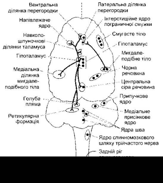
Fig. 15-8. Distribution in the brain of ß-endorphin- (left) and enkephalin-containing (right) neurons (reprinted with permission from Barchas JD et al: Behavioral neurochemistry: Neuroregulatory and behavioral states. Science 1978;200:964).
Other Transmitters
The association between GABA and anxiety, as well as the calming effects of benzodiazepines, was examined in Chapter 4. Besides enkephalins and ß-endorphin, other peptides evidently contribute to emotional and behavioral responses, though this matter requires further clarification.
Last update: 10/08/2026
Editorial and Educational Adaptation: This material has been compiled based on the primary/original source text. The project team performed an editorial review, corrected technical inaccuracies, structured sections, and adapted the content for an educational format.
What was processed:
- elimination of formatting defects (OCR errors, structural breaks, corrupted characters);
- editorial organization of content;
- standardization of terminology in accordance with academic sources;
- verification of factual statements against the original source text.
All mentions of the author, publication year, and origin of the primary text have been preserved in accordance with the source.