Review of Medical Physiology - William F. Ganong 2002
Functions of the Nervous System
Neural Bases of Instinctive Behavior and Emotions
Motivation and Addiction
Self-Stimulation
If an animal is placed in a cage equipped with special levers or barriers, it will sooner or later happen to press them by chance. If a lever is connected to an electrode implanted in a specific area of the Brain (the pleasure center), pressing it triggers stimulation of this region, causing the animal to stay close by and press the lever repeatedly. Over time, this activity occupies the majority of the animal's time; some animals may even forgo food and drink for its sake, pressing the lever until they collapse from exhaustion. For instance, rats press the lever 5,000–12,000 times per hour, and monkeys up to 17,000 times. Conversely, if the electrode is implanted in certain other areas (the aversion center), animals avoid pressing the lever, and The stimulation of these regions acts as a powerful unconditioned stimulus for establishing avoidance conditioning.
The areas whose stimulation encourages repeated lever pressing are located within the medial Forebrain bundle, which extends from the frontal cortex through the Hypothalamus to the Midbrain tegmentum (Fig. 15-4). The most active pathway in this regard is the dopaminergic pathway running from the ventral tegmental area to The Nucleus accumbens (see below). The points whose stimulation elicits an avoidance reaction are located in the lateral part of the posterior hypothalamus, the dorsal areas of the midbrain, and the entorhinal cortex. Although these areas are sometimes adjacent to those that trigger repeated lever pressing, they constitute a distinct system. The reward-eliciting areas are more extensive than the aversion-eliciting ones. Research has shown that in rats, repeated lever pressing is associated with 35% of the brain, whereas the avoidance reaction occurs upon stimulation of 5%, with the remaining 60% being indifferent.
Undoubtedly, some rewarding effect of the stimulation encourages the animal to stimulate itself again and again, though it remains unclear what the animal actually feels during this process. There are reports of relevant experiments involving lever pressing in humans with chronically implanted electrodes. Most of these individuals suffered from Schizophrenia or Epilepsy, while a few had internal organ tumors accompanied by intractable pain. Like animals, humans repeatedly press the lever. When describing their state during the experiment, they typically report experiencing pleasure, relief from tension, and a sense of calm and relaxation. Occasionally, the sensation is described as "joy" or "ecstasy," while a few individuals with the highest rates of self-stimulation could not explain the reason for their repeated pressing. Conversely, when electrodes were placed in areas where stimulation triggers avoidance, subjects characterized their state as a range of sensations from vague apprehension to sheer terror. Therefore, rather than using various vivid terms, it is arguably more appropriate to refer to these respective brain systems as the reward (or pleasure) system and the punishment (or aversion) system.
Agents that block postsynaptic D2 dopamine receptors decrease The rate of self-stimulation, whereas dopamine agonists increase it. The primary site of localization for these receptors appears to be the nucleus accumbens.
Class="center">
Fig. 15-4. Brain areas whose stimulation triggers repeated lever pressing, projected onto a sagittal section of a rat brain. Areas of the highest self-stimulation rate are indicated by rectangles (modified from Routtenberg A: The reward System of the brain. Sci Am [Nov] 1978;239:154).
The studies outlined above provide the physiological foundation for the assertion that behavior is driven not only by the reduction or avoidance of unpleasant sensations, but also—and primarily—by pleasure, as occurs when the brain's reward system is stimulated. Such stimulation provides powerful motivation for navigating a learning maze or performing other tasks. Evaluating these facts is crucial for understanding addictive behaviors, the classical drive-reduction theory of behavioral motivation involving the attenuation and enhancement of vitality, and dependency.
Addiction
Addictive behaviors—a compulsive, recurrent urge to consume substances despite adverse consequences—can arise under METABOLISM/18.html">The Influence of various factors. Unsurprisingly, this dependency is closely linked to the reward system, specifically the nucleus accumbens located at the Base of the striatum (Fig. 15-5) and the mesocortical dopaminergic Neurons, whose axons project from the midbrain to this nucleus and the frontal cortex (see below). Animals will press levers or perform other tasks until they receive an injection of the addictive substance via implanted catheters. The best-studied addictive substances include opiates such as morphine and heroin, as well as cocaine, ethyl alcohol, and nicotine. Although they all act on the brain through different pathways, they share the common property of increasing the concentration of dopamine acting on D3 receptors in the nucleus accumbens, thereby stimulating the brain's reward system. Over time, addictive substances induce tolerance, meaning that progressively larger amounts are required to achieve a state of gratification. Withdrawal leads to the onset of psychological and physical symptoms. The underlying mechanisms of tolerance and withdrawal are still not fully understood.
One of the hallmark features of addiction is the high tendency for relapse following Treatment. For opiates, for example, the relapse rate During the first year post-treatment reaches as high as 80%. Relapses are frequently triggered by visual cues, sounds, and contexts previously associated with The Use of the addictive substances. Excitatory glutamatergic fibers project from the medial prefrontal cortex, hippocampus, and amygdala to the nucleus accumbens, which can generate the neural activity that precipitates relapse under the Influence of the aforementioned environmental cues and memory.
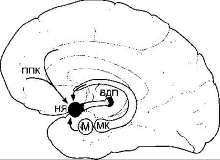
Fig. 15-5. Key brain regions involved in The Development of addiction. The ventral tegmental area (VTA) is connected via the mesocortical dopaminergic system to the nucleus accumbens (NA). The medial prefrontal cortex (mPFC), hippocampus (H), and amygdala (A) send excitatory glutamatergic inputs to the nucleus accumbens.
Last update: 10/08/2026
Editorial and Educational Adaptation: This material has been compiled based on the primary/original source text. The project team performed an editorial review, corrected technical inaccuracies, structured sections, and adapted the content for an educational format.
What was processed:
- elimination of formatting defects (OCR errors, structural breaks, corrupted characters);
- editorial organization of content;
- standardization of terminology in accordance with academic sources;
- verification of factual statements against the original source text.
All mentions of the author, publication year, and origin of the primary text have been preserved in accordance with the source.