Human Anatomy - H. I. Koliadenko 2009
The Study of the Viscera (Splanchnology)
Urogenital System
Urinary Organs
The URINARY AND REPRODUCTIVE Organs are combined into a single Urogenital System because they are closely interrelated both morphologically and functionally.
The Urinary Organs include the Kidneys, Ureters, Urinary Bladder, and Urethra (Fig. 109).
The Kidney is a paired, bean-shaped organ of a reddish-brown color, weighing 120–200 g, 10–12 cm long, 5–6 cm wide, and up to 4 cm thick. The anterior, posterior, and lateral surfaces are convex, while the medial surface is concave. On the medial surface is the renal hilum, through which the renal artery and nerves enter, and the renal vein, Ureter, and Lymphatic vessels exit. Externally, the kidney is covered by a fibrous capsule, over which a layer of fat is deposited, forming the adipose capsule. The largest accumulation of fat is found in the renal hilum. Upon incision, the capsule is easily peeled off. From above, the kidney is covered by a thin Connective Tissue fascia. The upper pole of the kidney is called the superior pole, and the lower one, the inferior pole. A small cap-shaped Adrenal gland rests on the upper pole of the kidney. The kidneys are located on the posterior wall of the Abdominal cavity on both sides of the spine at the level of the XII thoracic and I–II lumbar vertebrae. The right kidney lies 2–3 cm lower than the left; it contacts the Liver, transverse colon, and duodenum, whereas the left kidney contacts the Spleen, Stomach, Pancreas, and jejunum.
The kidney is fixed in the abdominal cavity by the anterior and posterior walls of the renal fascia, Blood Vessels, the adipose tissue surrounding the kidney, and intra-abdominal pressure.
A frontal section of the kidney (through the renal pelvis and its lateral margin) reveals that it consists of the renal cortex (5–7 mm thick), which has a reddish-brown color, and the renal medulla, which is slightly lighter (Fig. 110). However, it should be noted that the renal medulla has a heterogeneous coloration. It consists of 16–20 cone-shaped pyramids with their bases directed toward the renal cortex and their apexes toward the renal pelvis. Two to three pyramids, fusing together, form a larger pyramid. There are 7–8 such pyramids in the kidney. At the end of each, a papilla is formed, which is surrounded by a minor renal calyx, while 2–3 such calyces form a major renal calyx. By fusing together, the major calyces form the renal pelvis—a cone-shaped cavity with thin walls.
Class="center">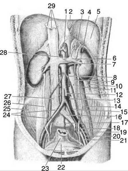
Fig. 109. Urinary organs:
1 — visceral artery; 2 — Esophagus; 3 — adrenal gland; 4 — kidney; 5 — Diaphragm; 6 — renal vein; 7 — renal artery; 8 — internal testicular (ovarian) artery; 9 — internal testicular (ovarian) vein; 10 — quadratus lumborum Muscle; 11, 26 — ureter; 12 — inferior mesenteric artery; 13 — Abdominal Aorta; 14 — psoas major muscle; 15 — left common iliac artery; 16 — left common iliac vein; 17 — median sacral artery; 18 — left hypogastric vein; 19 — left hypogastric artery; 20 — external iliac artery; 21 — external iliac vein; 22 — urinary bladder; 23 — rectum; 24 — common iliac artery and vein; 25 — right iliacus muscle; 27 — INFERIOR VENA CAVA; 28 — SUPERIOR VENA CAVA; 29 — hepatic Veins
The renal cortex penetrates into the renal medulla through extensions known as renal columns. The pyramids are permeated by A large number of renal tubules that open through numerous orifices at the apex of the pyramid.
The renal artery enters the renal hilum and branches into smaller Arteries that pass between the renal pyramids to their bases (Fig. 111). These are the interlobar arteries. At the base of each pyramid, they divide into two arcuate arteries. The arcuate arteries branch into numerous interlobular arteries. These give rise to afferent arterioles that divide into capillaries, forming the vascular (Malpighian) glomerulus. Its capillaries are exclusively of arterial origin, as they do not transition into venous capillaries, unlike in all other PARTS OF THE body. These vessels are referred to as the renal "wonderful net" (rete mirabile). The glomerular capillaries reunite into an arteriole, which is now called the efferent arteriole. The efferent arteriole, in turn, branches down to capillaries that transition into venous capillaries. These capillaries surround the nephron tubules (Fig. 112). The efferent arteriole is narrower than the afferent one.
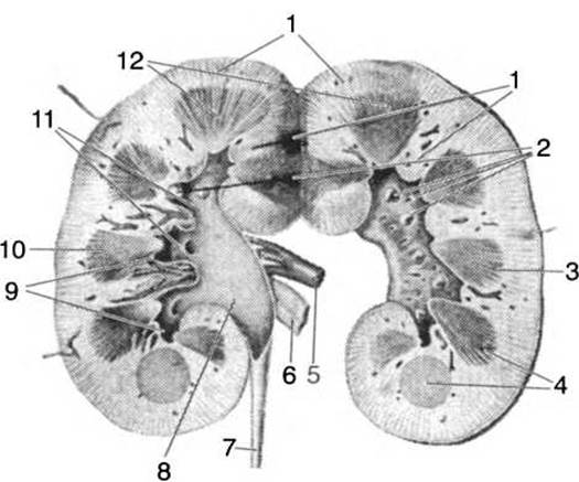
Fig. 110. Section of the kidney (diagram):
1 — renal columns; 2 — renal papillae; 3, 4, 10 — renal pyramids; 5 — renal artery; 6 — renal vein; 7 — ureter; 8 — renal pelvis; 9, 11 — renal calyces; 12 — renal cortex
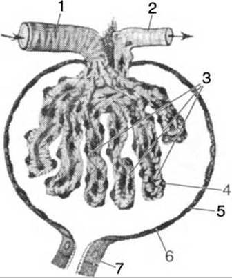
Fig. 111. Diagram of The Structure of the renal corpuscle:
1 — afferent arteriole; 2 — efferent arteriole; 3 — glomerular capillaries; 4 — Cells of the inner wall of the glomerular capsule; 5 — outer wall of the glomerular capsule; 6 — glomerular capsule space; 7 — beginning of the renal tubule
The Malpighian glomeruli are located in the renal cortex. Each glomerulus is surrounded by a double-walled capsule (Bowman's capsule), which is formed by the invagination of the blind end of the renal tubule inward. The inner layer adheres to the vascular glomerulus, while the outer layer continues into the renal tubule. A small space—the capsular space—is formed between the inner and outer layers of the capsule. The capsule walls consist of a single
layer of flat epithelial cells. The capsule gives rise to the proximal convoluted tubule of the first order, which is localized in the renal cortex. From it, a straight tubule extends into the renal medulla, reaches the pyramid, and upon reaching its middle, forms a loop (Loop of Henle) that ascends back to the renal cortex, where the tubule becomes convoluted again and is called the Distal convoluted tubule of the second order. This tubule transitions into the straight collecting renal duct. Several ducts, uniting together, form the papillary ducts, which pass through the pyramid and open at its apex.
The Malpighian vascular glomerulus, Bowman's capsule, and The system of renal tubules constitute the functional unit of the kidney—the nephron. Each kidney contains over 1 million nephrons, which can only be observed under a Microscope.

Fig. 112. Diagram of the nephron:
1 — glomerular capsule (Bowman's); 2, 4 — proximal convoluted tubules; 3 — distal convoluted tubule; 5 — collecting duct; 6 — straight tubule; 7 — loop of Henle
The kidney is not only an excretory organ but also an endocrine gland. At the transition point of the ascending limb of Henle's loop of the nephron into the distal part of the nephron tubule, between the afferent and efferent arterioles of the glomerulus, A large accumulation of nuclei is concentrated in the tubule wall, and the basement membrane is absent. This region of the distal segment is called the macula densa. In the areas of the walls of the afferent and efferent arterioles adjacent to the macula densa, beneath the endothelial cells, there are special granule-filled juxtaglomerular cells that produce the protein renin, which participates in the Regulation of Blood pressure, as well as the renal erythropoietic factor, which stimulates erythropoiesis.
Urine filtration from the blood takes place in the renal corpuscle, which comprises the vascular glomerulus and the capsule; this is where the so-called primary urine is formed.
Only one-third of the nephrons function simultaneously, while the others remain in reserve. Therefore, if necessary, The Human Body can survive with just one kidney, which simply increases somewhat in size. However, if the adipose capsule covering the kidney disappears or diminishes, or if the anchoring apparatus of the kidney weakens, such a kidney can change its previous position, a condition known as a floating (or mobile) kidney.
The ureters are cylindrical muscular tubes, 4–5 mm in diameter and 30–35 cm long. They originate from the renal pelves, run along the posterior abdominal wall, lie adjacent to the anterior surface of the psoas Muscles, reach the lesser pelvis, and, upon approaching the urinary bladder, pierce its wall obliquely to open into its cavity. The wall of the ureters consists of three layers: an inner mucous membrane (mucosa), a middle muscular layer, and an outer connective tissue layer (adventitia).
The urinary bladder (vesica urinaria, Fig. 113) is situated in the cavity of the lesser pelvis behind the Pubic Symphysis. It serves as a reservoir for urine delivered from the kidneys by the ureters. The average capacity of the urinary bladder is 750 cm3. The bladder consists of a body, an apex directed upward and forward, and a fundus (base). Posterior to the urinary bladder lie the Uterus and Vagina in females, and the rectum in males.
The interior of the urinary bladder is lined with a mucous membrane formed by transitional epithelium. As it fills with urine, the bladder expands due to the unfolding of the mucosal folds. The mucosa forms folds across its entire surface, with the exception of a small triangular area located on the floor of the bladder.
The muscular layer consists of three strata of smooth muscle cells. The inner and outer layers are longitudinally arranged, while the middle layer is circular. The outer layer is composed of connective tissue and is covered superiorly and posteriorly by the Peritoneum. The wall thickness of an empty urinary bladder is approximately 15 mm, decreasing to 2–3 mm when distended.
The ureteral orifices open at the corners of the trigone of the urinary bladder, piercing its wall obliquely. The mucous membrane forms a valve-like structure that ensures urine flows in only one direction—into the urinary bladder. Retrograde flow of urine is prevented by the oblique course of the ureters as they enter the bladder.
Urethra. The urethra originates from the inferior corner of the trigone on the floor of the urinary bladder and is guarded by two sphincters: an involuntary internal sphincter composed of smooth muscle, and a voluntary external sphincter formed by striated muscle fibers.
The female urethra is a tube approximately 3.5 cm long, lined with a mucous membrane. It opens into the Vestibule of the vagina, and its external orifice is encircled by a voluntary muscular sphincter.
The male urethra is a tube approximately 18–20 cm in length. It begins at the internal urethral orifice of the urinary bladder and terminates at the external orifice at the tip of the glans Penis. The urethra is lined with a mucous membrane containing glands that secrete mucus to lubricate the canal. The urethra is divided into three parts: prostatic, membranous, and spongy.
The prostatic part of the urethra begins at the internal orifice, perforates the Prostate Gland, and traverses its substance almost vertically. The smooth muscle fibers of the prostate form the involuntary internal sphincter.
The membranous part of the urethra is short (1–2 cm). Its striated muscles form the external voluntary urethral sphincter. This portion of the canal is firmly anchored to the pubic bones.
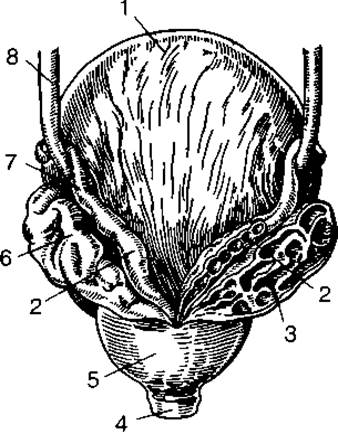
Fig. 113. Urinary bladder, prostate gland, and Seminal Vesicles:
1 — urinary bladder; 2 — ampulla of Ductus deferens; 3 — section of Seminal Vesicle; 4 — membranous part of urethra; 5 — prostate gland; 6 — seminal vesicle; 7 — ductus deferens; 8 — ureter
The spongy ( penile) part of the urethra is the longest (15–20 cm), runs along the corpus cavernosum, and terminates at the external urethral orifice.
Last update: 08/08/2026
Editorial and Educational Adaptation: This material has been compiled based on the primary/original source text. The project team performed an editorial review, corrected technical inaccuracies, structured sections, and adapted the content for an educational format.
What was processed:
- elimination of formatting defects (OCR errors, structural breaks, corrupted characters);
- editorial organization of content;
- standardization of terminology in accordance with academic sources;
- verification of factual statements against the original source text.
All mentions of the author, publication year, and origin of the primary text have been preserved in accordance with the source.