Human Anatomy - G. I. Koliadenko 2009
The Doctrine of Viscera (Splanchnology)
Respiratory System (Respiratory Organs)
The Respiratory system in The Human Body unites the Organs responsible for circulating inhaled air (enriched with oxygen) and exhaled air (saturated with carbon dioxide). This process, essential for the body's viability, constitutes METABOLISM/2.html">THE CONCEPT OF respiration. Oxygen from the air diffuses from the alveoli into the surrounding capillary Blood and is transported to the body Cells, while carbon dioxide—produced in the cells As a result of The oxidation of organic substances—passes from them into the blood. Carbon dioxide is carried by the blood to the Lungs, from where it is expelled through the respiratory tract during exhalation.
The respiratory apparatus includes the Nasal cavity with Paranasal Sinuses, the nasal part of the Pharynx, the Larynx, the Trachea, the Bronchi, and the lungs (see color insert, Fig. III).
The nasal cavity (cavum nasi) is formed by the nasal bones and cartilages. The nasal bones connect with the Frontal bone in the upper part of the Nose. The lateral surfaces of the nasal cavity—namely the nasal walls, the nasal alae, and partially the nasal septum—are formed of Cartilage. The internal surface of the nose is lined with a mucous membrane composed of pseudostratified ciliated epithelium. The nasal mucosa is rich in Blood Vessels. Its epithelial cover contains goblet cells and glands that secrete mucus. Through the beating of the epithelial cilia, the mucus, along with dust particles and foreign bodies, is cleared outward from the nasal cavity.
The nasal cavity has right and left halves that do not communicate with each other. Each half, in turn, consists of three nasal meatuses: superior, middle, and inferior. The sphenoid sinus opens into the superior meatus; the frontal, maxillary, and ethmoid sinuses open into the middle meatus; and the nasolacrimal duct opens into the inferior meatus. Posteriorly, the nasal cavity connects via the choanae with the nasopharynx, while anteriorly it opens outward through the nostrils.
Inhaled air entering the nasal cavity through the nostrils is warmed, moistened, and filtered of dust. Therefore, nasal breathing is much healthier than Mouth breathing.
The mucous membrane of the superior nasal meatuses is called the olfactory region because it contains olfactory bulbs with receptor cells emerging onto the epithelial surface. The middle and inferior nasal meatuses constitute the respiratory region.
The larynx (larynx, Figs. 105, 106) Functions as an organ of both respiration and articulated speech. It is located in the anterior neck region at the level of the IV, V, and VI cervical vertebrae. Superiorly, the larynx is connected to the Hyoid bone by the thyrohyoid ligament, and inferiorly it connects to the trachea.
The Skeleton of the larynx is formed by hyaline cartilages—the thyroid, cricoid, arytenoid, cuneiform, and corniculate cartilages—as well as the elastic epiglottis cartilage. The internal surface of the laryngeal cartilages is lined with a mucous membrane covered by pseudostratified ciliated epithelium containing numerous mucous glands.
Class="center">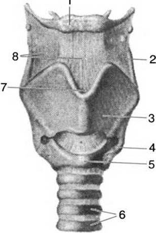
Fig. 105. Ligaments and cartilages of the larynx (anterior view):
1 — hyoid bone; 2 — superior horn of thyroid cartilage; 3 — thyroid cartilage; 4 — inferior horn of thyroid cartilage; 5 — arch of cricoid cartilage; 6 — tracheal cartilages; 7 — thyroid notch; 8 — thyrohyoid membrane
The thyroid cartilage (cartilago thyreoidea) is unpaired and forms almost the entire anterior and lateral walls of the larynx. It consists of two quadrilateral laminae that meet anteriorly to form an angle. In men, this prominence is known as the Adam's apple, projecting on the anterior surface of the neck. The posterior corners of the laminae form the superior and inferior horns. The superior horns are considerably larger and directed toward the greater horns of the hyoid bone, whereas the inferior horns articulate with the cricoid cartilage. The upper edge of the cartilage features a central notch above the Adam's apple, from which the thyrohyoid ligament originates. The superior horns connect to the hyoid bone via lateral thyrohyoid ligaments. The lower edge of the cartilage connects to the cricoid cartilage through the cricothyroid ligament and the cricothyroid joint, which allows slight tilting of the thyroid cartilage relative to the cricoid.
The cricoid cartilage (cartilago cricoidea) resembles a signet ring with a wide lamina in shape. It encircles the larynx anteriorly, while its lamina lies on the posterior surface. The upper margin of the cricoid lamina bears two articular surfaces for articulation with the arytenoid cartilages.
The arytenoid cartilage (cartilago arytenoidea) is paired and resembles two three-sided pyramids; each articulates by its base with the lamina of the cricoid cartilage, forming a joint with a vertical axis of rotation. This joint permits rotational movement around a vertical axis passing through the joint, as well as a sliding motion of the arytenoid cartilage forward and backward. The vocal process projects anteriorly from the Base of the arytenoid cartilage into the laryngeal cavity, while the muscular process projects posteriorly.
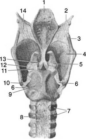
Fig. 106. Ligaments and cartilages of the larynx (posterior view):
1 — epiglottis; 2 — greater horn of hyoid bone; 3 — superior horn of thyroid cartilage; 4 — thyroid cartilage; 5 — arytenoid cartilage; 6 — cricothyroid joint; 7 — tracheal cartilages; 8 — membranous wall of trachea; 9 — lamina of cricoid cartilage; 10 — inferior horn of thyroid cartilage; 11 — cricoarytenoid joint; 12 — muscular process; 13 — vocal process; 14 — thyrohyoid membrane
A layer of elastic tissue lies between the laryngeal mucosa and the cartilages, forming the elastic cone between the inner angle of the thyroid cartilage and the arytenoid cartilages. Vocal cords, composed primarily of elastic fibers, extend from the upper thickened edges of this cone. The vocal cords attach to the vocal process of the arytenoid cartilages. Due to the Adam's apple, the length of the vocal cords is greater in men, measuring 22—24 mm, compared to 15—18 mm in women. Consequently, the male voice is lower, while the female voice is higher.
The epiglottis (epiglottis) is shaped like a tree leaf. Its narrowed lower part is connected by ligaments to the middle of the inner surface of the thyroid cartilage, while another attachment links it to the hyoid bone. The epiglottis closes the entrance to the larynx during the act of swallowing.
The larynx contains numerous small, paired striated Muscles. Those fixing it to the hyoid bone are termed extrinsic muscles, whereas those incorporated within the larynx itself are intrinsic muscles. Among the intrinsic muscles are the cricothyroid muscles, located externally on the lateral surface of the larynx. By contracting, they pull the thyroid cartilage away from the arytenoid cartilage, thereby tensioning the vocal cords and shortening them.
The internal Muscles of the larynx are referred to as vocal muscles, which include the posterior cricoarytenoids. They originate on the posterior surface of the cricoid lamina and insert into the muscular process of the arytenoid cartilage. When these muscles contract, they pull the muscular process posteriorly and medially; this causes the vocal processes to diverge, the rima glottidis to widen, and the vocal cords to tense.
The lateral cricoarytenoid muscles extend from the lateral part of the cricoid arch to the muscular process of the arytenoid cartilage. Upon contraction, they rotate the arytenoid cartilages inward, which promotes narrowing of the rima glottidis and relaxation of the vocal cords.
The transverse and oblique arytenoid muscles attach to the posterior surface of the arytenoid cartilages. By contracting, these muscles narrow the posterior part of the rima glottidis.
The aryepiglottic and thyroepiglottic muscles originate from the lateral margin of the epiglottis and insert into the cartilages—the former into the arytenoid, and the latter into the thyroid. By contracting, these muscles alter THE POSITION OF the epiglottis: the aryepiglottic muscles tilt it downward to close the laryngeal entrance, whereas the thyroepiglottic muscles lift it to open the entrance.
The laryngeal cavity is divided into upper (known as the vestibule), middle, and lower compartments. The boundary of the upper compartment is formed by the vestibular folds created by the laryngeal mucosa. In the middle compartment, situated between the vestibular folds and the underlying vocal folds of the mucous membrane, lies the vocal apparatus proper. The vocal folds of the mucosa cover the vocal processes, bounding the rima glottidis. The vocal folds are covered by stratified squamous epithelium. Blind recesses known as laryngeal ventricles are formed on the right and left sides between the vestibular and vocal folds. The lower part of the larynx is called the subglottic cavity.
Sound is produced in the larynx through the oscillatory Movements of the vocal cords, which result from the contraction of the laryngeal muscles during expiration. Spoken language is the result of Changes in the shape of the rima glottidis and the tension of the vocal cords. Voice timbre depends on the length of the vocal cords, Resonance within the laryngeal, pharyngeal, and oral cavities, as well as the paranasal sinuses of the Skull bones.
The trachea (trachea, Fig. 107) is a tube 11—13 cm in length, featuring 16—18 incomplete cartilaginous rings whose posterior ends are connected by a connective-tissue membrane composed of smooth Muscle. Together, they form a continuous membranous wall adjacent to the Esophagus. This wall can slightly indent into the lumen of the trachea during the passage of food into The Stomach. Adjacent tracheal cartilages are interconnected by annular ligaments.
The internal surface of the trachea is lined with a mucous membrane covered by ciliated columnar epithelium, which contains goblet cells and glands that secrete mucus.
After the age of 40, the tracheal cartilages begin to undergo ossification (calcification).
At the level of the IV—V thoracic vertebrae, the trachea bifurcates into the right and left bronchi. This division is known as the tracheal bifurcation. The left bronchus branches off from the trachea at an almost right angle, whereas the right bronchus is more vertically inclined downward. Consequently, foreign objects entering the trachea (especially in young children) invariably penetrate the right bronchus. Structurally, the bronchi resemble the trachea: they consist of cartilaginous half-rings connected by a connective-tissue membrane that forms the posterior wall. The right bronchus is wider but shorter than the left—about 3 cm in length, whereas the left bronchus is longer and narrower, measuring 4—5 cm. These bronchi are termed primary bronchi. At the pulmonary hilum, they divide into secondary, tertiary, and higher-order bronchi. As the order of division increases, the diameter of the bronchi decreases. This branching network of bronchi is referred to as the bronchial tree. As they branch further, the bronchi lose their cartilage; the finest bronchial branches (about 1 mm in diameter) are called bronchioles.

Fig. 107. Respiratory tracts:
1 — thyroid cartilage; 2 — cricoid cartilage; 3 — tracheal cartilages; 4, 11 — esophagus; 5 — bifurcation of the trachea; 6 — main left bronchus; 7 — left pulmonary artery; 8, 9, 12, 13 — bronchial branches; 10 — aorta; 14 — right pulmonary artery; 15 — main right bronchus
The bronchioles enter the pulmonary lobules, which are separated from one another by delicate layers of Connective Tissue. The mucous membrane of the bronchi is lined with ciliated epithelium containing mucous glands. In addition to these, the bronchioles contain distinct secretory cells (Clara cells) that produce Enzymes which break down surfactant (a protein substance) that prevents alveolar collapse.
Bronchioles transition into alveolar ducts, the walls of which feature numerous outpouches—pulmonary alveoli, or air sacs—which represent the terminal structures of the respiratory tract. The alveolar walls are extremely thin, composed of elastic fibers covered on the luminal side by a simple squamous epithelium, with blood capillaries located on the outer side of the elastic fibers. Gas exchange between the alveolar air and the blood takes place in the alveoli. It is estimated that the number of alveoli in both lungs reaches 700 million, with a total surface area of 100—120 m2. The total respiratory surface area of the lungs is 75 times greater than the surface area of the human body.
The lungs (pulmones, Fig. 108) are a massive organ comprising the right and left lungs. They fill almost the entire thoracic cavity (4/5 of its volume) and are cone-shaped, with the base resting against the Diaphragm and the apex extending beyond the Ribs to project 20—30 mm above the clavicle. The body of the lung lies between the apex and the base. The surface adjacent to the ribs, bearing impressions, is termed the costal surface.
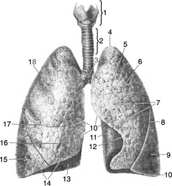
Fig. 108. Lungs:
1 — larynx; 2 — trachea; 3 — bifurcation of the trachea; 4 — apex of the lung; 5 — costal surface; 6 — superior lobe of the left lung; 7 — interlobar notch; 8 — inferior lobe of the left lung; 9 — anterior margin of the lung; 10 — base of the lung; 11 — posterior margin of the lung; 12 — cardiac notch; 13 — diaphragmatic surface; 14 — inferior margin of the lung; 15 — inferior part of the right lung; 16 — middle lobe of the right lung; 17 — interlobar surfaces; 18 — superior lobe of the right lung
The lower part of the lung (the base) adjacent to the diaphragm is called the diaphragmatic surface, while the part of the lung facing the corresponding surface of the opposite lung is termed the medial surface. The medial surface is also referred to as the mediastinal surface (facing the mediastinal organs).
The right lung is larger than the left; it is divided by two planes (horizontal and oblique) into three lobes: superior, middle, and inferior. The left lung is smaller in size and is divided by an oblique plane into two lobes: superior and inferior. The lobes, in turn, are subdivided into bronchopulmonary segments. There are 11 segments identified in the right lung and 10 in the left. Each segment is supplied by a segmental bronchus and a corresponding branch of the pulmonary artery. The Medial surface of the left lung features the cardiac fossa, and its anterior margin bears the cardiac notch.
The mass of the lungs is 0.5—0.6 kg, and their color is grayish-pink. On the mediastinal part of the medial surface is the hilum of the lung, which transmits the bronchus, pulmonary artery, and nerves supplying the lungs and bronchi, while two Pulmonary Veins and pulmonary Lymphatic vessels, bound together by connective tissue, emerge from it. These structures form the ROOT of the lung, by which it is anchored within the thoracic cavity.
The structural unit of the lungs is the pulmonary lobule, which has the shape of an irregular pyramid with its base facing the outer surface of the lungs and its apex directed inward. The lobules are separated from one another by connective tissue.
The functional unit of the pulmonary lobule is the acinus. A pulmonary lobule comprises 16—18 acini. The acinus, or secondary lobule, is formed by terminal bronchioles (each marking the beginning of an acinus) that branch into alveolar (or respiratory) bronchioles and their corresponding alveolar ducts, sacs, and alveoli.
The inflation of the alveoli during inspiration and the Prevention of their collapse during expiration are facilitated by alveolar surface tension, which is maintained by surfactant secreted by the alveolar walls. Surfactant prevents fluid from entering the alveoli and performs a protective function. It is composed of Phospholipids, Proteins, and Glycoproteins.
The lungs are permanently filled with air. In men, the lungs can hold 5—6 liters of air. During quiet breathing, a person exchanges about 0.5 liters of air with each respiratory cycle. During physical exertion, this volume increases to 3—3.5 liters. Even the lungs of a deceased person remain filled with air.
The lungs of a stillborn child contain no air because, within the mother's womb, the fetus receives oxygen directly from the maternal Organism.
Pleura. Each lung is enclosed by the pleura, a thin, glistening serous membrane consisting of two layers: the visceral (pulmonary) and parietal pleura. The pulmonary pleura tightly covers the lung tissue on all sides. Near the lung root, the pulmonary pleura transitions into the parietal pleura. Between these two layers lies the pleural cavity, which contains a small amount (about 20 ml) of serous fluid that reduces friction between the pleural membranes during breathing. The parietal pleura is subdivided into costal, diaphragmatic, and mediastinal parts, which line the corresponding Organs of the thoracic cavity. The spaces between the pleural folds that are not occupied by the lungs are called pleural recesses: the costodiaphragmatic, phrenicomediastinal, and costomediastinal recesses.
The Mediastinum refers to the cavity located between the medial surfaces of the right and left lungs. By convention, a plane passing through the anterior margin of the pulmonary hilum divides the mediastinum into the anterior and posterior mediastinum. The anterior mediastinum contains The Heart with its large outgoing vessels (the ascending aorta, pulmonary trunk, SUPERIOR VENA CAVA), the Thymus gland, Lymph Nodes, and nerves; the posterior mediastinum contains the esophagus, the trachea with the primary bronchi, the descending part of the aorta, the Thoracic duct, the azygos and hemiazygos veins, the vagus nerves, the sympathetic trunks, and lymph nodes.
Last update: 08/08/2026
Editorial and Educational Adaptation: This material has been compiled based on the primary/original source text. The project team performed an editorial review, corrected technical inaccuracies, structured sections, and adapted the content for an educational format.
What was processed:
- elimination of formatting defects (OCR errors, structural breaks, corrupted characters);
- editorial organization of content;
- standardization of terminology in accordance with academic sources;
- verification of factual statements against the original source text.
All mentions of the author, publication year, and origin of the primary text have been preserved in accordance with the source.