Human Anatomy, Part 1 - K. A. Dyubenko, A. K. Kolomiysev, Yu. B. Chaykovsky 2002
Special Part
Osteology, osteologia [the science of bones] – General Osteology
Structure of Tubular Bones
Bones are composed of compact bone, substantia compacta, and spongy bone, substantia sponqiosa s. [trabecularis]. The compact bone is located at the periphery and appears as a whitish plate that forms the diaphyses of long bones. In the superficial zone of the compact bone of long bone diaphyses, There is a layer of outer circumferential lamellae, stratum lamellarum generałom externum, followed by the osteonal layer, stratum osteonarum, and subsequently the layer of inner circumferential lamellae, stratum lamellarum generaliom internum.
The outer circumferential lamellae do not form complete rings around the bone but are instead overlapped by subsequent layers of lamellae. Perforating, or Volkmann's canals (Fig. 17), run through this region, housing Blood Vessels. These bone canals transition into larger nutrient canals, canalis nutriens, which open onto the bone surface via nutrient foramina, foramen nutriens (LNA).
The inner circumferential lamellae are better developed where the compact bone bounds the medullary cavity. Between the outer and inner layers of circumferential lamellae lies a system of lamellae that forms the bulk of the cortical substance of the diaphysis, namely the osteonal layer.
The osteonal layer can be visualized as a system of parallel cylinders, with the spaces between them filled with interstitial lamellae (Fig. 18). In this layer, bone lamellae are arranged concentrically around blood vessels. Such systems consisting of 5–20 concentrically arranged bone lamellae are called osteons, or Haversian systems. The canals located in the center of the osteon, through which blood vessels pass, are called osteonal canals, canales osteoni, or Haversian canals. Osteons serve as the structural unit of the compact bone of long bones, and each is separated from adjacent osteons by a cementing line.
Spongy bone is constructed of bony trabeculae and is predominantly found in the epiphyses of bones. The trabeculae are not arranged haphazardly, but rather perpendicular to the lines of compression and tension. They intersect at a 90° angle to form columns, and cross the long axis of the bones at a 45° angle (Fig. 19). The trabeculae are oriented with one end in the direction of the pressure force and the other abutting the compact bone, As a result of which forces are divided into two components, and the applied force is evenly distributed across the walls of the tubular bone.
In the middle of the diaphyseal region, the medullary cavity, cavum medullare, is formed; it is lined with a thin Connective Tissue membrane, the endosteum, endosteum, and filled with Bone Marrow, medulla ossea. There are two Types of bone marrow: red, medulla ossium rubra, and yellow, medulla ossium flava. The diaphyses and epiphyses of long bones, as well as all spongy and flat bones, contain predominantly Cytology/practical/86.html">Red bone marrow with a smaller amount of yellow bone marrow.
Red bone marrow is permeated by A large number of blood vessels containing sinusoid-type capillaries that facilitate the release of mature Blood Cells into the bloodstream. The total mass of red bone marrow accounts for 3.4–5.9% of total body weight. In fetuses and newborns, the bone marrow in all bones is red, but starting from 12–18 years of age, it is gradually replaced by yellow marrow. Its color is imparted by fat deposits. In cases of significant blood loss or blood system disorders, foci of hematopoiesis can form within yellow bone marrow.
As noted previously, the external surface of bones, with the exception of articular surfaces and tendon attachment sites, is covered by the periosteum, periosteum. The periosteum is a thin connective tissue membrane (100–2000 µm) consisting of two layers: an inner (osteogenic) and an outer (fibrous) layer. The periosteum is rich in blood vessels, Lymphatic vessels, and nerve elements. It contains two networks of lymphatic capillaries (V. N. Nadezhdin, 1951) (Fig. 20). A fine capillary network with a diameter of 0.01–0.02 mm is located in the osteogenic layer. The Superficial layer of the periosteum contains a network of larger, irregularly shaped capillaries with numerous lacunae. The periosteum is responsible for bone growth in thickness, its Nutrition, and healing after fractures.
Cell/15.html">Microscopy of bone histological sections reveals that the bulk of bone consists of Bone tissue formed by Collagen fibers, ground intercellular substance (oseomukoid), and Inorganic Compounds, among which calcium salts predominate. The latter provide bone hardness. Located between the bone lamellae within bone lacunae are the bodies of osteocytes, interconnected by their processes situated within bone canaliculi.
Class="center">
Fig. 17. Diagram of The Structure of a long bone (after W. Bergmann):
1 - osteones; 2 - substantia compacta; 3 - periosteum; 4 - trabeculae substantia spongiosa
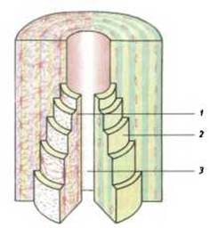
Fig. 18. Diagram of an osteon (after Braus): 1 - bone cells; 2 - interstitial substance; 3 - Haversian canal

Fig. 19. Architecture of the proximal epiphysis of the Femur (after F. Kiss and J. Szentágothai):
1 - trabeculae capitis; 2 - epiphysis caput femoris; 3 - linea epiphysialis; 4 - trabeculae colli; 5 - substantia spongiosa; 6 - substantia compacta; 7 - diaphysis corpus femoris; 8 - trabeculae trochanteris majoris; 9 - apophysis; 10 - linea apophysialis

Fig. 20. Blood and Lymphatic Vessels of the tibial periosteum (after V. N. Nadezhdin):
1 - deep network of lymphatic capillaries; 2 - superficial network of lymphatic capillaries; 3 - Water/144.html">Origin of the efferent lymphatic vessel
In images obtained by scanning Electron microscopy, the osteonal canals of compact bone are 300–350 µm long and may interconnect via various types of anastomoses (N. T. Gonchar-Zaikina, 1977). A perivascular space is formed between the blood vessel wall and the endosteum lining the osteonal canal. It contains connective tissue elements, osteoblasts, blood formed elements, and fibrous structures that weave into the canal wall on one side and into the wall of the vessel
located within it on the other. Thus, a unique framework is formed, suspending the blood vessel within the lumen of the canal.
Bones are dynamic structures that constantly renew themselves through remodeling driven by the interrelated processes of resorption and formation characteristic of living bone throughout human life. Remodeling is most intense During the first two years of life and during Puberty.
In the compact bone of an adult human, there is an average of 70% inorganic substances, 20% organic substances, and 10% water. In spongy bone, mineral components account for 35–40%, organic components for 50–55%, and water for 5–15%. Over 95% of the organic matter consists of the protein collagen. Bone collagen is characterized by a high content of amino groups. The organic matrix of bone—ossein—provides its shape and imparts elasticity.
Inorganic substances in bones are represented by various salts, the vast majority of which are calcium phosphate salts—60%, calcium carbonate—5.9%, magnesium sulfate—1.4%, and calcium fluoride, sodium carbonate, and sodium chloride—1%. In addition, bones contain Vitamins retinol, calciferol, and ascorbic acid. The ratio of organic to inorganic substances, as well as their internal architecture, determines the strength, resilience, and relative lightness of bone tissue.
Bone mineral salts easily dissolve in organic Solvents, a process known as decalcification. Following this Treatment, the organic matter remains within the bones, preserving their shape. When bone organic matter is burned, the original shape is also retained, yet the bones become brittle.
With age, The chemical composition of bones changes: the proportion of organic components decreases while inorganic ones increase. Alterations in bone chemical composition also occur in certain pathologies, such as Rickets, Osteomalacia, and marble bone disease (osteopetrosis), which can be accompanied by fractures.
Blood supply. Bones receive their blood supply from the branches of nearby arterial vessels—main, muscular, and fascial. Within the periosteum, these vessels form plexuses and networks, whose branches penetrate the dense bone layer through nutrient foramina (foramen nutriens), Volkmann's and Haversian canals, ultimately reaching the bone marrow. The vascular networks within the periosteal layers exhibit specific structural features and are interconnected by numerous anastomoses. According to M. G. Prives and N. B. Likhachyova (1955), long tubular bones have multiple sources of blood supply, the primary one being the main diaphyseal artery (a. diaphyseos princeps), which enters the tubular bone and divides into two branches: r. proximalis and r. distalis, along with additional diaphyseal Arteries (aa. diaphyseos accessoriae) that enter the bone at the ends of the diaphysis. The diaphyseal arteries nourish the diaphysis from the inside, whereas the cortical layer is supplied by Branches of the periosteal arteries (cortical arteries). Alongside diaphyseal arteries, long tubular bones are supplied by metaphyseal, epiphyseal, and apophyseal arteries. Short tubular bones with a single epiphysis (metacarpal and Metatarsal Bones) have a single source of blood supply—epiphyseal arteries that penetrate them from various directions. Bone Veins originate from the capillary network of the bone marrow. They form intraosseous plexuses before emerging onto the surface: in tubular bones, this occurs at the border between the epiphysis and diaphysis, and in short bones, near the articular surfaces.
Lymph drainage. Lymph drainage from the periosteum proceeds via both deep and superficial lymphatic pathways (D. A. Zhdanov, 1940).
Bone innervation. Bones are innervated by nearby nerves that form plexuses within the periosteum. Nerve fibers from these plexuses accompany the blood vessels through Volkmann's and Haversian canals, reaching all the way to the bone marrow.
Last update: 08/08/2026
Editorial and Educational Adaptation: This material has been compiled based on the primary/original source text. The project team performed an editorial review, corrected technical inaccuracies, structured sections, and adapted the content for an educational format.
What was processed:
- elimination of formatting defects (OCR errors, structural breaks, corrupted characters);
- editorial organization of content;
- standardization of terminology in accordance with academic sources;
- verification of factual statements against the original source text.
All mentions of the author, publication year, and origin of the primary text have been preserved in accordance with the source.