MEDICAL BIOLOGY, HUMAN ANATOMY, PHYSIOLOGY AND PATHOLOGY - Ya.I. Fedoniuk 2010
ANATOMY, PHYSIOLOGY, PATHOLOGY
CHAPTER 5. BLOOD AND LYMPH CIRCULATION. PATHOPHYSIOLOGY THEREOF
FUNCTIONAL ANATOMY OF THE LYMPHATIC AND IMMUNE SYSTEMS
LYMPHOID ORGANS
Organs where lymphocytes are formed and mature are called Lymphoid organs, and the tissue comprising such an organ is known as lymphoid tissue. Lymphoid organs include the Bone Marrow, Thymus, aggregated and Solitary lymphoid nodules of the digestive and respiratory systems, Lymph Nodes, and Spleen.
The bone marrow and thymus are central organs of hematopoiesis. The bone marrow contains stem Cells that undergo division and differentiation to become formed Blood elements: erythrocytes, leukocytes, and platelets. Stem cells also give rise to cells of The Immune System: lymphocytes and plasmocytes. The immune system integrates organs and Tissues that protect the body against genetically foreign cells or substances originating internally or entering from the outside. The Organs of the immune system produce immunocompetent cells that provide Immunity—resistance to substances with antigenic properties. The central organ of the immune system is the thymus, where stem cells carried by the bloodstream transform into T-lymphocytes (see chapters "Endocrine glands", "Hemocytopoiesis").
Lymph nodes (nodi lymphatici) are structures of various shapes (spherical, oval, bean-shaped, etc.), ranging from 1 to 20 mm in size, which participate in the body's immune responses and regulate lymph flow.
A lymph node features a depression on one side known as the hilum, through which Arteries and nerves enter, and Veins and efferent Lymphatic vessels exit (Fig. 5.43). Afferent vessels approach the node along its convex border. The lymph node is enclosed in a Cytology/practical/45.html">Dense Connective Tissue capsule, from which partitions (trabeculae) extend, dividing the node into lobules. The parenchyma of the node consists of a cortex and medulla.
Class="center">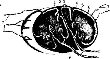
Fig. 5.43. Diagram of The Structure of a lymph node
Lymphoid tissue is quite sensitive to external factors. For instance, X-ray irradiation causes lymphocytes to perish rapidly.
The spleen (lien, splen) belongs to the lymphoid organs. It acts as a "graveyard for erythrocytes" because this is where their destruction takes place. Along with The Liver and Skin, the spleen can store a significant volume of blood (acting as a blood reservoir).
The spleen is located in the Abdominal cavity within the left hypochondrium at the level of the 9th–11th Ribs (Fig. 5.44).
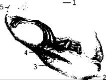
Fig. 5.44. Spleen; visceral surface
It exhibits diaphragmatic and visceral surfaces (the latter is in contact with The Stomach, left Kidney, and tail of the Pancreas). The visceral surface bears the hilum of the spleen, which is the site of entry and exit for Vessels and nerves. The spleen is covered by the Peritoneum on all sides (it is an intraperitoneal organ). Beneath the peritoneum lies a connective tissue capsule, from which trabeculae extend into the interior of the organ. The splenic pulp is situated between the trabeculae. The spleen pulp is formed by reticular tissue, within the meshes of which A large number of lymphocytes and erythrocytes are located. Red and white pulp are distinguished. The red pulp contains erythrocytes,
leukocytes, lymphocytes, and macrophages. The white pulp is formed by lymphoid nodules consisting of lymphocytes and other Cells of the lymphoid tissue. A central vein passes through each nodule (Fig. 5.45).
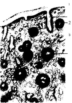
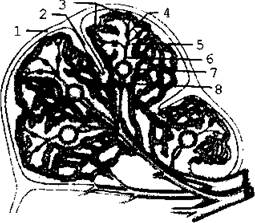
Fig. 5.45. Microscopic STRUCTURE OF THE spleen
Last update: 08/08/2026
Editorial and Educational Adaptation: This material has been compiled based on the primary/original source text. The project team performed an editorial review, corrected technical inaccuracies, structured sections, and adapted the content for an educational format.
What was processed:
- elimination of formatting defects (OCR errors, structural breaks, corrupted characters);
- editorial organization of content;
- standardization of terminology in accordance with academic sources;
- verification of factual statements against the original source text.
All mentions of the author, publication year, and origin of the primary text have been preserved in accordance with the source.