HUMAN MEDICAL BIOLOGY, ANATOMY, PHYSIOLOGY AND PATHOLOGY - Y.I. Fedoniuk 2010
ANATOMY, PHYSIOLOGY, PATHOLOGY
CHAPTER 5. THE PROCESS OF BLOOD AND LYMPH CIRCULATION. ITS PATHOLOGY
FUNCTIONAL ANATOMY OF THE LYMPHATIC AND IMMUNE SYSTEMS
LYMPHATIC VESSELS
The Lymphatic system is a part of The Vascular System. Lymph flows toward the large Veins OF THE neck, where it enters the venous Blood. The lymphatic system is a branched network of vessels with Lymph Nodes located along the course of Blood Vessels (Fig. 5.41).
Class="center">
Fig. 5.41. Human lymphatic system
Along with the Venous system, the lymphatic system ensures the absorption from Tissues of Water with dissolved substances, as well as colloidal solutions, Proteins, fats, and foreign particles (Bacteria, Cell breakdown products, etc.) that are not absorbed into the blood capillaries. In addition, lymphopoiesis takes place within it, which is of great importance for immunological reactions.
The lymphatic system begins with blind-ended endothelial tubes—capillaries that form networks within Organs. Capillaries are present in all organs except the Brain AND SPINAL cord, Eyeball, Inner ear, epithelial covering of the Skin and mucous membranes, Cartilage, splenic parenchyma, Bone Marrow, and Placenta. Lymphatic vessels originate from the capillaries and feature a significant number of Valves. These valves ensure the unidirectional flow of lymph. Lymphatic vessels emerging from organs proceed toward regional lymph nodes. Vessels originating from the Small Intestine carry lymph containing fat droplets, which give the lymph a milky appearance; therefore, these vessels are called lacteals. Thus, the lymphatic system comprises:
1) closed lymphatic capillaries that form a network within organs;
2) intraorgan vessels that emerge from the lymph capillary network and form their own network;
3) efferent vessels that exit the organ and are interrupted along their pathway by lymph nodes;
4) large lymphatic vessels that empty into lymphatic trunks. Lumbar, intestinal, subclavian, jugular, and bronchomediastinal trunks are distinguished. These trunks merge into two lymphatic ducts, which empty into the large veins of the neck (Fig. 5.42).
The Thoracic duct (ductus thoracicus) is formed in the Abdominal cavity by the convergence of two lumbar trunks. It passes through the Diaphragm, lies in the posterior Mediastinum, ascends into the neck region, and empties into the left venous angle. This duct collects lymph from the lower extremities, organs and walls of the pelvis, the abdominal and left thoracic cavities, the left upper extremity, and the left half of the HEAD and Neck.
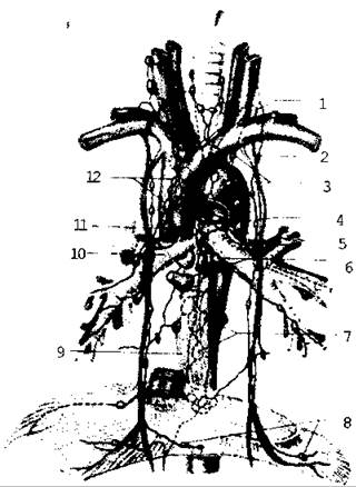
Fig. 5.42. Lymphatic ducts
Last update: 08/08/2026
Editorial and Educational Adaptation: This material has been compiled based on the primary/original source text. The project team performed an editorial review, corrected technical inaccuracies, structured sections, and adapted the content for an educational format.
What was processed:
- elimination of formatting defects (OCR errors, structural breaks, corrupted characters);
- editorial organization of content;
- standardization of terminology in accordance with academic sources;
- verification of factual statements against the original source text.
All mentions of the author, publication year, and origin of the primary text have been preserved in accordance with the source.