MEDICAL BIOLOGY, HUMAN ANATOMY, PHYSIOLOGY AND PATHOLOGY - Ya.I. Fedonyuk 2010
ANATOMY, PHYSIOLOGY, PATHOLOGY
SECTION 3. ANATOMICAL AND PHYSIOLOGICAL ASPECTS OF BODY FUNCTION AUTO-REGULATION
THE BRAIN
8. FOREBRAIN
8.1. Cerebral Cortex
The grey matter On the surface of the cerebral hemispheres is 3 to 5 mm thick and contains over 50 billion Nerve Cells. Based on morphological features, cortical Neurons are divided into pyramidal and non-pyramidal cells. Pyramidal cells have a characteristic pyramid shape, whereas non-pyramidal neurons comprise several distinct types: basket, stellate, fusiform, and others.
The cerebral cortex consists of six layers: molecular, external granular, pyramidal, internal granular, ganglionic, and the layer of polymorphic cells.
Neurocytes (neurons) and their processes within the cerebral cortex are arranged in indistinctly separated layers, or laminae. This layered arrangement of neurocytes is known as cytoarchitecture. Within the cortex, nerve Cell processes form distinct tangential bundles, known as stripes, located between the layers of nerve cells. The laminar arrangement of these tangential bundles of nerve fibers (stripes) within the cerebral hemispheres is referred to as myeloarchitecture (Fig. 3.28).
Class="center">
Fig. 3.28. Cytoarchitecture (a) and myeloarchitecture (b) of the cerebral cortex.
I.P. Pavlov regarded the cerebral cortex as a totality of cortical ends of analyzers.
Thus, the cortex can be schematically conceptualized as a collection of core regions belonging to various analyzers, interspersed with scattered elements of these same analyzers.
Modern science adheres to The Theory of dynamic Localization of Functions in the cortex, advanced by I.P. Pavlov. According to this theory, distinct areas of the cortex have different functional significance; however, there are no sharp boundaries between them.
Cells from one functional center extend into neighboring areas. In other words, each cortical nerve center has a core—a cluster of neurons specific exclusively to it—and a periphery consisting of less specialized nerve cells. The peripheral zones of adjacent centers overlap.
Therefore, let us examine the topography of some cortical ends of various analyzers (cores) in relation to the lobes and gyri of the human cerebral hemispheres.
1. The cortex of the postcentral gyrus houses the core of the general sensitivity analyzer (pain, Temperature, tactile, and proprioceptive). Specifically, the PROJECTION OF THE lower limbs and trunk is located in its upper part, the upper limbs below it, and the HEAD at the very bottom.
The primary function of the sensory cortex is the integrative critical evaluation of sensory information arriving from the thalamus. The cortex assesses the intensity of sensations and determines the spatial relationships of the stimulated body parts.
Because ascending sensory pathways cross over on their way to the cortex, a lesion in one hemisphere impairs sensation on the opposite side of the body.
2. The core of the motor analyzer is located in the precentral gyrus. The corticospinal tract originates here from Betz pyramidal cells. At the level of the Medulla Oblongata, this motor pathway decussates and terminates on the motor neurons of the anterior horns of the Spinal Cord, from where nerve fibers carry signals to the skeletal Muscles.
Due to the decussation of this tract, damage to one cerebral hemisphere results in paresis (partial loss of movement) or paralysis (complete loss of movement) of the contralateral half of the body. The morphofunctional Organization OF THE motor cortex has certain unique features. Centers for different Muscle groups are represented unequally and occupy varying areas. The largest regulatory area within the gyrus is dedicated to the Muscles of the head (Tongue, vocal apparatus, facial muscles) and the hand (Fig. 3.29).
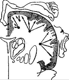
Fig. 3.29. Representation of The Human Body in the motor area of the cerebral cortex.
3. The core of the analyzer responsible for the conjugate turning of the head and eyes to the opposite side lies in the posterior PARTS OF THE middle frontal gyrus.
4. The core of the motor analyzer for written signs ("writing center") is located in the posterior parts of the middle frontal gyrus.
5. The motor speech area (Broca's center) is located in the posterior part of the inferior frontal gyrus on the left side.
6. The stereognosis analyzer center (responsible for tactile object recognition) lies within the superior parietal lobule.
7. The motor center for purposeful, coordinated movements acquired through practical activity and labor (the praxis center) is located in the supramarginal gyrus.
8. The visual speech center for written symbols (the reading center) is situated in the angular gyrus.
9. The cortical visual center is located along the "banks" of the calcarine sulcus within the lingual gyrus and cuneus. This area receives visual information transmitted via the optic tracts from the photoreceptors (rods and cones) of the retina.
10. The cortical auditory center is located in Heschl's gyrus (the middle portion on the Medial surface of the superior temporal
gyrus). In this cortical area, auditory signals arriving from the cochlea of the Inner ear via the auditory pathways are perceived as sounds distinguished by pitch, quality, and loudness.
11. The auditory speech center is located in the posterior part of the superior temporal gyrus (Wernicke's area).
12. The cortical centers for Olfaction and gustation are located in the uncus, dentate gyrus, and hippocampus.
Last update: 08/08/2026
Editorial and Educational Adaptation: This material has been compiled based on the primary/original source text. The project team performed an editorial review, corrected technical inaccuracies, structured sections, and adapted the content for an educational format.
What was processed:
- elimination of formatting defects (OCR errors, structural breaks, corrupted characters);
- editorial organization of content;
- standardization of terminology in accordance with academic sources;
- verification of factual statements against the original source text.
All mentions of the author, publication year, and origin of the primary text have been preserved in accordance with the source.