PLANT PHYSIOLOGY AND BIOCHEMISTRY
Lecture Notes
5. PHOTOSYNTHESIS
Plastid Pigments
Pigments are the most vital component of the photosynthetic apparatus. Research on pigments advanced significantly following the discovery of a fundamentally new analytical method by a Russian physiologist: adsorption Chromatography. A solution of substances is passed through a Column filled with an adsorbent. Substances with varying degrees of adsorption move through the column at different speeds, thereby separating from one another. This was the method used to first isolate the pigments of green leaves.
Plastid pigments belong to three classes of substances: chlorophylls, phycobilins, and carotenoids.
Chlorophylls
Structure and Composition
In 1817, French chemists P. Pelletier and J. Caventou isolated a green pigment from leaves and named it chlorophyll (from Greek for green leaf).
Polish biochemists M. Nencki and L. Marchlewski (1897) discovered that the core of the chlorophyll molecule, like the heme (prosthetic group) of Hemoglobin, is a porphyrin ring.
In 1914, German chemist R. Willstätter established the Elemental Composition of chlorophyll a (C55H72O5N4Mg) and chlorophyll b (C55H70O6N4Mg), while German biochemist H. Fischer fully deciphered the structural formula in the 1930s–40s. Artificial synthesis of chlorophyll was achieved in the 1960s.
Chlorophyll is an ester of the dicarboxylic acid chlorophyllin (C32H30ON4Mg(COOH)2), in which one carboxyl group is esterified with a methyl alcohol residue (CH3OH), and the other with a residue of the unsaturated monohydric alcohol phytol (C20H39OH).
Structural Formula of Chlorophyll
Four pyrrole rings (I–IV) are connected by methine bridges (α, β, γ, δ) to form a porphyrin core. The peripheral carbon atoms are numbered 1–10. The nitrogen atoms of the pyrrole rings interact with the Mg atom via four coordination bonds. The cyclopentanone ring (V) contains a chemically active carbonyl group at C9 and a methylated carboxyl group at C10. The side chain of the IV pyrrole ring consists of propionic acid, linked via an ester bond to the polyisoprenoid unsaturated alcohol phytol (C20H39OH).
Class="center">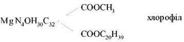
Carbon atoms 1, 3, 5, and 8 of the pyrrole rings bear methyl groups, the 2nd bears a vinyl group, and the 4th an ethyl group. The porphyrin ring is a system of nine pairs of alternating conjugated double and single bonds with 18 delocalized π-electrons. Chlorophyllide is the chlorophyll structure lacking the phytol tail.
Substitution of Mg with protons results in The formation of pheophytins.
All photosynthetic plants and cyanobacteria contain chlorophyll a; higher plants, green Algae, and euglenids contain chlorophyll b, brown and diatom algae contain chlorophyll c, and red algae contain chlorophyll d.
Biosynthesis of Chlorophylls
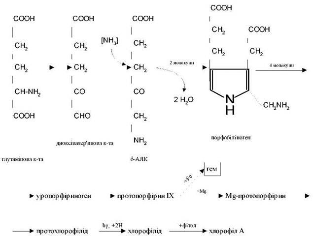
The First stage of chlorophyll biosynthesis in plants is the formation of δ-aminolevulinic acid (ALA) from C5-dicarboxylic acids.
Cyclization of two ALA molecules leads to the Formation of the pyrrole compound porphobilinogen. Four pyrrole rings assemble to form uroporphyrinogen, which is then converted into protoporphyrin IX. The subsequent pathway of protoporphyrin transformation can vary. With the involvement of iron, heme is formed, which is a component of Cytochromes, catalase, peroxidase, and hemoglobin. When Mg is incorporated, protochlorophyllide is formed, which is converted into chlorophyll a in the presence of light. All these processes occur within the METABOLISM/14.html">Chloroplasts.
PHYSICOCHEMICAL PROPERTIES OF Chlorophylls
In its solid state, chlorophyll a is an amorphous blue-black substance. The melting point of chlorophyll a is 117–120 °C. Chlorophylls are highly soluble in ethyl ether, benzene, chloroform, acetone, and ethyl alcohol; they are poorly soluble in petroleum ether and insoluble in Water.
The sharp absorption maxima of chlorophylls occur in the red (chl a: 660–663 nm; chl b: 642–644 nm) and blue (chl a: 428–430 nm; chl b: 452–455 nm) Regions of the spectrum (data for chlorophyll solutions in ethyl ether). Chlorophylls absorb orange and yellow light weakly and do not absorb green or infrared rays at all.
Absorption in the blue-violet region of the spectrum is due to The system of conjugated single and double bonds within the porphyrin ring. The intensity of absorption in the red region is linked to the hydrogenation of the C7-C8 double bond in the fourth pyrrole ring during the transition from protochlorophyllide to chlorophyllide, as well as the presence of Mg in the porphyrin core.
The positioning of absorption spectrum maxima is influenced by The Nature of the Solvents and the interaction of chlorophyll molecules with other pigments and organic substances.
Chlorophyll solutions in polar solvents exhibit bright fluorescence (luminescence) and are capable of phosphorescence (in the infrared part of the spectrum). Native chlorophyll fluoresces weakly.
These phenomena depend on the various pathways by which electronic excitation energy (E) is utilized.
As is well known, the most stable atomic states are those in which valence ē occupy the lowest energy levels and are distributed According to the Pauli exclusion principle (no more than 2 ē with antiparallel spins per orbital). This molecular state is referred to as the ground singlet (S0) energy state. When a molecule absorbs a light quantum, this energy causes an ē to transition to higher vacant orbitals. If the excited ē retains the same spin direction, the molecule is in a singlet excited state (S*); if the ē spin changes, such excitation is termed triplet (T*).
Absorption of a red light quantum by a chlorophyll molecule leads to a transition from the ground singlet energy state (S0) to the first singlet excited state (S*1); absorption of blue light with higher E causes the ē to transition to a higher orbital (S*2).
An excited chlorophyll molecule returns to the ground state through various pathways, including heat dissipation, fluorescence, and phosphorescence. Finally, the E of the excited state can be utilized for photochemical reactions. In the latter case, neither fluorescence nor phosphorescence is observed.
The structure of the chlorophyll molecule is evolutionarily optimized for its role as a photosensitizer in photochemical reactions. It contains 18 delocalized π ē, which makes the chlorophyll molecule highly excitable upon the absorption of light quanta.
Simultaneously, chlorophyll is capable of redox transformations. Chlorophyll dissolved in pyridine, under anaerobic conditions and light exposure, is reduced by ascorbic acid or other electron Donors. Once the light is turned off, the reaction reverses. Photoreduced chlorophyll, in turn, can reduce various electron acceptors (NAD+, riboflavin, quinone, Fe3+, oxygen). Thus, a chlorophyll molecule can act not only as a primary electron acceptor but also as its primary donor. Based on this, it can be concluded that due to its structural and physicochemical properties, the chlorophyll molecule is capable of performing 3 vital Functions:
1) selectively absorbing light E;
2) storing it in the form of electronic excitation E;
3) photochemically converting the E of excited electrons into the chemical E of photoreduced and photooxidized states.
Spatial Organization of chlorophyll:
the magnesium-porphyrin ring forms a hydrophilic plate positioned above the membranes;
the phytol chain is angled relative to the porphyrin ring, forming a hydrophobic pole that interacts with membrane structures.
Phycobilins
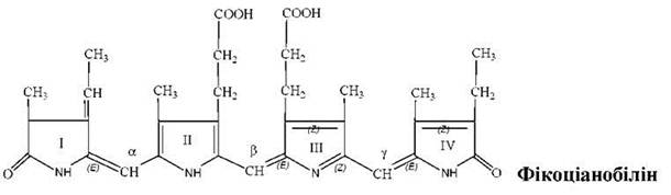
The most common representatives of phycobilins are phycoerythrobilins (predominant in red algae) and phycocyanobilins (predominant in cyanobacteria).
They belong to the Bile pigments known as bilins. These are open-chain tetrapyrroles lacking a metal ion. They contain a system of conjugated double and single bonds. The pyrrole rings are connected by methylene or methine bridges. Rings I and IV each contain one carbonyl group. They possess side chains: 4 methyl groups (C1, 3, 6, 7), a vinyl group (C2), an ethyl group (C8), and two propionic acid residues (C4, 5).
Phycobilins serve as the chromophore groups of phycobiliproteins (linked via covalent bonds). Phycobiliproteins are divided into 3 groups:
1) phycoerythrins - red Proteins (498-568 nm);
2) phycocyanins - blue-green proteins (585-630 nm);
3) allophycocyanins - blue proteins (585-650 nm).
The overall absorption spectrum is 490-650 nm (green and yellow-orange rays). They are water-soluble and localized in phycobilisomes—granules situated on the outer surface of photosynthetic lamellae.
Significance
The Light absorption maxima of phycobilins lie between the two absorption peaks of chlorophylls: in the orange, yellow, and green PARTS OF THE spectrum. This is due to the Optical Properties of water, which primarily absorbs long-wavelength rays. Consequently, light of different quality reaches different depths. At a depth of 36 m, red rays disappear completely; at 177 m, yellow rays vanish; at 322 m, green rays are absorbed; and by 500 m, even blue rays fail to penetrate. As a result, green algae predominantly inhabit the upper layers of the ocean, blue-green algae live deeper, and red algae reside at the greatest depths (a phenomenon known as chromatic complementary adaptation).
In algae, phycobilins serve as accessory pigments that function as a light-harvesting complex instead of chlorophyll. Approximately 90% of light Energy is transferred by them to chlorophyll a. All plants contain Phytochrome, a phycobilin that acts as a photoreceptor and performs regulatory functions.
Carotenoids
Carotenoids are fat-soluble pigments ranging in color from yellow and orange to red, present in the chloroplasts of all plants. They are also found in chromoplasts within non-green plant Tissues. They are synthesized by Bacteria and Fungi and consist of cyclic or acyclic Isoprenoids.
Structure and properties
Carotenoids are classified into three groups of compounds:
1) orange or red carotenes (С40Н56);
2) yellow xanthophylls (С40Н56О2, С40Н56О4);
3) carotenoid acids — products of carotenoid oxidation; for example, crocetin (С20Н24О4) features a shortened chain and two carboxyl groups.
Carotenes and xanthophylls are highly soluble in chloroform, benzene, carbon disulfide, and acetone. Carotenes are highly soluble in ethers and nearly insoluble in alcohols; xanthophylls exhibit the opposite solubility profile.
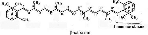
All carotenoids are polyene compounds. Carotenoids of the first two groups consist of eight isoprene residues (a chain of conjugated double bonds). Carotenoids can be acyclic (aliphatic), monocyclic, or bicyclic. The rings at the ends of carotenoid molecules are derivatives of ionone.
The acyclic carotenoid is lycopene. Cyclic α-carotene contains a β-ionone ring and an ε-ionone ring (С4=С5). β-carotene contains two β-ionone rings (С5=С6). Hydrolysis of β-carotene at the central double bond yields two molecules of retinol (vitamin A).
The primary carotenoids in Higher Plants and algae are β-carotene, lutein, violaxanthin, neoxanthin, and zeaxanthin.
Carotenoid synthesis begins with acetyl-CoA, proceeding through mevalonic acid and geranylgeranyl pyrophosphate to lycopene, which serves as the precursor for all other carotenoids. The absorption spectra of carotenoids are characterized by two bands in the violet and blue regions, ranging from 400 to 500 nm.
Like chlorophylls, carotenoids are non-covalently bound to Membrane Proteins and Lipids.
The Role of carotenoids in Photosynthesis
Carotenoids are essential Components of the pigment systems in all photosynthetic organisms. Their primary functions include:
1) participation in light absorption as accessory pigments;
2) Protection of the chlorophyll molecule from irreversible photooxidation;
They may also participate in oxygen exchange during photosynthesis.
The Significance of carotenoids becomes clear when we examine the distribution of energy in the total solar radiation spectrum at the Earth's surface, which peaks in the blue-cyan and green parts of the spectrum (480-530 nm). Model studies have demonstrated the high efficiency of light energy transfer from carotenoids to chlorophylls. Carotenes possess this capability, whereas xanthophylls do not.
Numerous experiments (biochemical in vitro studies and The Use of non-carotenoid mutants such as Chlamydomonas and corn) have proven the protective role of carotenoids. They shield chlorophyll molecules from destruction (oxidation).
In the light-sensitive "eyes" of unicellular organisms and at the tips of higher plant shoots, carotenoids facilitate the determination of light direction by creating contrast. Carotenoids also determine the color of petals and fruits in certain plants (though in most cases, this depends on vacuolar anthocyanins).
Last update: 07/08/2026
Editorial and Educational Adaptation: This material has been compiled based on the primary/original source text. The project team performed an editorial review, corrected technical inaccuracies, structured sections, and adapted the content for an educational format.
What was processed:
- elimination of formatting defects (OCR errors, structural breaks, corrupted characters);
- editorial organization of content;
- standardization of terminology in accordance with academic sources;
- verification of factual statements against the original source text.
All mentions of the author, publication year, and origin of the primary text have been preserved in accordance with the source.