MODERN BOTANY - P. RAVEN - 1990
SECTION IV. DIVERSITY
CHAPTER 17. VASCULAR CRYPTOGAMS
Phyla of Vascular Cryptogams
Three phyla of these plants — Rhyniophyta, Zosterophyllophyta, and Trimerophyta — flourished during the Devonian period and became extinct by its end, i.e., about 360 million years ago, with the rhyniophytes having appeared at least 420 million years ago in the Late Silurian. All these are relatively simple organisms in terms of Structure. The fourth phylum of spore-bearing plants, Progymnospermophyta (or progymnosperms), will be discussed in Chapter 18, as its representatives may have been the ancestors of gymnosperms and angiosperms (Fig. 17-7). In addition to extinct plants, we will discuss in this chapter the phyla Psilotophyta, Lycophyta, Sphenophyta, and Pterophyta, which have living representatives today.
Class="center">Fig. 17-7. Possible evolutionary pathway of vascular plants, starting from zosterophyllophytes and rhyniophytes. The duration of the Devonian period in the general timescale is enlarged, and connections to flowering plants are not shown. It remains unclear exactly which taxa gave rise to psilophytes and sphenophytes. Only a few fossil genera have been described that may be intermediate between rhyniophytes, which appeared in the Silurian period (about 420 million years ago), and
zosterophyllophytes, whose oldest remains are approximately 10 million years younger. The precise Nature of the relationships between these two groups and their common ancestor has yet to be established
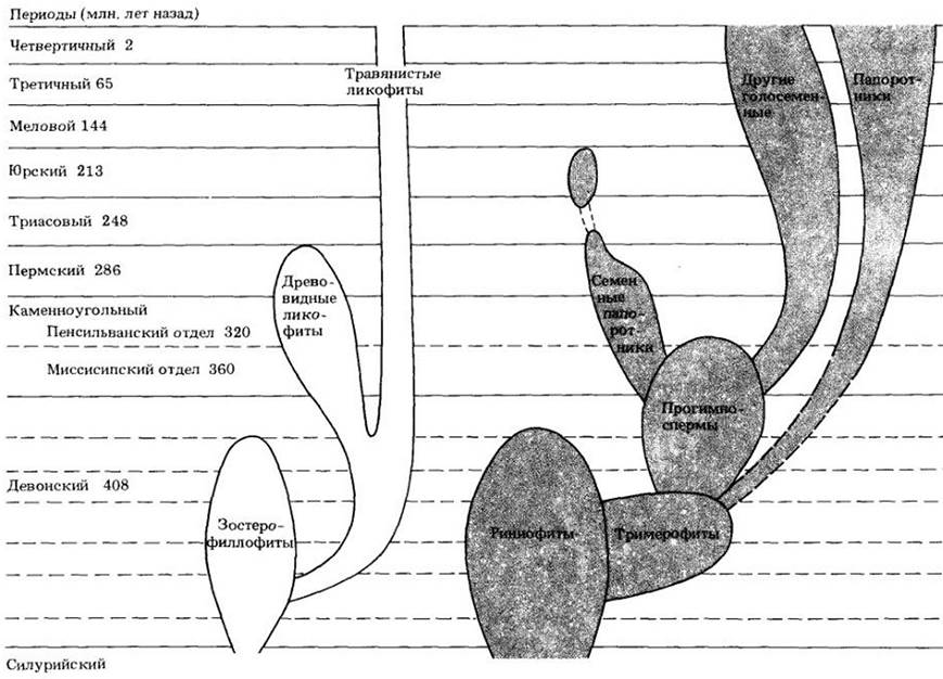
The overall pattern of the rise in floral diversity can be viewed as the successive dominance of four major groups, accompanied by the rapid displacement of previous dominants. In each case, as these groups assumed leading positions, they achieved high species richness. The primary contributors to this historical process were:
1. The first vascular plants with a simple and presumably ancestral Morphology relative to other groups. These include rhyniophytes, zosterophyllophytes, and trimerophytes (Fig. 17-8). These primitive forms dominated from the Late Silurian to the Middle Devonian, i.e., approximately 420 — 370 million years ago (see Fig. 17-1).
2. Ferns, lycophytes, sphenophytes, and progymnosperms. These more complexly structured plants dominated from the Late Devonian to the end of the Carboniferous period (see Figs. 17-9, 18-1), roughly 380 — 290 million years ago. The Appendix to the chapter provides An Overview of the plants of that era (see p. 307).
3. Seed plants, which appeared in the Late Devonian (at least 360 million years ago) and were already represented by numerous evolutionary lineages by the beginning of the Permian period. Gymnosperms predominated in terrestrial floras throughout most of the Mesozoic era, up until about 100 million years ago.
4. Flowering plants, whose oldest remains date back about 127 million years. They conquered the land within 20 — 30 million years and have maintained a dominant position ever since.
Phylum Rhyniophyta
This phylum includes the oldest vascular plants known to us in detail. They appeared in the Late Silurian (at least 420 million years ago) and became extinct in the Middle Devonian (about 380 million years ago). Even older vascular plants, whose fossil remains are at least 15 million years older, were probably similar to rhyniophytes. The phylum comprises spore-bearing plants consisting of simple, dichotomously branching axes with terminal sporangia. Differentiation into roots, stems, or leaves was absent, and the species were homosporous. The name of the phylum comes from the village of Rhynie1 in Scotland, near which the remains of these primitive plants have been exceptionally well preserved in chert deposits.
1In Latin, its name (Rhynie) is pronounced "Rhy-nee". — Ed. note.
Cooksonia, a rhyniophyte representative believed to have inhabited marshy lowlands, is the oldest known vascular plant (see Figs. 1-5, 17-1). Its slender, leafless aerial stems reached 6.5 cm in length; the sporangia were spherical. Although nothing is known about the lower parts of these plants, it is quite likely that Cooksonia possessed a rhizome, i.e., an underground stem from which aerial branches arose. Tracheids have been discovered in macerated stem fragments. Cooksonia became extinct by the Early Devonian, about 390 million years ago.
The best-known representative of the rhyniophytes is Rhynia (Fig. 17-8, A), presumably a wetland plant whose leafless, dichotomously branching stems arose from a rhizome bearing clusters of Water-absorbing rhizoids. The aerial stems of Rhynia, 20 — 50 cm long and 3 — 6 mm thick, were covered with a cuticle, possessed Stomata, and served as photosynthetic Organs.
Fig. 17-8. Ancient vascular plants. A. The rhyniophyte Rhynia major — one of the simplest known vascular plants. Its leafless stem branched dichotomously. Terminal sporangia released spores, likely through longitudinal splitting. B. In the genus Zosterophyllum and other zosterophyllophytes, sporangia aggregated into terminal spikes split along specific grooves around their distal margin. Larger than rhyniophytes, these plants generally exhibited dichotomous branching, much like the former, and were naked, spiny, or toothed. C. Trimerophytes are even larger and more complexly organized plants with a robust central axis and smaller lateral branches that branched dichotomously and often terminated in pairs of narrowed sporangia. The best-known genera in this group are Psilophyton and Trimerophyton. The figure shows a reconstruction of Psilophyton princeps. Individual specimens of Rhynia major reached heights of 0.5 m, while some trimerophytes exceeded 1 m (see also Fig. 17-1)
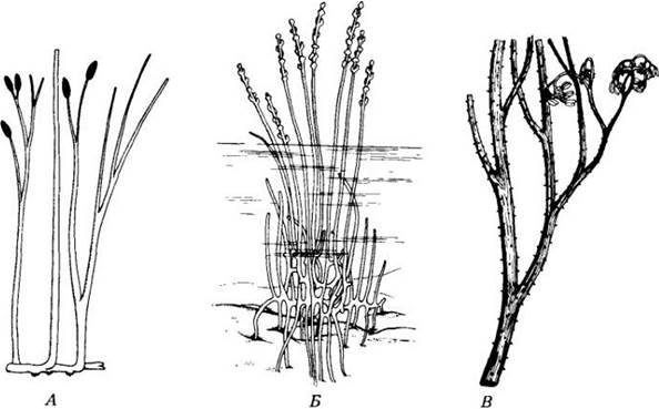
Fig. 17-9. Reconstruction of a Carboniferous swamp forest (see also Fig. 18-1)

Fig. 17-10. Psilotum nudum. A. Cross-section of the stem, showing the established Tissues. B. Protostele; xylem and phloem are visible

Fig. 17-11. Underground gametophyte of Psilotum nudum. The gametophytes are bisexual, meaning they bear both antheridia and archegonia

Fig. 17-12. A. Tmesipteris parva on the trunk of the tree fern Cyathea australis in New South Wales, Australia. B. Tmesipteris lanceolata from New Caledonia
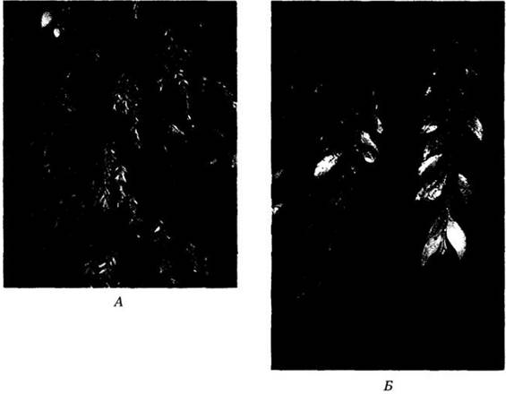
Fig. 17-13. Life Cycle of Psilotum. In this genus, dichotomously branching aerial shoots arise from rhizomes. Sporangia develop in groups of three in the axils of scale-like appendages on these shoots. Like all vascular plants, Psilotum is characterized by the alternation of heteromorphic generations with a dominant sporophyte. Spores are produced inside the sporangium As a result of Meiosis, initiating the gametophytic phase. Psilotum is homosporous; its spores give rise to bisexual gametophytes that bear both antheridia and archegonia simultaneously. To reach the egg Cell, the multiflagellated sperm requires water. Syngamy produces a zygote, from which the sporophyte generation develops. The embryo forms within the venter of the archegonium, with the young sporophyte obtaining nourishment from the gametophyte via its FOOT embedded in the gametophyte tissue. Eventually, the sporophyte detaches from the foot and begins an independent existence

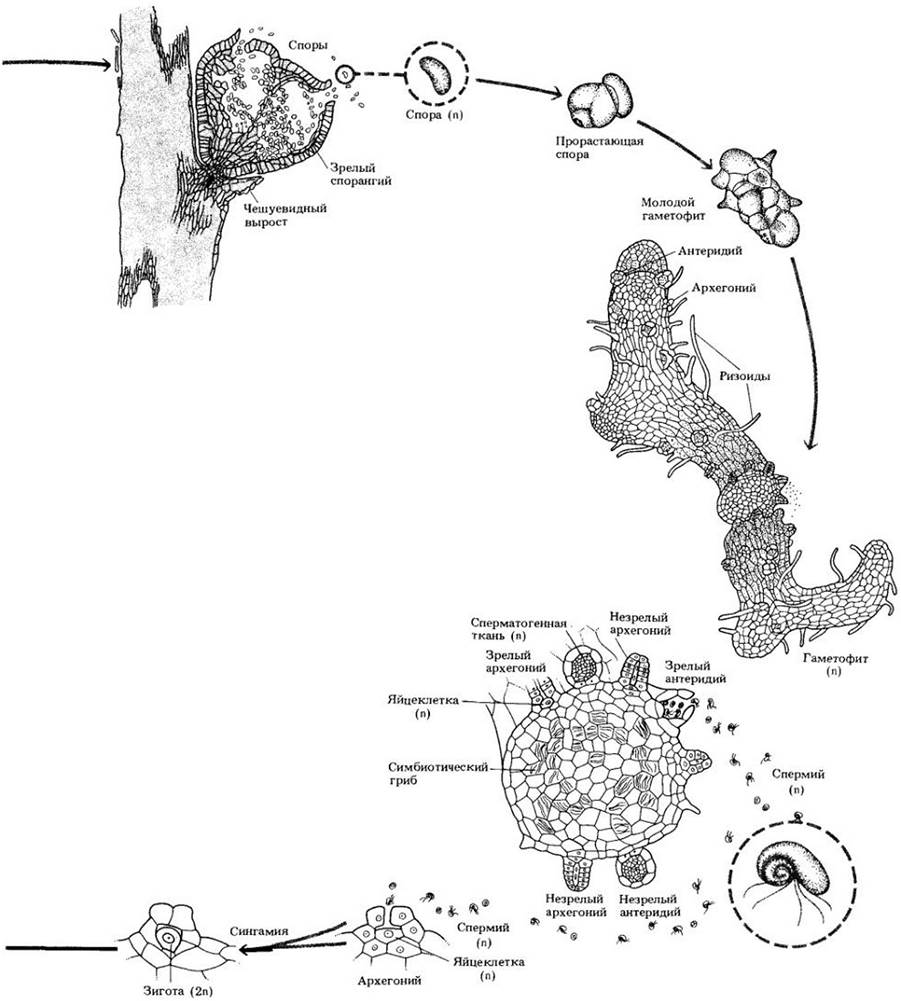
Fig. 17-14. A. Transverse section of the stem of Lycopodium complanatum showing mature tissues. B. Protostele of the same species, showing xylem and phloem. Both the stem and the ROOT of clubmosses are protostelic (see also Fig. 17-2)

Fig. 17-15. Lycopodium lucidulum, a clubmoss lacking a strobilus; the sporophylls are scattered among sterile microphylls
In its internal structure, Rhynia resembled modern vascular plants. A single layer of surface Cells—the epidermis—surrounded the assimilation tissue of the cortex, and the center of the axis consisted of a solid strand of xylem surrounded by two layers of cells, possibly phloem. Evidently, the xylem cells in the central part of the strand developed first, while those on its periphery developed last.
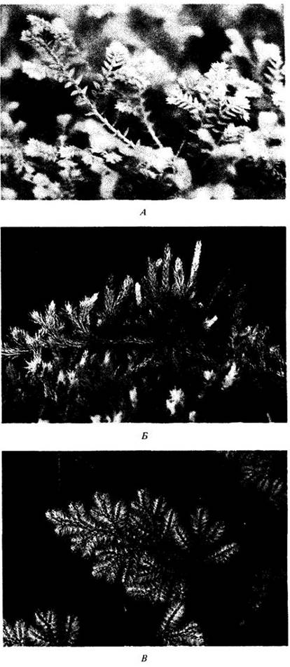
Fig. 17-16. A. Selaginella kraussiana is a prostrate, creeping plant. Adventitious roots arising from the stems are visible. B. S. rupestris with strobili. C. S. willdenowii, a plant of the Old World tropics found in Vietnam, Malaysia, and the Himalayas. This shade-tolerant species, with iridescent, metallic blue leaves, can climb trees up to a height of seven meters
Phylum Zosterophyllophyta
Fossil remains of representatives of this phylum of extinct vascular plants are found in early-to-late Devonian deposits dating from approximately 408 to 370 million years ago. Like rhyniophytes, they were leafless and dichotomously branched. Plants of this group likely inhabited aquatic environments. The aerial stems were covered with a cuticle, but only the uppermost branches possessed stomata, which may indicate that the lower branches were submerged in silt. It is suggested that in Zosterophyllum, these branches frequently gave rise to lateral divisions that forked into two axes, one growing upward and the other downward (Fig. 17-8, B). The downward-growing branches apparently served to anchor the plant, allowing it to spread laterally. Zosterophyllophytes got their name due to their general resemblance to the modern angiosperm Zostera, which grows in seawater and superficially resembles grasses.
Unlike rhyniophytes, the spherical or Kidney-shaped sporangia of zosterophyllophytes were positioned laterally on short stalks. The plants were homosporous. Although the Internal Structure of zosterophyllophytes is essentially the same as that of rhyniophytes, the xylem cells that reached maturity first were located at the periphery of the xylem strand, whereas those that developed last were in its center.
It can be considered almost certain that zosterophyllophytes are the ancestors of lycophytes. The sporangia of ancient lycophytes were also positioned laterally, and the xylem in both phyla developed centripetally (from the periphery to the center). Representatives of these two phyla differ markedly from rhyniophytes and trimerophytes.
Phylum Trimerophyta
This phylum probably originated directly from rhyniophytes, and its representatives apparently gave rise to ferns, progymnosperms, and quite possibly horsetails as well. Trimerophytes, which were larger and more complexly structured plants than rhyniophytes and zosterophyllophytes (Fig. 17-8, C), first appeared in the Early Devonian, about 395 million years ago, and became extinct by the end of the Middle Devonian, roughly 20 million years later, thus existing for a relatively short span of time.
Although trimerophytes are more evolutionarily advanced than rhyniophytes, they still lack leaves. Systems of lateral branches that repeatedly forked dichotomously were formed on the main axis. Like rhyniophytes and zosterophyllophytes, these plants were homosporous. Some of the smaller branches terminated in elongated sporangia, whereas others were exclusively vegetative. In Addition to a more complex branching pattern, trimerophytes possessed a more massive conducting strand than rhyniophytes, which, together with a robust cortex composed of thick-walled cells, probably provided support for the relatively large plant. As in rhyniophytes, the first xylem cells in trimerophytes formed in the center (centrifugal development). The name of the phylum comes from the Greek words "tri", "meros", and "phyton", meaning "three-part plant." This reflects the tripartite division of the secondary branches in the genus Trimerophyton.
Fig. 17-17. Life cycle of the clubmoss. Like Psilotum, this plant is homosporous, and therefore their life cycles are similar. Meiosis produces spores that give rise to bisexual gametophytes. In some clubmoss species, the latter are subterranean, and their normal growth, as shown in the illustration, requires the presence of a mycorrhizal fungus. Fertilization requires water, through which the biflagellated sperm swim to the archegonium. Syngamy produces a zygote. Embryo development takes place within the venter of the archegonium. The young sporophyte, sometimes attached to the gametophyte for an extended period, eventually becomes independent. In many clubmoss species, the sporophylls are aggregated into strobili, as seen in the figure


Fig. 17-18. Selaginella. A. Transverse section of the stem showing mature tissues. The protostele is supported in the center of the hollow stem by elongated cortical (endodermal) cells called trabeculae. Only fragments of these are visible in the figure. B. Close-up of the protostele

Fig. 17-19. Sporophyte of Isoetes muricata. Porcupine quill-like leaves, stem, and roots are visible

Phylum Psilotophyta
The phylum includes two surviving genera: whisk ferns (Psilotum) and Tmesipteris. Psilotum is widely distributed in tropical and subtropical regions; in the United States, it is found in Florida, Louisiana, Arizona, Texas, Hawaii, and Puerto Rico, as well as acting as a common greenhouse weed. The geographical range of Tmesipteris is restricted to Australia, New Caledonia, New Zealand, and other South Pacific islands. Both genera are very simply structured plants that bear a strong basic resemblance to rhyniophytes.
Psilotum is unique among living plants in lacking both roots and leaves. The sporophyte consists of a dichotomously branching aerial portion bearing small scale-like outgrowths and a branched subterranean rhizome system with numerous rhizoids (Fig. 17-11). Endomycorrhizal zygomycetes are present in the outer cortical Cells of the rhizomes. Psilotum possesses a protostele and consequently lacks leaf gaps (Fig. 17-10).
This plant is homosporous; spores are produced in sporangia located at the tips of short lateral branches. Upon germination, they give rise to bisexual gametophytes that resemble fragments of rhizomes (Fig. 17-11). Like the latter, the subterranean gametophyte harbors a symbiotic fungus and, in some cases, features conducting tissue. The multiflagellated sperm of Psilotum require water to reach the egg cell. Initially, the sporophyte is attached to the gametophyte by a foot—a structure that absorbs nutrients from the gametophyte. Later, it detaches from the foot, which remains embedded within the gametophyte.
Tmesipteris grows as an epiphyte on tree ferns and other plants (Fig. 17-12). Its leaflike appendages are larger than the scale-like outgrowths of Psilotum, but in other respects the two genera are quite similar.
The life cycle of Psilotum is illustrated in Fig. 17-13.
Phylum Lycophyta
The four living genera and approximately 1000 modern species of the phylum Lycophyta (lycophytes) represent an evolutionary Lineage known as far back as the Devonian period. Zosterophyllophytes can be considered the ancestors of lycophytes with almost complete certainty (see Fig. 17-8,5). Several orders belong to this phylum; at least three of them, now extinct, included trees that sometimes reached massive proportions. However, the three surviving orders of lycophytes consist exclusively of herbaceous plants. Both modern and fossil lycophytes possess microphylls, a leaf type that serves as the most distinctive feature of the phylum. Tree-like representatives of this group were among the dominant plants of the coal-forming forests of the Carboniferous period (see the appendix to this chapter and Fig. 18-1). Some of them formed structures similar to the seeds of modern seed plants. Most lineages of arboreal lycophytes became extinct by the end of the Paleozoic era, i.e., no later than 248 million years ago.
Lycopodium
Clubmosses (genus Lycopodium, see Fig. 10-7,5) are arguably the best-known modern lycophytes. Roughly 200 species are distributed from arctic regions to the tropics, although they rarely play a prominent role in plant communities. Most tropical forms are epiphytic and therefore inconspicuous, but several temperate species form carpets that are occasionally noticeable in the forest understory. Because these plants are evergreen, they are most easily spotted in winter.
The Lycopodium sporophyte consists of a branching rhizome that gives rise to aerial branches and adventitious roots. Both stems and roots are protostelic (Fig. 17-14). Microphylls are usually spirally arranged. Clubmosses are homosporous; sporangia are borne singly on the upper surface of fertile microphylls, termed sporophylls—modified leaves or leaf-like organs. In some species, sporophylls are intermixed with sterile microphylls and are nearly indistinguishable from them (Fig. 17-15). In others, non-photosynthetic sporophylls are aggregated into strobili at the tips of aerial branches (see Fig. 10-7,5).
Upon germination, clubmoss spores give rise to bisexual gametophytes, which, depending on the species, appear either as green, irregularly lobed structures or as branching, subterranean, non-photosynthetic bodies. Much like Psilotum and Tmesipteris, these underground gametophytes form a symbiotic association with a fungus. The development and maturation of archegonia and antheridia in clubmosses can sometimes take anywhere from 6 to 15 years. As the gametophyte continues to grow, a succession of sporophytes may arise from sequentially maturing archegonia.
Water is essential for fertilization; the biflagellated sperm swims through water to the archegonium and subsequently navigates through its neck. Following fertilization, the zygote develops into an embryo growing within the venter of the archegonium. The young sporophyte may remain attached to the gametophyte for an extended period, but eventually becomes independent.
The life cycle of Lycopodium is shown in Fig. 17-17.
Selaginella
Among extant lycophyte genera, spikemosses (genus Selaginella) comprise the largest number of species—around 700, found primarily in the tropics. Many inhabit moist environments, while some occur in desert regions, entering a state of dormancy during the driest part of the year. Among the latter is the so-called "resurrection plant," Selaginella lepidophylla, whose range extends from Mexico northward to Texas and New Mexico.
In their general Organization, the herbaceous sporophytes of Selaginella and Lycopodium are similar; both feature microphylls, and their sporophylls form strobili (Fig. 17-16). Unlike Lycopodium, Selaginella develops a small scale-like outgrowth called a ligule near the Base of the upper surface of each microphyll and sporophyll. Both stem and root are protostelic (Fig. 17-18).
Unlike homosporous clubmosses, spikemosses are heterosporous and produce unisexual gametophytes, which constitutes the most fundamental difference between the two genera. Each sporophyll bears a single sporangium on its upper surface. Megasporangia are produced on megasporophylls, whereas microsporangia are borne on microsporophylls. Both types of sporangia occur within the same strobilus.
Male gametophytes (microgametophytes) in Selaginella develop from microspores. Four microspores are produced via meiosis from each microspore mother cell. The gametophyte itself develops entirely within the microspore wall and lacks chlorophyll. When mature, it consists of a single prothallial (or vegetative) cell and an antheridium that produces numerous biflagellated sperm. The microspore wall must rupture to release them.
During The Development of the female gametophyte (megagametophyte), the megaspore wall splits open, and the region where archegonia are formed protrudes through the rupture. Evidence suggests that METABOLISM/14.html">Chloroplasts occasionally appear in the female gametophyte, although it is more likely that it derives nourishment primarily from reserves stored within the megaspore.
Water is required for sperm to reach the archegonia and effect fertilization. Gametophytes typically become detached from the strobilus prior to this. During Embryogenesis in both clubmosses and spikemosses, a structure known as a suspensor is formed. In Lycopodium and certain Selaginella species it remains inactive, but in other species of Selaginella it serves to "push" the developing embryo deep into the nutrient-rich mass of the female gametophyte. The growing sporophyte gradually emerges from the gametophyte tissues and achieves independence.
The life cycle of Selaginella is presented in Fig. 17-22.
Fig. 17-22. Life cycle of Selaginella, a heterosporous plant. Microsporangia and megasporangia are produced on the sporophyte within the same strobilus. Microspores forming in the microsporangia develop into male gametophytes, whereas megasporogenesis in megasporangia yields female gametophytes. Microspores and megaspores are shed close to one another on the ground, requiring sperm to swim only a very short distance through water to reach the egg. Each sporangium is located in the axil of a scale-like appendage, the ligule. In heterosporous plants like Selaginella, gametophyte development begins while still enclosed within the spore wall. Much like in seed plants, the young sporophyte develops embedded within the megagametophyte tissues, and the primary nutrient source for the growing embryo is the storage reserves of the megaspore. However, this process lacks the dormancy period characteristic of many seed plants, as well as the integuments that give rise to the seed coat


Isoetes
A remarkable representative of the clubmosses is Isoetes, commonly known as quillwort. The species of this genus are aquatic or grow in seasonally wet habitats that dry up at certain times of the year. The sporophyte of the quillwort consists of a short, fleshy subterranean stem (a corm or rhizophore) bearing porcupine quill-like microphylls on its upper surface and roots on its lower surface (Figs. 17-19). Each leaf Functions as a potential sporophyll.
Like Selaginella, the quillwort is heterosporous. Megasporangia are produced at the base of the megasporophylls, whereas microsporangia are borne on the microsporangia near the center of the plant (Figs. 17-20). Immediately above the sporangium of each sporophyll lies a small appendage called a ligula.
Fig. 17-20. Diagram of a vertical section through a quillwort. Leaves develop on the upper surface and roots on the lower surface of the short, fleshy underground stem. Some leaves (megasporophylls) bear megasporangia, while others (microsporophylls) bear microsporangia. The microsporophylls are located closer to the center of the plant.

One of the distinctive Features of the quillwort is the presence of a specialized cambium that produces secondary tissues within the corm. Toward the outside, it deposits only parenchyma, whereas toward the inside, it forms a peculiar vascular tissue composed of sieve elements, parenchymal cells, and tracheids in varying proportions.
In 1984, it was demonstrated that certain high-altitude tropical quillwort species possess a unique property: they obtain carbon for Photosynthesis not from the atmosphere, but from sediment. The leaves of these plants lack stomata, are covered with a thick cuticle, and undergo virtually no gas exchange with the atmosphere. They exhibit CAM-type photosynthesis (see p. 106), similar to some quillwort species that dry out during certain seasons of the year.
Phylum Sphenophyta
Like the lycophytes, the Sphenophyta (horsetails) date back to the Devonian period, but they reached their greatest Abundance and diversity later in the Paleozoic, about 300 million years ago. In the Late Devonian and Carboniferous periods, they were represented by calamites (see p. 307)—trees reaching up to 15 m in height, with trunks sometimes exceeding 20 cm in diameter. Today, sphenophytes are represented by a single herbaceous genus, Equisetum (horsetail), which includes 15 species (Fig. 17-21).
Fig. 17-21. A horsetail species with nearly chlorophyll-free fertile shoots that differ dramatically in appearance from the vegetative ones. A. Fertile shoots, each with a terminal strobilus; whorls of scale-like leaves are present at each node. B. Vegetative shoots.
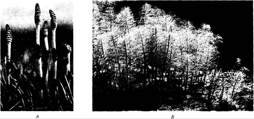
Horsetail species are widespread in damp or marshy places, near rivers, and along forest edges (Fig. 17-21). They are easily recognized by their distinctly jointed stems and rough texture. Small, scale-like leaves, simple in structure but likely representing reduced megaphylls, are arranged in whorls at the nodes. Branches, when present, arise laterally from the nodes, alternating with the leaves. The internodes (the stem segments between adjacent nodes) feature ridges that are stiffened by the deposition of silica in the epidermal cells. Because of this abrasiveness, horsetails were used for scouring pots and pans, particularly during the European colonization of America, earning them the historical name "scouring rushes." Roots are adventitious, emerging from the nodes of the rhizomes.
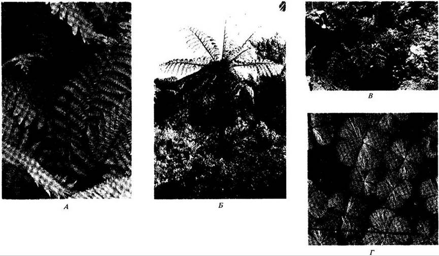
Fig. 17-25. Fern diversity. A. Osmunda cinnamomea. B. The tree fern Dicksonia squarrosa from New Zealand. C. The limestone-dwelling species Notholaena neglecta, found in Texas, Arizona, and New Mexico (USA). D. The heterosporous aquatic fern Marsilea.
The aerial shoots of horsetails, which sometimes die back during unfavorable seasons, arise from perennial, branching rhizomes. The aerial stem has a complex Anatomical Structure (Fig. 17-23). In mature plants, a hollow central pith in the internodes is surrounded by a ring of smaller channels, known as carinal canals, each associated with a strand of primary xylem and primary phloem.
Fig. 17-23. Stem anatomy of a horsetail. A. Cross section showing mature tissues. B. An isolated vascular strand with xylem and phloem.

Fig. 17-26. Life cycle of a horsetail. In broad outline, it is identical to the life cycles of Psilotum, clubmosses, and homosporous ferns. Meiosis occurs within sporangia located along the margins of umbrella-shaped structures called sporangiophores, which are aggregated into strobili. Upon spore maturation, elaters attached to the spore wall presumably aid in their dispersal from the dehiscing sporangium. The gametophytes are green, free-living, and either bisexual (as shown in this illustration) or male. Multiflagellated sperm require water to reach the egg cell. In horsetails, as in other seedless vascular plants, embryo development takes place within the venter of the archegonium, and the young sporophyte remains attached to the gametophyte by a foot. Eventually, the sporophyte detaches from it and becomes an independent Organism. Unlike other seedless vascular plants, the mature horsetail sporophyte features jointed, ribbed stems.

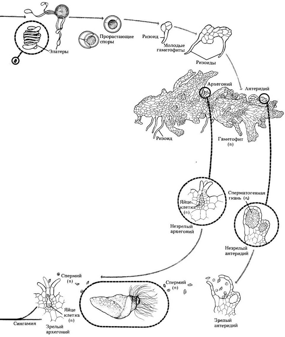
Fig. 17-27. Anatomical structure of fern rhizomes. A. Adiantum. Cytology/practical/72.html">Cross section of the rhizome, showing a siphonostele. Note the large leaf gap. B. Cross section through the vascular region of the tree fern Dicksonia. The phloem consists primarily of sieve elements, and the xylem consists exclusively of tracheids.

Horsetails are homosporous plants. Sporangia are produced in groups of five to ten along the margins of small, umbrella-like structures called sporangiophores, which are gathered into a terminal strobilus at the apex of the stem (see Figs. 17-21A and 17-26). The fertile stems of some species contain little chlorophyll, differ sharply in appearance from vegetative stems, and often appear earlier in the spring (see Fig. 17-21). In other species, strobili are formed at the tips of vegetative stems (see Fig. 10-7). When the numerous spores mature, the sporangia shrink and split along their inner surface, releasing them. Elaters arising from the outer layer of the spore wall are tightly coiled when moist and uncoil when dry, a mechanism believed to play a role in spore dispersal.
The gametophytes of horsetails are green, free-living, and mostly about the size of a pinhead. They develop primarily on silt that was recently inundated with water and is therefore rich in nutrients. The gametophytes (Figs. 17-24) reach sexual maturity in 3 to 5 weeks and are either bisexual or male. In bisexual gametophytes, archegonia develop earlier than antheridia, which increases the likelihood of cross-fertilization. The sperm are multiflagellated, and water is required for them to reach the egg cells. Several egg cells can be fertilized simultaneously on a single gametophyte, subsequently developing into embryos, i.e., young sporophytes.
Fig. 17-24. Bisexual horsetail gametophyte with male and female gametangia. Compare this with the 400-million-year-old gametophyte from Scotland shown in Fig. 16-2.

The life cycle of horsetails is shown in Fig. 17-26.
Fig. 17-28. Convolutely coiled leaves of Osmunda cinnamomea. Although formerly used for food, they are now strongly suspected to be somewhat toxic.

Fig. 17-29. Sori—clusters of sporangia on the lower surface of fern leaves. A. In Dennstaedtia punctilobula and other species of this genus, the sori are naked. B. In bracken (Pteridium aquilinum), shown in the figure, as well as in Adiantum, the sori are located along the margins of the leaf blade, which reflex to cover them. C. In the evergreen woodland fern Dryopteris marginalis, the sori, also situated near the leaf margins, are completely covered by kidney-shaped indusia. D. In Onoclea sensibilis, the sporangia are enclosed within spherical lobes of specialized leaves that differ markedly from the sterile leaves. The sporangial walls of many ferns consist of only a single cell layer and possess specialized dehiscence mechanisms. However, in some primitive groups of this phylum, the walls are multilayered and rupture along a single line of thin-walled cells.
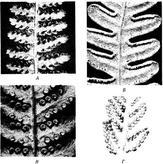
Fig. 17-30. The homosporous fern Cyrtomium falcatum. Transversal section of a leaf with a sorus on the lower surface. Sporangia at various Selection/3.html">Stages of development are covered by an umbrella-shaped indusium.

Phylum Pterophyta
Representatives of this phylum (ferns) are relatively abundant in the fossil record, dating back to the Carboniferous period (see the appendix to this chapter and Fig. 18-1) and surviving to the present day, with some groups occurring as early as the Devonian period. About two-thirds of the approximately 12,000 modern species grow in the tropics, while the remaining third inhabits temperate Regions of the globe, including desert areas. Ferns far surpass all other groups of modern seedless vascular plants in number of species.
Representatives of this phylum are quite diverse in their life forms and habitats (Fig. 17-25). Some differ greatly in appearance from the ferns with which we are most familiar; for example, Salvinia, an aquatic plant with entire leaves up to 2 cm long. More "fern-like" are the floating aquatic species of the genus Azolla (see Fig. 26-16), which play an important role in agricultural and natural ecosystems because symbiotic nitrogen-fixing cyanobacteria inhabit cavities at the base of their leaves. At the other extreme are tree ferns (Fig. 17-25, B), such as those of the genus Cyathea, with individuals exceeding 24 m in height and leaves over 5 m long. Although their trunks can reach more than 30 cm in diameter, the tissues here are entirely primary in origin. A cambium is known only in Botrychium (moonwort), a relatively small herbaceous plant.
Most temperate woodland ferns possess fleshy, siphonostelic rhizomes (Fig. 17-27) that produce new flushes of leaves each year. The roots are adventitious, arising from the rhizomes at the leaf bases. The leaves, or fronds, are megaphylls and constitute the most conspicuous part of the sporophyte. Their surface-to-volume ratio indicates that they are much more efficient photosynthetic organs than the microphylls of lycophytes. Ferns are the only seedless vascular plants with megaphylls. Fronds are typically compound, meaning the blade is divided into leaflets (pinnae) attached to an extension of the leaf stalk called the rachis, or petiole. In almost all ferns, young leaves are circinate in the bud (Fig. 17-28), exhibiting what is known as circinate vernation. This is caused by the lower surface of the frond growing faster than the upper surface during early development, a process regulated by the hormone auxin produced by young leaflets on the inner side of the "fiddlehead."
With the exception of a few genera, all ferns are homosporous. Sporangia are arranged in various ways on the lower surface of ordinary leaves, on modified fronds, or on specialized axes (Fig. 17-29). They are usually aggregated into clusters called sori. In many genera, sori are covered by specialized outgrowths of the leaf called indusia, which may wither as the sporangia mature (Fig. 17-30). At this time, mature spores—formed from spore mother cells via meiosis—are released into the environment thanks to the abrupt rupture of cells in a region of the sporangium called the stomium. The sporangia are stalked, and the wall of each bears a specialized layer of cells with unevenly thickened walls, the annulus. The contraction of the annulus causes the cells of the stomium to tear, and its subsequent sudden snapping back discharges the spores in a catapult-like manner.
Heterospory among modern ferns is known only in two specialized aquatic groups (see Fig. 17-25, D); some extinct fern taxa were also heterosporous.
The spores of most homosporous ferns give rise to free-living, bisexual gametophytes. The gametophyte begins its development as a tiny, pale-green, alga-like chain of cells called a protonema. This then develops into a flat, Heart-shaped, thalloid structure, the prothallus, with numerous rhizoids in the center of its lower surface. Antheridia and archegonia also form on its lower surface. Antheridia usually appear earlier, mostly among the rhizoids, while archegonia develop later near the notch at the anterior end of the gametophyte. This temporal difference in The formation of the two gametangial types promotes cross-fertilization in ferns. Both homosporous and heterosporous Representatives of the phylum require water for the multiflagellated sperm to swim to the egg cells.
In the Early stages of its development, the embryo—that is, the young sporophyte—receives nutrients from the gametophyte via a foot. However, it grows very rapidly and soon becomes an independent organism, by which time the gametophyte withers and dies.
The life cycle of a homosporous fern is illustrated in Fig. 17-31.
Fig. 17-31. Life cycle of the homosporous fern Polypodium. Following meiosis, spores are produced within the sporangia; upon dispersal, they develop into gametophytes, which in most species are green and nutritionally independent. Many of these are only a single cell layer thick, more or less heart-shaped, with an apical notch; others are thicker and may be irregular in shape. Specialized cellular filaments (rhizoids) extend from the lower surface of the gametophyte into the substrate.
On the lower surface of the gametophyte are flask-shaped archegonia with an expanded lower portion embedded in the gametophytic tissue. Their necks consist of several tiers of cells. Antheridia are also located on the lower surface of the gametophyte and are surrounded by a sterile jacket layer. Within them, numerous spirally coiled, multiflagellated sperm are produced. When mature and in the presence of adequate moisture, the antheridia burst, releasing the sperm, which swim into the neck of the archegonium. Fertilization takes place within the venter, and the resulting zygote immediately begins to divide. The young embryo grows and differentiates directly into the adult sporophyte, receiving nourishment from the gametophyte for a short time before soon beginning to photosynthesize actively enough to maintain an independent existence. Once the young sporophyte has rooted in the soil, the gametophyte disintegrates.


Last update: 07/08/2026
Editorial and Educational Adaptation: This material has been compiled based on the primary/original source text. The project team performed an editorial review, corrected technical inaccuracies, structured sections, and adapted the content for an educational format.
What was processed:
- elimination of formatting defects (OCR errors, structural breaks, corrupted characters);
- editorial organization of content;
- standardization of terminology in accordance with academic sources;
- verification of factual statements against the original source text.
All mentions of the author, publication year, and origin of the primary text have been preserved in accordance with the source.