MODERN BOTANY - P. RAVEN - 1990
SECTION III. GENETICS
CHAPTER 8. CHEMISTRY AND HEREDITY
The Nature of DNA
In 1951, American geneticist James D. Watson arrived in England, where he initiated a collaboration with Francis Crick at the Cavendish Laboratory in Cambridge. Watson and Crick were among the scientists convinced that DNA, rather than protein, constituted the primary substance of heredity. In Watson's words, DNA is 'the most golden of all molecules.'
Watson and Crick based their investigations into the Organization OF THE DNA molecule partly on METABOLISM/2.html">THE CONCEPT OF genes—the chromosomal units of heredity—that had already emerged by that time. They recognized that for DNA to function as genetic material, it had to fulfill at least four requirements.
1. It must transfer Genetic information from Cell to Cell and from generation to generation; furthermore, it must carry a vast Amount of Information. (Just think how many instructions must be contained within the set of genes that direct The Development of an elephant, a tree, or even a bacterium.)
2. It must duplicate prior to each Cell Division, much like Chromosomes do; moreover, it must replicate with high fidelity. (From accumulated data on mutation rates in humans, for instance, we know that any given human Gene must copy itself without the slightest error, on average, over millions of years.)
3. On the other hand, a gene must occasionally change, or mutate. (When a gene changes—that is, when an error occurs—it is the 'error' that must be copied rather than the original sequence. This is a crucial property, likely unique to all living things, because without the capacity to copy 'errors,' evolution by natural Selection could not occur.)
4. There must exist a mechanism for 'reading' the accumulated information and translating it within the living Organism.
Watson and Crick fully understood that the DNA molecule could serve as the genetic material only if its size, conformation, and complexity were proven sufficient to encode and replicate the vast amount of information required by living organisms.
In their work, Watson and Crick utilized previously acquired biochemical data on DNA, including the following findings:
1. The DNA molecule is very large, as well as long and thin.
2. Its three components (a nitrogenous base, a sugar, and a phosphate) form NUCLEOTIDES, as illustrated in Fig. 3-19.
3. Rosalind Franklin and Maurice Wilkins at King's College London studied the X-Ray Diffraction patterns of DNA preparations and discovered that its long molecules consist of regularly repeating units, which appear to be arranged in a helical Structure.
4. As shown in Table 8-1, The ratio of nucleotides containing adenine to those containing thymine is 1:1, which is precisely the same as the ratio between nucleotides containing guanine and cytosine.
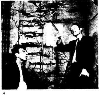
Watson and Crick conducted no experimental research of their own, but instead synthesized all available knowledge regarding Introduction/20.html">DNA Structure. Drawing upon all the aforementioned experimental data, they attempted to build a physical metal model of DNA consistent with the Physical and Chemical evidence of its structure (Fig. 8-4, A).
Class="center">Fig. 8-4. A. Watson (left) and Crick with their metal model of DNA. B. Schematic diagram of the DNA molecule proposed by Watson and Crick. Left: a side view of the DNA molecule indicating the vertical axis. The molecule consists of polynucleotide chains wound into right-handed helices and intertwined to form a double helix. Its average diameter is 2 nm (20 Å). The two chains forming the helix are composed of nucleotides containing deoxyribose residues (8). The sugar of each nucleotide is linked via a phosphate group (P) to the sugar of the adjacent one. The regular alternation of sugars and phosphates forms the backbone of the molecule. The sugars of each helix project toward the interior of the cylinder. The Base Pairs (indicated by bold horizontal lines) occupy the central region of the cylinder (dashed rectangles on the cross-sections to the right). The base pairs are formed by thymine (T) and adenine (A) at level A, and cytosine (C) and guanine (G) at level B. The bases are joined by Hydrogen Bonds. The circles drawn with dashed lines indicate the outer boundary of The Double Helix as viewed from its end. The bases are stacked at intervals of 3.4 Å and rotated by 36° at each step. Thus, there are 10 base pairs per helical turn. As a result of this rotation, when viewed from the side, the base pairs appear as lines of varying length depending on the viewing angle.

From their perspective, the most critical question was how the Chemical Structure of DNA reflects its biological function. Reflecting on their research, Watson wrote: 'In moments of pessimism, we often feared that the correct structure might turn out to be boring—that is, completely devoid of meaning.' In reality, however, The structure of DNA proved to be extraordinarily fascinating.
The Double Helix
By piecing together various data, Watson and Crick concluded that DNA is not a single helix, as many Proteins are, but rather a giant, tightly intertwined double helix. Imagine the handrail of a spiral staircase forming a single helix. If the staircase is twisted into a helix while the steps remain perpendicular to its outer edges, you obtain a rough model of a double helix.
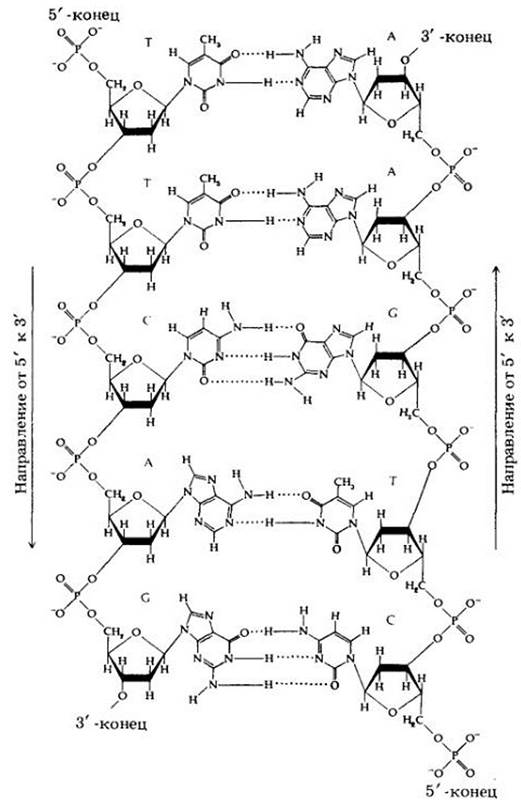
In the DNA molecule, the 'rails' consist of alternating deoxyribose and phosphate molecules (see Figs. 8-4, B and 8-5), while the 'steps' are formed by nitrogenous bases—adenine (A), thymine (T), guanine (G), and cytosine (C)—with one base attached to each sugar-phosphate unit, and each step comprising a pair of bases. The bases are linked by hydrogen bonds, relatively weak chemical bonds that play a vital role in establishing the secondary and tertiary structures of proteins (Fig. 8-5).
According to Wilkins's measurements, the distance between the 'rails' is 2 nm. If two Purines were to pair, the distance between them would be too great, whereas two Pyrimidines would not reach each other. Everything falls into place, however, when a purine pairs with a pyrimidine. Therefore, each base pair—a step of the spiral staircase—is always composed of one purine and one pyrimidine (see Fig. 8-5). For this reason, the ratio of purines to pyrimidines in a DNA molecule is always equal to 1:1.
Watson and Crick noted that The nucleotide sequence of each strand in the double helix can be arbitrary, such as ATGCGTACATT, and so forth. Since a DNA molecule can be several thousand nucleotides long, an enormous variety in the nucleotide sequence can be expected. The number of base pairs ranges from 5,000 in the simplest virus to 5,000,000,000 in the 46 Human chromosomes. If the DNA from a single human cell were stretched out linearly, the strand would reach a length of 1.5 m; The amount of information it contains would fill 600,000 printed pages averaging 500 words each, which is equivalent to a library of 1,000 books. In short, the DNA molecule is truly capable of storing the necessary genetic information.
Fig. 8-5. Double-stranded structure of a section of a DNA molecule. Each nucleotide consists of a sugar (deoxyribose), a phosphate group, and a nitrogenous base (a purine or a pyrimidine). Note the repeating sugar-phosphate-sugar-phosphate backbone that forms the framework of the molecule. Each phosphate group is attached to the 5' carbon atom of one sugar and the 3' carbon atom of the sugar in the adjacent nucleotide, giving the strand two distinct ends: 5' and 3'. The bridges formed by the phosphate groups between nucleotides run in opposite directions; that is, the strands are antiparallel (compare this figure with Fig. 8-4, B).
A Molecule That Copies Itself
The most fascinating discovery came when Watson and Crick attempted to join two DNA strands: they found that not only can purines not bond with purines, nor pyrimidines with pyrimidines, but adenine can pair exclusively with thymine, and guanine with cytosine. Only these base pairs form hydrogen bonds; adenine cannot form them with cytosine, nor guanine with thymine.
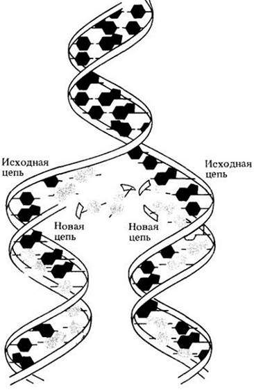
Let us look back at Fig. 8-1. The Watson-Crick model simply and logically explains the base composition of DNA. Perhaps the most important property of the model is that the two strands are complementary, meaning each contains a sequence of bases that is the counterpart of the other. When a DNA molecule "replicates," it simply "unzips"; the hydrogen bonds between the nitrogenous bases break (Fig. 8-6). The two strands separate, and new ones are synthesized along each template. If a thymine is present in the parent strand, only adenine will attach to it, just as guanine pairs exclusively with cytosine. Thus, a complementary strand is formed along each original strand, resulting in two exact copies of the parent DNA. In this way, the Discovery of the DNA structure helped answer a fundamental long-standing question: how hereditary information is duplicated and passed down from generation to generation.
Fig. 8-6. The DNA molecule shown here is in The process of Replication. The parent strands unwind as the hydrogen bonds between the bases break. (For clarity, the bases are shown out of plane.) Each parent strand serves as a template upon which a new complementary strand is built from nucleotides available within The Cell.
Regarding this one of the greatest mysteries of all time, Watson and Crick wrote in their brief initial paper: "It has not escaped our notice that the specific pairing we have postulated immediately suggests a possible copying mechanism for the genetic material." In 1962, 9 years after publishing their original hypothesis, Watson, Crick, and Wilkins were awarded the Nobel Prize, highlighting the immense significance of their research.
It was subsequently demonstrated that due to its extraordinary length and structural complexity (the double helix), the DNA replication process occurs in multiple enzyme-catalyzed steps. Specific Enzymes are required to separate and unwind the DNA strands and to properly align the complementary bases. The mechanism by which these enzymes operate paves the way for understanding DNA replication.
As the Chemical Nature of this process became clearer, additional "complexities" emerged—the ends of the two strands in the double helix are distinct. In each strand, the phosphate group linking two deoxyribose molecules is attached to one sugar at the 5' position (the fifth carbon atom of the deoxyribose ring) and to the adjacent sugar at the 3' position (the third carbon atom of the deoxyribose ring). Because of this sugar-phosphate-sugar linkage, each strand has 5' and 3' ends (see Fig. 8-5).
Furthermore, since DNA Synthesis proceeds in the 3'-to-5' direction on one strand, it runs in the opposite direction on the other strand. Thus, the two DNA strands are antiparallel (see Fig. 8-5). Strand synthesis in the 3'-to-5' direction is continuous, with nucleotides added one by one sequentially, whereas Synthesis of the other strand is discontinuous, being built from a series of short fragments that are subsequently joined to form a continuous strand, with each fragment synthesized in the reverse direction. Currently, more than 14 different enzymes have been identified in the bacterium Escherichia coli that participate in various Stages of DNA replication, and duplication of the genetic material is even more complex in eukaryotes.
Last update: 07/08/2026
Editorial and Educational Adaptation: This material has been compiled based on the primary/original source text. The project team performed an editorial review, corrected technical inaccuracies, structured sections, and adapted the content for an educational format.
What was processed:
- elimination of formatting defects (OCR errors, structural breaks, corrupted characters);
- editorial organization of content;
- standardization of terminology in accordance with academic sources;
- verification of factual statements against the original source text.
All mentions of the author, publication year, and origin of the primary text have been preserved in accordance with the source.