Medical Genetics - V. M. Zaporozhan 2005
Diagnostic Methods for Hereditary Disorders
Molecular Genetic Methods (DNA Diagnostics)
Stages of DNA Diagnostics Using the Polymerase Chain Reaction
Today, one of the primary Methods of DNA Diagnostics is the Polymerase Chain Reaction (PCR). The technique was developed in 1983 by K. Mullis (USA), for which he was awarded the Nobel Prize in Chemistry in 1993.
The method is based on the in vitro Amplification (i.e., multiple reduplication) of a specific DNA region ranging from several dozen to a thousand or more Base Pairs. This makes it possible to obtain A large number of copies of the desired DNA sequence within 3.5 hours. Identifying such a large quantity of DNA copies subsequently does not pose a major challenge. Any DNA isolated from various sources can serve as a suitable template. The main advantages of the PCR method are as follows:
— amplification of the target DNA region even if the analyzed sample is insufficiently purified or represents a complex mixture of molecules. Such samples include Blood, urine, exudates, sputum, and other biological fluids, as well as bacterial cultures;
— amplification of DNA present in a sample in extremely small quantities. Essentially, the Starting Material for PCR can be as little as a single Cell or even a single molecule. This is crucial when testing air or Water samples for pathogenic disease agents, or human tissue fragments for forensic investigations;
— The ability to store research material for long periods due to the inherent Stability of the DNA molecule.
The following stages of DNA diagnostics using the polymerase chain reaction are distinguished.
1. DNA extraction. Amplification does not require a large amount of template DNA; even small fragments of poorly purified DNA are suitable, and in principle, a single molecule may suffice. Typically, DNA is isolated from biological material through cell lysis followed by the removal of protein components. To increase the sensitivity of the reaction, DNA can be sorbed onto ion-exchange resins.
2. Polymerase chain reaction, which allows for the selective amplification (multiple reduplication) of the target DNA region by millions of times.
A prerequisite for performing PCR is knowledge of The nucleotide sequence of the amplified DNA region, as the specific Selection of this region is achieved by hybridizing the template DNA with two artificially synthesized primers. Primers are oligonucleotide DNA sequences 15 to 30 bp in length, complementary to the 3'-ends of the amplified region on the sense and antisense DNA strands, respectively. Thus, the distance between the primers determines the length of the synthesized DNA fragments.
Essentially, the PCR method mimics the natural process of DNA reduplication within Cells. The reaction mixture is supplemented with the patient's DNA, four types of DNA NUCLEOTIDES (dNTPs — deoxynucleoside triphosphates), and a thermostable DNA polymerase (Taq polymerase) that maintains its activity at high temperatures. This enzyme was isolated from Bacteria living in hot springs (Thermus aquaticus). The optimal operating Temperature for the enzyme is +72 °C.
The reaction takes place in a special buffer solution with specific concentrations of potassium, chlorine, and magnesium ions, and a precise pH level.
The polymerase chain reaction proceeds cyclically. Each cycle consists of three phases (Fig. 10.7):
1) Denaturation (melting) — the mixture is heated to 90–95 °C. This breaks the Hydrogen Bonds connecting the two DNA strands, converting the DNA into a single-stranded form;
2) Hybridization (annealing) — the mixture is cooled to 45–60 °C, allowing the primers to bind to their complementary DNA regions;
3) synthesis — the mixture is heated again to 72 °C, the thermostable DNA polymerase becomes active, and the daughter DNA strand is synthesized. The DNA polymerase extends the nucleotide chain complementary to the template DNA, with DNA Synthesis proceeding from the primer binding site in the 3'→5' direction. The primer is thereby incorporated into the newly synthesized nucleic acid region. In subsequent cycles, the newly synthesized DNA molecules serve, in turn, as templates for the analogous synthesis of new copies. Since the synthesis of each of the two antiparallel DNA strands begins at the primer hybridization site, this site defines the boundary of the synthesized region.
Class="center">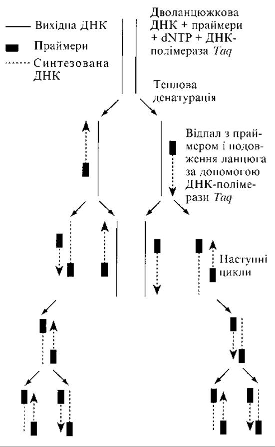
Fig. 10.7. Polymerase chain reaction (exponential increase in the number of DNA copies)
Assuming that the reaction mixture contained a single DNA molecule with the region to be reduplicated, 2 molecules are obtained after the first cycle, 4 after the second cycle, and so on. In other words, the number of DNA copies increases exponentially.
The number of such cycles in PCR typically ranges from 25 to 30. As a result, the number of DNA copies increases by millions of times.
The mixture is placed in a specialized device known as a thermal cycler (amplifier), which automatically carries out the temperature changes required for the reaction.
Advantages of PCR:
— a significant (2- to 10-fold) reduction in labor and time costs;
— high accuracy, with a fundamental impossibility of obtaining false-positive results;
— the ability to conduct studies on extremely small volumes of material (a few microliters);
— simplicity in preparing material for analysis; virtually any tissue or bodily fluid can be used.
Performing PCR diagnostics requires strict adherence to all Sanitary and hygienic standards to prevent sample contamination with foreign DNA, specially equipped laboratories, and trained personnel.
3. Analysis of the obtained results. Further analysis of the amplified DNA fragments involves examining Specific features of the amplified product. Fragments with normal and mutant sequences may differ in electrophoretic mobility; therefore, gel Electrophoresis of the amplified fragments is frequently performed (10.4.5.2). At the same time, electrophoresis of control DNA fragments is carried out. Ethidium bromide (a DNA-staining dye) is added to the gel. The DNA fragments migrate a certain distance. The areas of the gel containing DNA fragments will emit an orange glow under ultraviolet illumination. A match between the bands of the control and test fragments indicates the presence of the target Gene. The gel can be photographed or its image transferred to a computer screen (Fig. 10.8).
To identify the amplified fragments, sequencing methods as well as the allele-specific oligonucleotide assay can be used. This method is based on the hybridization of amplified DNA fragments with labeled oligonucleotides complementary to either the normal or mutant DNA sequence. An example of this approach is The Use of DNA Microarrays.
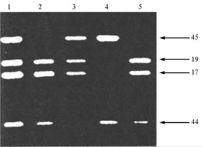
Fig. 10.8. Direct DNA Diagnosis of Duchenne muscular dystrophy using multiplex polymerase chain reaction. Four exons of the dystrophin gene were simultaneously amplified in each of the subjects tested. Exons 17, 19, 44, and 45 are indicated by arrows. Lane 1 represents the control, showing all four exons. Lanes 2–5 represent patients with Duchenne muscular dystrophy featuring various dystrophin gene deletions (lanes 2 and 5 show a deletion of exon 45, lane 3 shows a deletion of exon 44, and lane 4 shows deletions of exons 17 and 19)
Last update: 11/08/2026
Editorial and Educational Adaptation: This material has been compiled based on the primary/original source text. The project team performed an editorial review, corrected technical inaccuracies, structured sections, and adapted the content for an educational format.
What was processed:
- elimination of formatting defects (OCR errors, structural breaks, corrupted characters);
- editorial organization of content;
- standardization of terminology in accordance with academic sources;
- verification of factual statements against the original source text.
All mentions of the author, publication year, and origin of the primary text have been preserved in accordance with the source.