BIOLOGY Volume 3 - A Guide to General Biology - 2004
20. EXCRETION AND OSMOREGULATION
20.5. The Human Kidney
20.5.5. Selective Reabsorption in the Proximal Convoluted Tubule
Humans produce about 125 ml of filtrate per minute, which amounts to approximately 180 liters per day. Since actual urine output averages only about 1.5 liters, it is clear that the vast majority of the filtrate must be reabsorbed. Indeed, out of the 125 ml of glomerular filtrate (GF) produced per minute, 124 ml is reabsorbed, with 80% of this amount occurring in the proximal convoluted tubule.
During ultrafiltration, substances essential for life are removed from the Blood along with Metabolic waste products. The function of the nephron is to selectively reabsorb those substances that are still needed, either directly by the Cells or to maintain the homeostatic composition of Body Fluids. In addition, further excretory products can be actively secreted into the tubules from the surrounding capillaries.
Thus, Urine Formation involves three key processes: ultrafiltration, selective reabsorption, and secretion.
Analysis of Fluid in the Nephron
To determine the composition of fluid from different PARTS OF THE nephron, micro-pipettes are typically used. By taking samples with these pipettes, a complete picture can be obtained of what happens to the GF in various Regions of the nephron. The flow rate of the filtrate is also measured using the polysaccharide inulin as a marker. Inulin is not produced by The Human Body; when injected into the bloodstream, it is filtered into the nephron. It is neither reabsorbed nor secreted there, so its concentration increases in proportion to The amount of Water reabsorbed. As the water volume decreases, the flow rate of the GF drops.
In the example illustrated in Fig. 20.21, the flow rate of the GF in Bowman's capsule is set at 100 arbitrary units for convenience of comparison. This value is called the GF flow rate index and serves as an indicator of the water volume in the GF. For instance, if the index drops from 100 to 40, it indicates that 60% of the water has been reabsorbed. Using the figures given in Fig. 20.21, one can calculate the reabsorption rates for fluids of various compositions flowing through the nephron. Try to answer the following questions in order. If you find it difficult, check the corresponding explanation at the end of the book before moving on to the next question.
Class="center">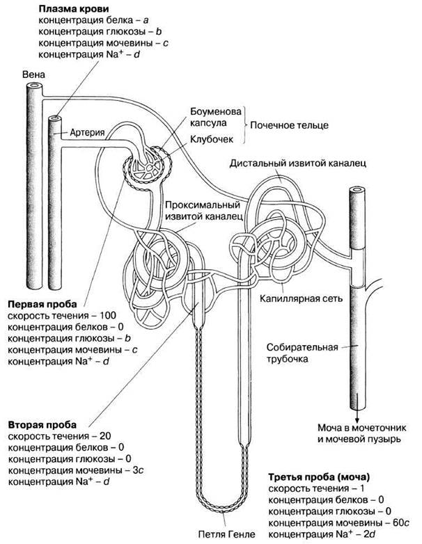
Fig. 20.21. Diagram of The Structure of a nephron and part of its blood capillary network. (From Biological Science, Study Guide, 1970, p. 373.)
20.2. How does the concentration of dissolved substances change as fluid passes from the blood into Bowman's capsule? Explain your answer.
20.3. On the way from Bowman's capsule to the end of the proximal convoluted tubule, the flow rate index drops from 100 to 20. What percentage of water is reabsorbed into the blood by this part of the nephron?
20.4. By the end of the proximal convoluted tubule, only 20%, or one-fifth, of the incoming water remains. Consequently, the concentration of all dissolved substances should increase fivefold, unless something happens to them. However, the concentration of urea, for example, increases only threefold—which is 3/5 or 60% of the expected level. This means that only 60% of the urea remains, while 40% of it is reabsorbed. Based on this diagram, calculate what percentage of glucose and sodium ions are reabsorbed in the proximal convoluted tubule.
20.5. How do the concentrations of water, sodium ions, and urea change along the path from the end of Mouth/proximal convoluted tubule to the end of the collecting duct?
20.6. What is the total percentage of water and sodium ions reabsorbed between Bowman's capsule and the end of the collecting duct?
STRUCTURE OF THE Proximal Convoluted Tubule
The proximal convoluted tubule is the longest (14 mm) and widest (60 µm) part of the nephron. Through it, the GF travels from Bowman's capsule into the Loop of Henle. The tubule wall consists of a single layer of cuboidal epithelium, the cells of which are densely covered on their inner side with microvilli forming a brush border (Fig. 20.22). At the Base of the epithelial cells—that is, on the outer surface of the convoluted tubule—lies the basement membrane; here, Cell/30.html">The Plasma Membrane forms a complex system of folds called basal channels, thereby increasing the surface area of the cells. Adjacent cells fit tightly together only near the lumen of the tubule, while along the rest of their length they are separated by narrow intercellular spaces. Together with the basal channels, these spaces form the so-called basolateral labyrinth, which is filled with tissue fluid connecting the nephron epithelium to the surrounding capillary network. The Cells of the proximal convoluted tubules are rich in Mitochondria, which are concentrated near the basement membrane to supply ATP to The Active Transport systems embedded within it (Fig. 20.23). Cytology/cytology/93.html">ELECTRON MICROGRAPHS OF these cells are shown in Fig. 20.22.
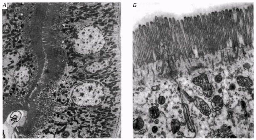
Fig. 20.22. A. Electron micrograph of the cuboidal epithelium of the proximal convoluted tubule; ×7,000. B. Microvilli (brush border) of the proximal convoluted tubule epithelium at a higher magnification; ×18,000.

Fig. 20.23. Structure and function of the proximal convoluted tubule.
Selective Reabsorption in the Proximal Convoluted Tubule
The cells of the proximal convoluted tubule are adapted for reabsorption due to the following structural features:
1) a vast absorptive surface area provided by microvilli and the basolateral labyrinth;
2) numerous mitochondria;
3) the close proximity of adjacent capillaries.
The proximal convoluted tubules reabsorb over 80% of the glomerular filtrate, including all glucose, Amino Acids, Vitamins, and Hormones, along with approximately 80% of sodium chloride and water. The reabsorption mechanism is as follows.
1. Glucose, amino acids, and ions diffuse from the filtrate into the cells of the proximal convoluted tubule, from which they are actively transported by plasma membrane transport systems into the intercellular spaces and labyrinth clefts via membrane-embedded carrier Proteins.
2. From the intercellular spaces and labyrinth, they diffuse into the highly permeable peritubular capillaries and are removed from the nephron.
3. The continuous removal of all these substances from the cells of the proximal convoluted tubule establishes a diffusion gradient between the filtrate in the tubular lumen and the cells. Driven by this gradient, a continuous influx of new molecules enters the cells, is subsequently actively transported into the intercellular spaces and labyrinth clefts, and the cycle continues. We will examine this mechanism in greater detail below using The transport of sodium and glucose as an example (Fig. 20.24).

Fig. 20.24. A. Selective reabsorption of sodium in the proximal convoluted tubule. B. Selective reabsorption of glucose in the proximal convoluted tubule.
As a result of the active uptake of sodium and other ions, the Osmotic Pressure of the filtrate decreases, and an equivalent amount of water passes into the permeable capillaries via osmosis. The bulk of dissolved solutes and water is extracted from the filtrate at a fairly constant rate. This process produces a filtrate within the tubule that is isotonic to the Blood Plasma of the peritubular capillaries.
Approximately 40–50% of urea is also reabsorbed from the filtrate by diffusion, entering the peritubular capillaries and returning to the general Circulatory system. This urea is neither required by the body nor harmful to it; the remaining urea is excreted in the urine.
Small protein molecules entering the nephron during ultrafiltration are removed in the proximal convoluted tubule via pinocytosis occurring at the base of the microvilli. They end up inside pinocytotic vesicles, to which Lysosomes attach. Lysosomal hydrolytic Enzymes break down The polypeptide chains into amino acids, which are either utilized by the tubular cells themselves or diffuse into the peritubular capillaries.
Finally, unwanted substances, such as creatinine and a certain amount of urea, are actively secreted from the blood capillaries into this segment of the nephron. These substances are transported from the interstitial fluid bathing the tubules into the tubular filtrate and are excreted in the urine.
Last update: 06/08/2026
Editorial and Educational Adaptation: This material has been compiled based on the primary/original source text. The project team performed an editorial review, corrected technical inaccuracies, structured sections, and adapted the content for an educational format.
What was processed:
- elimination of formatting defects (OCR errors, structural breaks, corrupted characters);
- editorial organization of content;
- standardization of terminology in accordance with academic sources;
- verification of factual statements against the original source text.
All mentions of the author, publication year, and origin of the primary text have been preserved in accordance with the source.