BIOLOGY Volume 3 - A Guide to General Biology - 2004
20. EXCRETION AND OSMOREGULATION
20.5. The Human Kidney
20.5.4. Ultrafiltration
The first step in Urine Formation is the ultrafiltration of Blood, which takes place in the renal corpuscle. Ultrafiltration is filtration under pressure. In this case, hydrostatic pressure is provided by the blood, which enters the glomerulus under high pressure from The Heart via the aorta, renal artery, and afferent arteriole (Fig. 20.11). The glomerulus is a capillary network surrounded by Bowman's capsule (Fig. 20.15, D). The diameter of its capillaries is much smaller than that of the afferent arteriole, which increases the blood pressure within them. Water and small dissolved molecules are "squeezed" out of the capillaries and pass through the epithelium of Bowman's capsule into its lumen. Large molecules, such as Proteins, as well as Blood Cells, remain in the blood. The Structure of the glomerulus and Bowman's capsule is entirely adapted to their function—blood filtration—as illustrated in Figs. 20.16–20.19. The filter consists of three layers, shown in detail in Figs. 20.16, B and 20.19.
Class="center">
Fig. 20.16. A. STRUCTURE OF THE renal corpuscle. Top: afferent and efferent arterioles. The outer surface of the glomerular capillaries is surrounded by specialized epithelial cells called podocytes. As a result, the capillaries themselves are not visible, but their outlines can be discerned. (Just as a glove conceals the fingers while revealing their general shape—the glove is equivalent to the podocytes, and the fingers to the capillaries.) Bottom: the beginning of the proximal convoluted tubule, whose wall is formed by cuboidal epithelium with microvilli (brush border) (after: L. C. Junqueira & J. Carneiro (1980) Basic Histology, 3rd ed., Lange Medical Publications). B. Detailed structure of podocytes and a capillary in longitudinal section. It can be seen that the podocytes loosely interdigitate with their processes (pedicels), much like interlocking fingers, leaving slit pores between them that allow the glomerular filtrate to pass into Bowman's capsule.
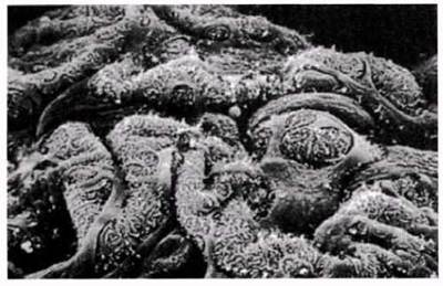
Fig. 20.17. Scanning electron micrograph of podocytes; ×900. Their surfaces feature numerous processes of varying sizes, which limit the size of molecules capable of passing through the renal corpuscle filter.

Fig. 20.18. Diagram of the structure of the renal (Malpighian) corpuscle, showing typical Features of the glomerulus and Bowman's capsule.
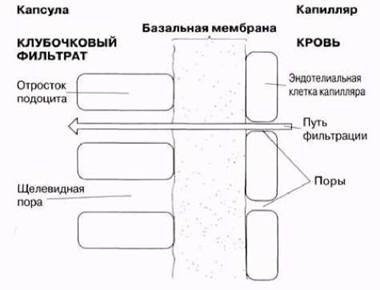
Fig. 20.19. Schematic diagram of the pathways through which plasma is filtered from the glomerular capillary into Bowman's capsule.
1. Capillary endothelium. This is an extremely thin layer of cells perforated by numerous pores approximately 10 nm in diameter. Pores account for about 30% of the Capillary Wall area and are far too large to retain Plasma Proteins.
2. Capillary basement membrane. All epithelial cells rest on a basement membrane, which is a non-cellular layer consisting of an interwoven meshwork of fibers, particularly Collagen. Water and small molecules easily pass through this "sieve," whereas blood cells are retained. Protein molecules are also too large to pass through; furthermore, they are repelled by the negative electrical charge of the basement membrane fibers.
3. The epithelium of Bowman's capsule is formed by cells specially adapted for filtration—podocytes (from the Greek podos, meaning FOOT), which bear numerous surface processes. The processes of adjacent cells interdigitate loosely, as shown in Figs. 20.16 (A and B) and 20.17, leaving slit pores of ~25 nm (Fig. 20.19) through which the filtrate passes.
Approximately 20% of the plasma is filtered into the capsule. Of the three layers listed, the basement membrane acts as the primary filter. The fluid that enters the capsule is called the glomerular filtrate (GF) and is chemically similar to Blood Plasma. It contains glucose, Amino Acids, Vitamins, electrolytes, nitrogenous waste products (mainly urea, along with small amounts of uric acid and creatinine), certain Hormones, and water. Blood leaving the glomeruli has a lower water potential due to an elevated plasma protein concentration, but its hydrostatic pressure is reduced.
Factors affecting the Glomerular Filtration Rate (GFR)
The pressure driving fluid filtration out of the glomeruli depends not only on the hydrostatic pressure of the blood but also on the opposing pressure of the GF (Fig. 20.20). If these two latter forces were equal, they would cancel each other out. In reality, the hydrostatic pressure of the GF is much lower than the corresponding blood pressure, remaining just slightly above zero. GFR is also influenced by the osmotic potential on both sides of the filter. Water tends to move down this potential gradient—that is, toward the region with a more negative potential (higher solute concentration). As blood flows from the afferent to the efferent arteriole, it loses water and small dissolved molecules while retaining plasma proteins, whose concentration consequently increases by about 20%. This leads to a drop in its osmotic and water potentials, thereby tending to reduce the GFR. However, taking all these factors into account, the GFR remains positive, and fluid continues to move from the glomerulus into Bowman's capsule. The more diluted the blood relative to the GF, the higher the filtration pressure and GFR.
The GFR can be increased by raising blood pressure or by dilating the afferent arteriole, thereby reducing its resistance to blood flow into the glomerulus. A third regulatory mechanism involves increasing resistance in the efferent arteriole by constricting it.
The dilation and constriction of Blood Vessels are regulated by both neural and hormonal mechanisms.

Fig. 20.20. Direction and magnitude of the forces determining filtration pressure in human renal glomeruli.
Last update: 06/08/2026
Editorial and Educational Adaptation: This material has been compiled based on the primary/original source text. The project team performed an editorial review, corrected technical inaccuracies, structured sections, and adapted the content for an educational format.
What was processed:
- elimination of formatting defects (OCR errors, structural breaks, corrupted characters);
- editorial organization of content;
- standardization of terminology in accordance with academic sources;
- verification of factual statements against the original source text.
All mentions of the author, publication year, and origin of the primary text have been preserved in accordance with the source.