Biochemistry - The Chemical Reactions of Living Cells, Volume 3 - D. Metzler 1980
Metabolism of Nitrogen-Containing Compounds
Serine and Glycine
Porphobilinogen, Porphyrins, and Related Compounds
In 1946, Shemin and Rittenberg [73a] described one of the first successful Applications of radioactive tracers to The Study of METABOLISM. This classic work demonstrated that the carbon atoms of the porphyrin ring in the heme molecule originate from such simple compounds as acetate and Glycine. As we now know, acetate is converted into succinyl-CoA within The Tricarboxylic Acid Cycle. In the mitochondrial matrix of animal Cells, succinyl-CoA condenses with glycine to yield δ-aminolevulinic acid [equation (8-20)] [73b], which is subsequently converted into porphobilinogen (Ch. 10, Sec. B, 1), the immediate precursor of Porphyrins. By degrading the 14C-labeled porphyrins derived from labeled acetate and glycine, Shemin and Rittenberg established the isotope labeling pattern in the pyrrole ring shown in Fig. 14-13 for porphobilinogen. The black circles designate those atoms that originally derived from the methyl carbon of the acetate molecule (it should be recalled that the acetyl groups of acetyl-CoA pass through the tricarboxylic acid cycle more than once, so the label from the acetate methyl groups enters both the 2- and 3-positions of succinyl-CoA). The atoms marked by open circles originate predominantly from the carboxyl carbon of acetate and to a lesser extent from the methyl carbon. Atoms marked with asterisks derive from glycine, whereas unlabeled carbon atoms originate from the carboxyl carbon of acetate.
Class="center">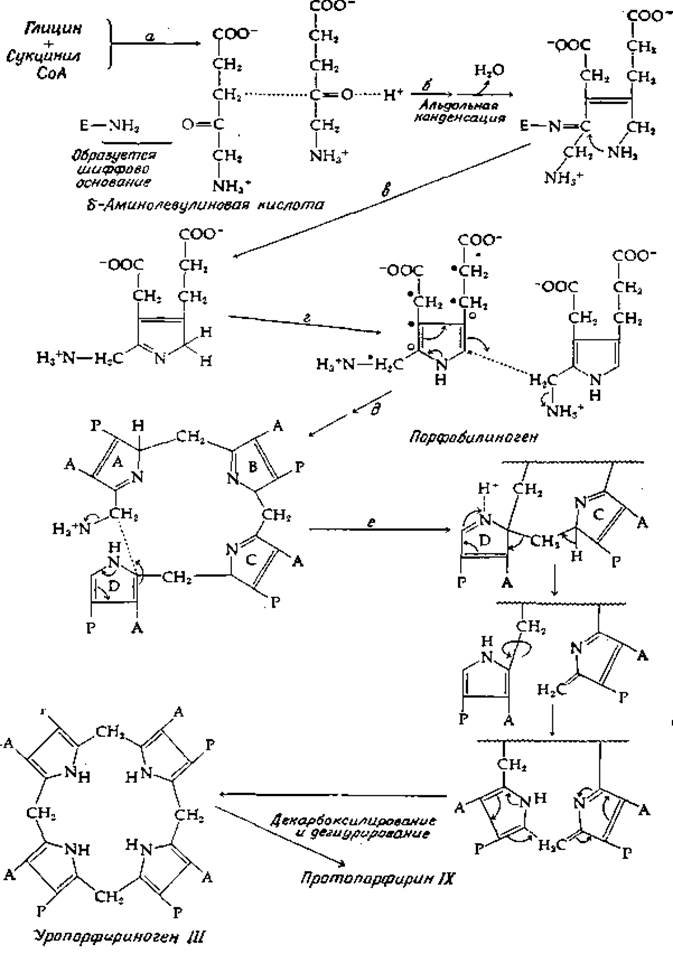
FIG. 14-13. Biosynthesis of porphyrins from glycine and succinyl-CoA.
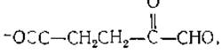
Plants utilize a different pathway. The entire 5-carbon Skeleton of α-ketoglutarate is incorporated intact into δ-aminolevulinate. The process presumably begins with an intramolecular oxidation-reduction reaction (possibly following the prior conversion of α-ketoglutarate to a thioester) analogous to the reverse reaction catalyzed by glyoxalase I (Ch. 7, Sec. L). The product of this reaction is presumably γ,δ-dioxovalerate, which undergoes reductive amination to yield δ-aminolevulinate [74].
As illustrated in Fig. 14-13, The conversion of two molecules of δ-aminolevulinic acid into porphobilinogen is a multistep reaction beginning with an aldol Condensation (stage b). The enzyme catalyzing this reaction is believed to form a Schiff base with the carbonyl group of one of the substrate molecules, as indicated in the figure [75, 76]. The aldol condensation is followed by dehydration to establish a double bond between two carbon atoms, and ring closure (stage c) proceeds via successive shifts of the imine linkage similar to those described previously (Vol. II, p. 228). Finally, tautomerization (stage d) is required to yield porphobilinogen. The condensation of porphobilinogen molecules leading to porphyrins involves two Enzymes: porphobilinogen deaminase and uroporphyrinogen III cosynthetase. The deaminase catalyzes stage d (Fig. 14-13). Ammonia is eliminated in the process, though this does not necessarily occur via the direct displacement reaction shown in the figure; an electron shift from a neighboring nitrogen within the same ring may also be operative. One can readily envision a fourfold repetition of such a condensation to yield the symmetrical precursor of the uroporphyrin I molecule (Fig. 10-1). In the presence of the cosynthetase, a different type of reaction takes place. Note that the five-membered ring of porphobilinogen features a symmetrical arrangement of double bonds. Consequently, the condensation reaction can involve either of the carbon atoms situated in the α-position relative to the ring nitrogen. The series of condensation reactions (Fig. 14-13, stage e) is followed by tautomerization, ring Cleavage, and reclosure such that the arrangement of the carboxymethyl and carboxyethyl side chains corresponds to that of uroporphyrinogen III. A subsequent series of decarboxylation and oxidation steps [77] leads directly to protoporphyrin IX.
The insertion of a ferrous ion into protoporphyrin requires a dedicated enzyme, protoheme ferro-lyase (ferrochelatase) [78, 79]. This enzyme has been shown to be tightly bound to The inner mitochondrial membrane of animal cells, plant Chloroplasts, and bacterial chromatophores. Although Fe2+ is typically the sole metal ion incorporated into the porphyrin, Yeast accumulates appreciable amounts of the [Zn2+]-protoporphyrin chelate, and a Cu2+ complex is also known (Ch. 10, Sec. B, 1).
a. Chlorophyll [80, 81]
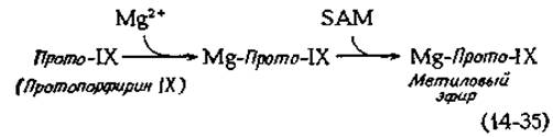
The initial step in the conversion of protoporphyrin IX into chlorophyll is likely the insertion of Mg2+. This reaction occurs negligibly on its own and requires catalytic assistance. This is followed by the methylation of the carboxyethyl side chain of ring III [equation (14-35)].
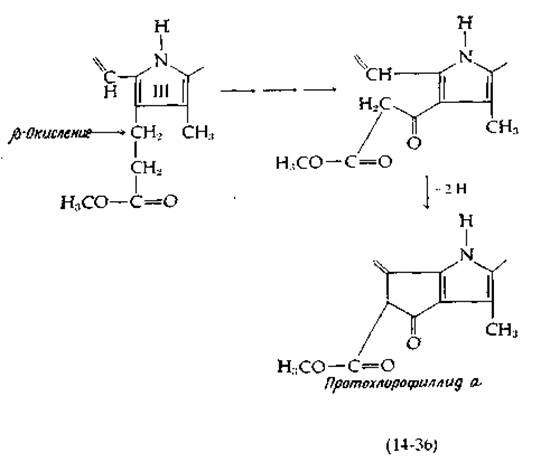
The remaining steps in chlorophyll biosynthesis entail the saturation of the vinyl group at ring IV, the closure of ring V, and the attachment of the phytyl residue (see Fig. 13-19 for the structures of chlorophylls). Ring closure at ring V follows the β-oxidation of a three-carbon side chain, as shown in equation (14-36), which is succeeded by oxidative ring closure to protochlorophyllide a.
The latter couples with phytol (probably via The intermediate formation of phytyl pyrophosphate) to generate chlorophyll a. Chlorophyll b is presumably derived from chlorophyll a, whereas bacteriochlorophylls are synthesized from chlorophyllide a, with the phytyl groups being attached subsequent to the reduction of ring IV.
b. Corrins
The biosynthesis of vitamin B12 and related corrins entails ring contraction coupled with the elimination of the methene bridge between rings A and D of porphyrins (Box 8-E). It is logical to assume that the methyl group at C-1 of the corrin ring originates from the same carbon atom that forms the methene bridge in porphyrins (one can readily visualize a modified condensation reaction wherein ring closure at stage e (Fig. 14-13) occurs via nucleophilic attack on the C=N bond of ring A). However, 13C NMR data rule out this possibility. When vitamin B12 synthesis was conducted in the presence of 13C-methyl-labeled Methionine, Analysis of the product revealed that the 13C label was incorporated into seven methyl groups. All the "extra" methyl groups situated along the molecular periphery as well as the one at C-1 were found to be labeled [82]. Other experiments established that uroporphyrinogen III serves as the precursor of vitamin B12. This implies that the macrocycle must first close in the conventional manner and subsequently reopen between rings A and D, accompanied by the extrusion of the methylene bridge carbon [83]. Alternative mechanisms have also been proposed [83a].
Box 14-B
Iron Metabolism
Iron is one of the most abundant elements in the Earth's crust, with its concentration in typical soils reaching ~4%. The Functions of iron in living cells are numerous and diversea-c. The total iron content in Bacteria and Fungi averages ~1 mmol/kg, whereas in animal Tissues it is generally lower. Of the 3 to 5 g of iron present in The Human Body, 70% is localized in erythrocytes, where the overall iron concentration is ~20 mM. In other tissues, the total iron concentration is merely ~0.3 mM, primarily accounted for by various storage forms. The total content of all iron-containing enzymes amounts to ~0.01 mM. Although average concentrations appear low, iron is concentrated within oxidative enzymes located in membranes, and consequently, local concentrations can be significantly higher. Remarkably, a specific group of anaerobic bacteria—namely, lactic acid bacteria, which entirely lack oxygen-responding enzymes—appears to be completely devoid of both Iron and copper. In all other organisms, iron is an absolute requirement.
In the tissues of humans and other animals, as well as in green plants and fungi, a substantial fraction of iron exists as ferritin, a reddish-brown, Water-soluble proteind-f. Ferritin functions as a soluble, non-toxic, and readily accessible storage depot for Fe(III). Ferritin is a somewhat unusual protein: its iron content ranges from 17% to 23%, sequestered as a central, dense core of hydrated ferric oxide that occupies a space roughly 7 nm in diameter. This core is surrounded by a protein shell composed of 24 subunits arranged with cubic Symmetry, much like the architecture shown in Fig. 8-17. The outer diameter of the particle is ~12 nm. Apoferritin has a Molecular Weight of 445,000, and each subunit possesses a molecular weight of 18,500. A fully loaded ferritin molecule (containing up to 23% Fe) houses over 2,000 iron atoms packed in a quasi-crystalline lattice. The core of the molecule is readily discernible under an Electron microscope, making ferritin a frequent marker in microscopic studies. Another iron storage form, hemosiderin, apparently consists of ferritin aggregates associated with additional iron. Upon the administration of excessive iron loads, hepatic hemosiderin deposition can reach toxic levels.
A formidable challenge confronts all organisms due to the relative insolubility of ferric hydroxide and other iron-reserve compounds. Consequently, iron is frequently chelated and shuttled from one organic Ligand (most commonly a protein) to another with minimal exposure to free Fe3+. Typical formation constants for Fe2+ chelates lie between those characteristic of Mn2+ and Co2+ complexes (Ch. 4, Sec. B, 8, b; Table 4-2). For instance, log K1 for the Fe2+-EDTA chelate is 14.3. As expected, Fe3+, owing to its smaller ionic radius and higher charge, binds more tenaciously (log K1 = 25.0). Of fundamental biochemical significance is the preference of Fe3+ for oxygen-donor ligands.
Conversely, Fe2+ exhibits a stronger affinity for nitrogen-donor ligands.
It is also noteworthy that Fe3+ bound to oxygen ligands readily exchanges with other ferric ions present in the medium, whereas Fe3+ bound to nitrogen-containing ligands—particularly heme—exchanges extremely slowly. This property may hold profound physiological significance for iron-transport Proteins and iron-containing enzymes.
When the extracellular iron concentration is maintained at a sufficiently high level (e.g., 50 μM or greater for E. coli), bacteria and other microorganisms assimilate iron without difficulty. However, under conditions of low environmental iron, specialized compounds known as siderophoresg are deployed to enhance iron solubility and mediate its cellular uptake. For example, at iron concentrations of 2 nM or lower, E. coli and related enteric bacteria secrete large quantities of the specialized chelating agent enterobactin (Fig. 2-39). The exceptionally stable complex formed between this compound and Fe3+ is translocated across the bacterial membrane by a dedicated transport system. Within The Cell, enterobactin is cleaved by an esterase, and the subsequent release of iron is facilitated by its reduction to Fe2+. A number of siderophores incorporate hydroxamate groups within their iron-binding sites
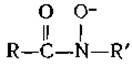
These include the peptide ferrichrome (produced by certain bacilli). Notably, oxygen atoms likewise provide the coordination bonds to iron in these compounds. In ferrichrome, Fe3+ is bound with extreme stabilityh; the formation constant log K for the complex between Fe3+ and the free trihydroxamate ligand is 29.
On average, the human daily diet contains ~15 mg of iron, of which about 1 mg is absorbed by the body. This is usually sufficient to compensate for minor iron losses, primarily excreted through Bile. The human body apparently lacks a mechanism for eliminating excess iron; iron content is regulated solely by The rate of its intake. This rate increases in women during Pregnancy and in young women during menstrual bleeding (to replace iron lost with Blood). Excessive amounts of iron can be highly toxic. The mechanism regulating iron absorption remains unclear, but it has been established that once inside the body, Fe3+ binds to transferrin—a protein with a molecular weight of 80,000 containing two iron-binding sites. A companion anion binds along with each Fe3+ ion. Chicken transferrin appears to be identical to conalbumin, the iron-binding protein found in chicken egg white. On the other hand, lactoferrin, a red iron-binding protein present in milk, differs from blood transferrin in its Amino Acid Sequence. Iron-binding proteins found in Body Fluids are sometimes grouped together under the generic name siderophilins.
The primary function of transferrin is The transport of iron within the body, but it can also serve as a buffer that regulates iron uptake; possibly, iron absorption through the intestinal mucosa is regulated by the degree of iron saturation of transferrin in the blood. For The transfer of iron from transferrin to heme, which occurs in immature Blood Cells formed in the Bone Marrow, Fe3+ must be reduced to Fe2+. Reduction of the ferri-ion is probably also necessary for its release from ferritink. The mechanisms of these processes have not been established, but it is known that ascorbic acid or Glutathione can act as reducing agents. The ferri-ion in Hemoglobin (methemoglobin) is reduced by an NADH-dependent enzyme (Supplement 10-A). Along with this, Fe2+ apparently must sometimes undergo oxidation to Fe3+ under the action of a copper-containing ferroxidase (ceruloplasmin; Supplement 10-B). Once in the body, iron is meticulously retained. For example, As a result of the daily destruction of 9 billion erythrocytes, 20–25 mg of iron is released, almost all of which is reused or reserved in the body.
a Neilands J. B., ed., Microbial Iron Metabolism, Academic Press, New York, 1974.
b Jacobs A., Worwood M., eds., Iron in Biochemistry and Medicine, Academic Press, New York, 1974.
c O'Dell B. L., Campbell B. J., Compr. Biochem., 21, 179–265 (1970).
d Harrison P. M., Hoy T. G., in: Inorganic Biochemistry (G. L. Eichhorn, ed.), Vol. 1, pp. 253–279, Elsevier, Amsterdam, 1973.
e Hoare R. J., Harrison P. M., Hoy T. G., Nature (London), 255, 653–654 (1975).
f Massover W. H., Cowley J. M., PNAS, 70, 3847–3851 (1973).
g Neilands J. B., in: Inorganic Biochemistry (G. L. Eichhorn, ed.), Vol. 1, pp. 167–202, Elsevier, Amsterdam, 1973.
h Rosenberg H., Young I. G., in: Microbial Iron Metabolism (J. B. Neilands, ed.), pp. 67–82, Academic Press, New York, 1974.
i Aisen P., in: Inorganic Biochemistry (G. L. Eichhorn, ed.), Vol. 1, pp. 280–305, Elsevier, Amsterdam, 1973.
k Cavill I., Worwood M., Jacobs A., Nature (London), 256, 328–329 (1975).
b. Porphyria [84–86]
The human body does not use all the porphobilinogen it produces; normally, small amounts of it are usually excreted in the urine, mainly as coproporphyrins (Ch. 10, Sec. B, 1). There are inherited and acquired disorders in which blood porphyrin levels are elevated and significantly larger amounts are excreted in the urine (porphyria). There are cases where porphyria is mild and accompanied by almost no symptoms, but in other cases, intensely fluorescent free porphyrins are deposited in the Skin beneath the stratum corneum, accompanied by photosensitization and leading to skin ulceration. In the most severe cases, the excreted porphyrins impart a wine-red color to the urine. Patients develop severe neurological disorders. A number of other symptoms are also observed1). In one form of congenital porphyria, large amounts of uroporphyrin I are excreted in the urine. The biochemical defect in this case apparently amounts to a deficiency of cosynthetase, which is necessary for The formation of protoporphyrin IX. Another form of porphyria is caused by the Excessive production of δ-aminolevulinic acid in the Liver. It has been suggested that such patients might be treated by the administration of benzoate or p-aminobenzoate [87]. The rationale for this approach is to divert glycine metabolism toward the synthesis of hippuric acid (Supplement 9-A) or its p-amino derivative, thereby reducing the rate of porphyrin synthesis.
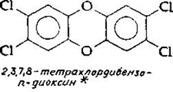
In some mild forms of porphyria, the intake of certain medications can trigger an acute attack of the disease. Drugs and other chemicals sometimes induce porphyria by stimulating the overproduction of δ-aminolevulinic acid synthetase. Compounds that produce this effect include hexachlorobenzene and tetrachlorodibenzo-p-dioxin. The latter is one of the most potent known agents inducing synthetase formation [88].
1) A vivid Description of the manifestations of this disease, which are believed to have been observed in King George III of England, is given by McAlpine and Hunter [84].
* The frequent presence of this dioxin as an impurity in the herbicide 2,4,5-trichlorophenoxyacetic acid (2,4,5-T) has caused great concern. It is quite possible that the dioxin in question is the most toxic of all known small molecules: upon ingestion, the LD50 of this compound for guinea pigs is only 1 µg per 1 kg of body weight [88]. Furthermore, it is a potent teratogenic agent (causing fetal developmental abnormalities).
a. Bile pigments
The enzymatic degradation of heme is an important metabolic process if only because it releases iron, which is reused by the body. Some of the heme Catabolic pathways are shown in Fig. 14-14. It is believed that oxygenation (hydroxylation) initially targets the α-methene carbon (between rings A and B). The hydroxylated product is cleaved with the release of carbon monoxide. The reactions are catalyzed by microsomal hydroxylases [89–90a]. In experiments using 18O2, it was demonstrated that the resulting ring-opened tetrapyrrole biliverdin contains two 18O atoms, while CO2 contains one 18O atom. Reduction and oxidation reactions of biliverdin lead to the formation of A large number of various non-cyclic tetrapyrroles.
Biliverdin, the initial ring-opening product, is reduced to bilirubin, which is transported to the liver as a complex with serum albumin. In the liver, bilirubin is converted into glucuronides [equation (12-12)] formed by the glycosylation of propionic acid side chains. Many of the bilirubin conjugates enter the bile. In the intestine, they are hydrolyzed back to free bilirubin, which is reduced by intestinal bacteria to urobilinogen, stercobilinogen, and mesobilirubinogen. These compounds are colorless, but are easily oxidized by oxygen to urobilin and stercobillin. Some of the urobilin and other bile pigments re-enter the bloodstream and are excreted in the urine, giving it its familiar, characteristic yellow hue.
The yellow skin color observed in jaundice may be caused by excessive heme destruction (e.g., resulting from excessive hemolysis), the liver's inability to form bilirubin conjugates, or various factors preventing The entry of heme breakdown products into the intestinal tract.
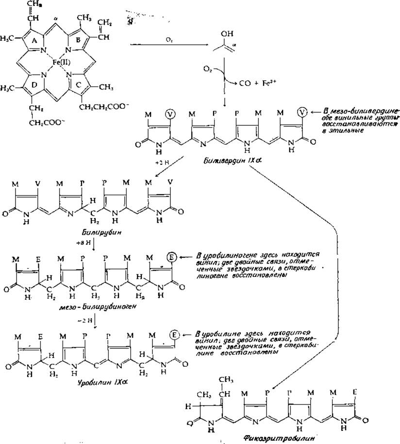
We have already mentioned the acyclic tetrapyrroles of Algae and the Phytochrome chromophore (Fig. 13-22). All of them derive from phycoerythrobilin, which is related to biliverdin, as shown in Fig. 14-14.
Last update: 06/08/2026
Editorial and Educational Adaptation: This material has been compiled based on the primary/original source text. The project team performed an editorial review, corrected technical inaccuracies, structured sections, and adapted the content for an educational format.
What was processed:
- elimination of formatting defects (OCR errors, structural breaks, corrupted characters);
- editorial organization of content;
- standardization of terminology in accordance with academic sources;
- verification of factual statements against the original source text.
All mentions of the author, publication year, and origin of the primary text have been preserved in accordance with the source.