Biochemistry: The Chemical Reactions of Living Cells, Volume 3 - D. Metzler 1980
Metabolism of Nitrogen-Containing Compounds
Cysteine and Sulfur Metabolism
Cysteine is not only an essential component of Proteins, but it is also one of the key compounds through which inorganic sulfur is initially incorporated into organic molecules. Consequently, it participates in a multitude of metabolic processes. Autotrophic organisms carry out the sequential reduction of sulfate to sulfite and further to sulfide (H2S). It is precisely these reduced sulfur compounds that are incorporated into organic matter. Animals, by contrast, utilize preformed organic sulfur compounds produced by autotrophs; within the animal Organism, these compounds enter an active oxidative metabolic pathway during which they are degraded, and the sulfur is re-oxidized to sulfate.
Class="center">
FIG. 14-15. Pathways of cysteine Biosynthesis AND Catabolism and several Other Aspects of sulfur METABOLISM. Solid arrows indicate major biosynthetic pathways, and dashed arrows represent more specialized pathways. The same style of arrows illustrates The conversion of Methionine to cysteine and The breakdown of cysteine in animal Tissues.

FIG. 14-16. Special heme (siroheme) isolated from the sulfite reductase of E. coli Cells.
Certain aspects of inorganic sulfur metabolism were already discussed in Chap. 10. For instance, we examined the reduction of sulfate to H2S by sulfate-reducing Bacteria (Chap. 10, Sec. E, 2, b). In plants and E. coli cells, the assimilatory sulfate reduction pathway also involves The formation of adenylylsulfate as one of the early steps (step a, Fig. 14-15; see also equation (10-38)). Sulfate-reducing bacteria are capable of reducing adenylylsulfate directly to sulfite [equation (10-38), step b], whereas in E. coli, the assimilatory pathway proceeds via the formation of 3'-phospho-5'-adenylylsulfate (PAPS); the function of this compound as "active sulfate" was already discussed in Chap. 11 (Sec. B, 3). The reduction of PAPS to sulfite (Fig. 14-15, step d) is catalyzed by an NADPH-dependent enzyme.
The reduction of sulfite to sulfide in E. coli is catalyzed by sulfite reductase—a large protein (mol. wt. 670,000) composed of four molecules of bound riboflavin phosphate (FMN), four molecules of FAD, 20–21 iron atoms, 14–15 labile sulfur atoms, and 3–4 molecules of a specialized heme [91]. The enzyme utilizes NADPH as a reductant and transfers electrons via its own intramolecular Electron Transport Chain to the specialized heme, which contains an isobacteriochlorin-type tetrahydroporphyrin ring (with adjacent pyrrole rings reduced; Fig. 14-16) [92]. This enzyme protein appears to have a subunit composition of a8ß4, where a is a flavoprotein with a Molecular Weight of ∼54,000, and ß is an iron-sulfur-containing hemoprotein with a molecular weight of ∼60,000 [93]. The same pathway has been found in Chlorella, although another pathway of sulfate reduction appears to be more prominent in this alga [94]. Adenylylsulfate transfers its sulfonyl group to the thiol group of a carrier [equation (14-37), step a].

The resulting thiosulfonate is reduced by the action of a ferredoxin-dependent reductase. Finally, from the —S—S--group of the reduced carrier, the sulfide group is transferred directly into the newly forming cysteine molecule via a ß-substitution reaction analogous to the one described in the following paragraph.
Cysteine is formed from sulfide and Serine following the Acetylation of the latter by The transfer of an acetyl group from acetyl-CoA (Fig. 14-15, step e). This reaction is a standard Pyridoxal phosphate-dependent ß-substitution reaction (Fig. 8-7) and is catalyzed by cysteine synthetase [94a]. A similar enzyme is utilized by certain microorganisms to synthesize homocysteine by the direct incorporation of a sulfide ion into either O-succinylhomoserine or O-acetylhomoserine, as indicated by the dashed arrows in the lower left corner of Fig. 14-15. In E. coli, however, the primary pathway for methionine formation undoubtedly proceeds via cystathionine, as reflected in equation (8-22) and indicated by the solid arrows on the left side of Fig. 14-15.
In animals, the reverse process—the conversion of methionine to cysteine—plays a vital role. Animals are incapable of synthesizing cysteine via the direct incorporation of sulfide; therefore, cysteine must either be obtained preformed in the diet or be synthesized from dietary methionine. The latter process occurs to a limited extent, which makes cysteine an essential dietary requirement for infants. The steps involved in the formation of cysteine from methionine are shown in Fig. 14-15 by the vertical dashed arrows. The reaction sequence begins with the conversion of methionine to S-adenosylmethionine and subsequently to homocysteine via the metabolic pathway shown in Fig. 14-9 (steps g–i). Homocysteine then reacts with serine under the action of cystathionine-β-synthase, and cystathionine is cleaved into cysteine and a-ketobutyrate by the action of a "γ-cleaving enzyme" present in animal tissues (Fig. 14-9, bottom left). The reaction types are illustrated in Fig. 8-7. This pathway bears some similarity to the reverse process described by equation (8-22).
Cysteine can undergo catabolism or be incorporated into various biosynthetic pathways. Simple Cleavage of cysteine, as observed in certain bacteria [95], involves a pyridoxal-phosphate-dependent a,ß-elimination reaction yielding H2S, Pyruvate, and ammonia (Fig. 14-15, reaction g; Fig. 8-6, reaction b). Another pathway involves Transamination to yield ß-mercaptopyruvate (Fig. 14-15, step h). The latter can undergo reductive cleavage to pyruvate and sulfide. An interesting enzymatic ß-substitution reaction (involving pyridoxal phosphate) leads to the conversion of cysteine into ß-cyanoalanine—a factor present in certain plants that causes lathyrism in animals (Supplement 11-B) [96].
A quantitatively significant catabolic pathway of cysteine in animals involves its oxidation to cysteine sulfinate (Fig. 14-15, reaction i); this is a dual hydroxylation process utilizing O2, NADPH or NADH, and Fe2+. Cysteine sulfinate can be further oxidized to cysteic acid, the decarboxylation of which yields taurine. Taurine is a component of Bile salts (Fig. 12-16) and likely acts as a neurotransmitter (Chap. 16, Sec. B, 4). It probably performs some other specific function in retinal photoreceptor cells as well. For cats, it appears to be an essential amino acid [96a]. Taurine can be reduced to isethionic acid, a component of neural tissue. Cysteic acid can also be formed via an alternative pathway—from O-acetylserine and sulfite (reaction m, Fig. 14-15)—whereas taurine can be generated through the decarboxylation of cysteine sulfinate to hypotaurine followed by its subsequent oxidation (reaction n).
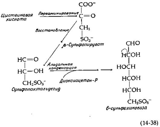
An interesting metabolic process occurring in Chloroplasts is the conversion of cysteic acid into a sulfolipid (Fig. 2-32, Table 13-2). The probable reaction sequence begins with transamination to yield ß-sulfopyruvate, which is subsequently reduced (presumably to sulfolactaldehyde), followed by an aldol Condensation with dihydroxyacetone phosphate, as outlined in equation (14-38); isomerization of the product yields 6-sulfoquinovose, a sugar moiety incorporated into the sulfolipid [96b].
Returning to cysteine sulfinate, its transamination yields ß-sulfinylpyruvate—a compound that readily loses SO2 via a spontaneous reaction analogous to the decarboxylation of oxaloacetate (reaction o, Fig. 14-15). In animals, this is presumably one of the major pathways for the removal of sulfur from Organic compounds. However, for excretion from the body, sulfite must first be oxidized to sulfate. The enzyme sulfite oxidase belongs to an expanding group of proteins known to contain molybdenum (Supplement 14-A). Sulfite oxidase also contains a cytochrome b5-type domain; electrons pass directly from it to cytochrome c in the Mitochondrial Electron Transport chain. The profound physiological importance of this enzyme in humans is underscored by a case report of an infant lacking sulfite oxidase who failed to excrete urinary sulfates and suffered from severe neurological disorders [97].
Another reaction, which generally plays a minor role in animals but can become significant in cases of sulfite oxidase deficiency, is the oxidative condensation of two sulfite molecules to form thiosulfate (reaction p, Fig. 14-15). Thiosulfate participates in a fascinating reaction catalyzed by an enzyme with the eccentric name rhodanese, found in the Liver, which catalyzes the Displacement of the sulfite ion from the thiosulfate molecule by a cyanide ion [equation (14-39)]. This reaction results in the detoxification of cyanide ions.

Animal tissues contain small amounts of organic hydrosulfide derivatives, including thiocysteine.

Thioglutathione and the more oxidized thiotaurine belong to this same class of compounds. Thiocysteine can be generated through the action of the cystathionine-cleaving enzyme on cystine. In this reaction, thiocysteine is eliminated, while the remaining portion of the cystine molecule yields pyruvate and ammonia.
The majority of the sulfate produced in the organism is excreted unchanged in the urine, but a significant portion is esterified with Oligosaccharides and Phenolic Compounds. In this case, of course, the sulfonyl group is transferred from PAPS [equation (11-4)]. Many readers (perhaps ~40%) may have noticed that after eating asparagus, their urine develops a distinctive, pungent odor. These genetic "skunks" excrete S-methyl thioacrylate and related compounds, but the exact Nature of the plant metabolite from which these products are derived has not yet been established [98].

Last update: 06/08/2026
Editorial and Educational Adaptation: This material has been compiled based on the primary/original source text. The project team performed an editorial review, corrected technical inaccuracies, structured sections, and adapted the content for an educational format.
What was processed:
- elimination of formatting defects (OCR errors, structural breaks, corrupted characters);
- editorial organization of content;
- standardization of terminology in accordance with academic sources;
- verification of factual statements against the original source text.
All mentions of the author, publication year, and origin of the primary text have been preserved in accordance with the source.