Biochemistry - Chemical Reactions in the Living Cell, Volume 1 - D. Metzler 1980
How molecules join together
Self-assembly of macromolecular structures
While the elongation of bacterial flagella (Supplement 4-B) can be readily envisioned As a result of subunits passing through a central channel, understanding how more complex structures, such as Muscle sarcomeres, are formed is quite difficult. Significant progress has recently been achieved in this area, largely through observations of bacteriophage self-assembly. Fig. 4-8 illustrates the assembly of a filamentous phage from hydrophobic protein molecules and DNA as an example. Protein subunits are synthesized and stored in the bacterial membrane. Small a-helices fit easily into the membrane and can remain there until a DNA molecule enters the membrane. Although the exact initiation mechanism of self-assembly remains unclear, each subunit appears to possess a DNA-binding site. The surface Regions of the subunits exhibit hydrophobic properties, and through mutual interaction, the subunits spontaneously "coat" the DNA surface. Upon completion of assembly, the hydrophobic groups of the "rod" become sequestered within the interior of the Structure. It is hypothesized that the groups located on the outer surface of the viral particle possess hydrophilic properties, and that The formation of such a hydrophilic "rod" serves as the driving force responsible for the automatic extrusion of the phage from the membrane [39]. Bacterial pili may be extruded via a similar mechanism; they form very rapidly and can presumably retract back into the bacterial membrane.
A striking example of self-assembly is the assembly process of T-even phages (Supplement 4-D) [101–103]. The results of meticulous genetic analysis (Ch. 15, Sec. G.2) have demonstrated that HEAD formation requires at least 18 genes, tail formation requires 21 genes, and tail fiber formation requires 7 genes. Most of these genes encode Proteins that are directly incorporated into the mature virion, whereas several genes determine specific Enzymes essential for the assembly process. Mutant virus strains capable of synthesizing all structural proteins except one have been isolated. In this case, all synthesized proteins accumulated inside the host bacterial Cell without aggregating. However, upon The addition of the missing protein (synthesized by a bacterium infected with a virus of a different strain), the rapid assembly of fully functional Viral Particles took place. These and other findings led to the Conclusion that proteins attach to the growing structure in a strictly defined sequence, where the binding of one protein establishes the binding site for the next.
Supplement 4-D
T-Even Bacteriophages
One of the most fascinating objects observable under an Electron microscope are the T-even bacteriophages (T2, T4, and T6), which infect E. coli Bacteriaa-c. While the mode of entry for many Viruses into Cells remains unknown, T-even phages are a rare exception. These particles act as a kind of "molecular syringe," piercing The Cell wall of the host bacteria and injecting their DNA. The viral particle, measuring 200 nm in length and having a mass of ~255∙106 daltons, contains approximately 130∙106 daltons of DNA within its elongated icosahedral head, which measures 100×70 nm. The surface of the bacteriophage head is constructed from ~840 identical protein molecules with a Molecular Weight of 45,000 (encoded by Gene 23), organized into 140 hexamers, and ~55 molecules of another protein forming 11 pentamersd. The head contains at least nine other proteins, including three major internal proteins that enter the bacterium along with the DNA.
Class="center">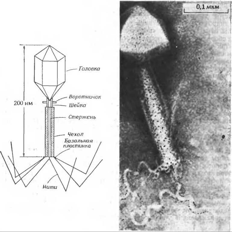
Phage with a contractile tail infecting Bacillus subtilis. (Courtesy of A. S. Tikhonenko.)
The phage tail features an internal core with a 2.5 nm lumen, which is wide enough to allow a DNA molecule to pass through it into the bacterium. The sheath surrounding the core (with a mass of 8∙106 daltons) is built from 144 protein subunits of ~55,000 molecular weight, assembled into 24 rings, each consisting of six subunits. The sheath is capable of contracting, reducing its length from 80 to 30 nm, thereby driving the core through the bacterial wall. At the end of the tail lies a hexagonal baseplate bearing a series of short spikes as well as six long fibers, each composed of six subunits with a molecular weight of ~100,000. Among the 10 proteins forming the baseplate, a specific enzyme, Lysozyme, was discoverede. In addition to these components, the viral particle contains A number of small molecules. For instance, approximately 30% of the negatively charged DNA groups are neutralized by Polyamines—putrescine and spermidine (Ch. 14, Sec. B.4). The tail baseplate also contains six molecules of the coenzyme 7,8-dihydropteroylhexaglutamate (Ch. 8, Sec. L).
How does bacterial infection by a T-even phage occur? The process begins with the attachment of the tail fibers to specific receptor sites on the bacterial surface. This triggers a series of Conformational Changes in the fibers, baseplate, and sheath. Concurrently, lysozyme is released from the baseplate and degrades the bacterial wall. Sheath contraction initiates at the baseplate and propagates toward the base. Once the core penetrates the bacterium, the DNA is rapidly injected into the host cell.
During contraction, the sheath subunits rearrange to form a structure comprising 12 larger rings, each consisting of 12 subunitsf. A distinctive mutual interpenetration (intercalation) of subunits takes place. The strictly defined directionality and irreversible nature of this structural transition distinguish the shortening of the phage tail from Muscle contraction. It is quite plausible that the protein subunits within the phage sheath exist in an unstable, high-energy state, and that the energy stored during assembly is subsequently utilized to drive the contraction process.
a Wood W. B., Edgar R. S., Sci. Am., 217, 60–72 (Jul. 1967).
b Mathews C. K., Bacteriophage Biochemistry, Van Nostrand-Reinhold, Princeton, New Jersey, 1971.
c Cummings D. J., Couse N. C., Forrest C. L., Adv. Virus Res., 16, 1–41 (1970).
d Branton D., Klug A., JMB, 92, 559–565 (1975).
e Not to be confused in its catalytic properties with the better known lysozyme of egg white (Fig. 2–9 and Chapter 7, Section C, 4, a).
f Moody M. F., JMB, 80, 613–635 (1973).
The assembly process of the phage tail has been studied in detail. Six copies of each of three proteins assemble in a sequence to form a hexameric "bushing" (Fig. 4-26). Concurrently, seven other proteins associate with one another to form a wedge-shaped structure. Six such wedges then assemble around the bushing to form a hexagonal baseplate. Only after this do two additional proteins attach to the surface of the baseplate, activating it to initiate the assembly of the tail core. Upon completion of this process, the tail sheath assembles from structural units, and only once the tubular structure reaches the required length does the head protein attach to its apex. Subsequently, the "head" is formed above the tail, followed by the attachment of the tail fibers, which are assembled independently.
It is difficult to conceive how each stage of this complex assembly process lays the groundwork for the subsequent stage. Nevertheless, the available data are remarkably compelling. The structure of each newly added protein likely remains stable only until a specific protein binds to it. The energy of interaction is sufficient to induce a conformational change in another region of the protein molecule, exposing a site complementary to the binding region of the next protein. Remarkably, every protein in the baseplate appears to possess this capacity for self-activation.

FIG. 4-26. Sequential stages in the assembly of the T4 bacteriophage tail. Numbers correspond to gene numbers on the T4 phage chromosome map (see Fig. 15-19). The letter P next to a number indicates that the respective protein is directly incorporated into the tail. The absence of the letter P signifies that the products of those genes are not incorporated into the tail and presumably play a catalytic role [101, 102].
Protein conformation changes induced by interactions with other proteins are not restricted to such exotic entities as contractile phages. This principle undoubtedly plays a critical role in the self-assembly of microtubules, muscular myofibrillar assemblies, and many other more labile yet equally vital cascade systems involving Protein-Protein Interactions, such as the Blood clotting system (Fig. 6-16). Membrane formation is likewise driven by self-assembly, a process that must incorporate receptors capable of reliably responding (though the precise mechanism remains unclear) to chemical signals from the environment. Viewing all these phenomena from a unified perspective reveals a fundamental commonality between the protein-protein interactions leading to small protein aggregates and the complex events occurring within intact cells in response to Hormones and other external stimuli.
1. Note: This problem utilizes the dissociation constant rather than the formation constant, which was primarily used in this chapter. The apparent dissociation constant Ka for the H2PO4- ion at 25°C and a total phosphate concentration of 0.05 M is 1.380∙10-7 (corresponding to the National Bureau of Standards buffer, which is a solution of 0.025 M KH2PO4 and 0.025 M NaH2PO4) (see Bates R. G., Determination of pH, 1964).
a. Calculate the negative logarithm of Ka (i.e., pKa). Plot this value on the curve of pKa versus μ (Fig. 3-1).
b. Convert the acid dissociation constant expression into its logarithmic form:

where a is the fraction of acid molecules in the ionized form, b. Suppose you wish to prepare a buffer with pH 7.00 at 25 °C using anhydrous KH2PO4 (mol. wt. 136.09) and Na2HPO4 (mol. wt. 141.98). If you place 3.40 g of KH2PO4 into a 1 L volumetric flask, how much anhydrous Na2HPO4 must be weighed out before adding Water to the mark to obtain a solution with the required pH value? If you wish to obtain a buffer with pH 7±0.01, what must be the weighing precision for the salts? Note: A buffer with an exact pH value can be prepared much faster by this method than by titrating the acid component of the buffer solution with NaOH to the required pH value.
2. The apparent pKa value for a 0.1 M formic acid solution at 25°C is 3.7.
a. Concentrated HCl is added to 1 L of a 0.1 M sodium formate solution until the pH value reaches 1.9. What will be the concentrations of the formate ion and unionized formic acid in the resulting solution?
б. Calculate the hydrogen ion concentration.
в. How many equivalents of HCl had to be added to 1 L of a 0.1 M sodium formate solution to obtain a pH of 1.9, i.e., in problem (a)?
3. 0.01 mol of Glycine was placed into 100 mL volumetric flasks, and the amounts of HCl or NaOH indicated below were added. The flasks were filled with water to the mark and the resulting solutions were thoroughly mixed, after which their pH was measured. Based on the obtained pH values, calculate the pKa values for the carboxyl and amino groups, making as many independent pK calculations as the available data permit. Recall that at low pH values, the concentration of free hydrogen ions must be taken into account (see problem 26).
|
flasks |
HCl, mol |
NaOH, mol |
pH |
|
1 |
0.010 |
1.71 |
|
|
2 |
0.009 |
1.85 |
|
|
3 |
0.006 |
2.25 |
|
|
4 |
0.002 |
2.94 |
|
|
5 |
0.002 |
9.00 |
|
|
6 |
0.004 |
9.37 |
|
|
7 |
0.005 |
9.60 |
4. Using the pKa values obtained in problem 3, plot the theoretical titration curve showing the dependence of the number of equivalents of H+ and OH- reacting with 1 mole of glycine on pH. Note that the shape of such a curve is independent of pKa. Plot similar curves for glutamic acid (whose pKa values are 2.19, 4.25, and 9.67), Histidine (pKa values are 1.82, 6.00, and 9.17), and Lysine (pKa values are 2.18, 8.95, and 10.53).
Compare the curve you plotted for glycine with the curve obtained by adding 1 N acid or base to a solution* containing 0.01 mol of glycine per 100 mL of water. Also, compare your curves with those for glycine given in other textbooks.
5. The equations for the reversible reactions between aliphatic amines and formaldehyde are given below.

Indicate (qualitatively) how the titration curves discussed in problem 4 would change if the solution contained 9% formaldehyde.
6. The pKa values for amino acid side chains in proteins sometimes differ slightly from the values given in Table 2-2. Most often, such deviations occur in the case of groups "buried" within the structure, i.e., inaccessible to the solvent, or for groups located very close to other ionized groups. Try to explain the following facts:
a. The unusually high pKa value for the carboxyl group of an aspartic acid residue (Ch. 5, Sec. B.4.a).
б. The unusually low pKa value for the carboxyl group of an aspartic acid residue.
в. The unusually high pKa value for the phenolic group of a Tyrosine residue.
7. Rewrite equations (4-20) through (4-25) using dissociation constants. It would be convenient to designate them as K1, K2, Ka, etc., but to avoid confusion, it is better to use the designations K1d, K2d, Kad, etc.
8. a. Describe two ways to determine or estimate the tautomeric ratio R [equation (4-21)].
б. Calculate the pKc value for equation (4-21) and compare it with the pKb* value and the corresponding pK value for phenol. Analyze the differences.
9. A molecule has two identical binding sites for Ligand X. The Free energy of interaction between ligands bound to the same molecule, e, is defined as The change in the free energy of ligand binding to the molecule caused by the binding of the first ligand to a neighboring site. Show that if the fractional saturation  , then the equation characterizing the binding isotherm implies that

10. Fig. 4.4 shows the hydrogen ion binding curve for succinate. Estimate from this the value of e and the Microscopic Binding Constants.
11. A linear molecule has a very large number of identical binding sites for ligand X. The free energy of interaction between ligands bound to adjacent sites is equal to e. The interaction between ligands that are not nearest neighbors is considered negligibly small. If the binding constant for a site located adjacent to unoccupied sites is denoted by Kr, the expression for the binding isotherm will have the form

[J. Applequist, J. Chem. Ed. in press (1977).]
Show that at  the relation given above implies that

12. The binding of adenosine to poly(U) was studied by equilibrium dialysis (Huang, Ts’o, JMB, 16, 523, 1966). The table below gives the fractions of occupied sites on the poly(U) molecules, i.e., The values of  at various molar concentrations of free adenosine (A) in solution at 5 °C. Determine the true association constant for the binding of adenosine to poly(U) and the free energy of interaction between adjacent adenosines, assuming that the interaction between non-nearest-neighbor adenosines can be neglected. Do neighboring molecules attract or repel each other?

Last update: 06/08/2026
Editorial and Educational Adaptation: This material has been compiled based on the primary/original source text. The project team performed an editorial review, corrected technical inaccuracies, structured sections, and adapted the content for an educational format.
What was processed:
- elimination of formatting defects (OCR errors, structural breaks, corrupted characters);
- editorial organization of content;
- standardization of terminology in accordance with academic sources;
- verification of factual statements against the original source text.
All mentions of the author, publication year, and origin of the primary text have been preserved in accordance with the source.