Human Biochemistry, Volume 2 - Murray R. 1993
Structure, Function, and Replication of Information Macromolecules
Regulation of Gene Expression
Regulation of Gene Expression in Prokaryotes
Lac Operon
In 1961, François Jacob and Jacques Monod described the now-classic operon model. Their concept relied heavily on studying lactose METABOLISM regulation in the bacterium E. coli. Today, the molecular mechanism governing The regulation of genes involved in lactose metabolism is the best understood. The enzyme ß-galactosidase hydrolyzes lactose into galactose and glucose (Fig. 41.2). The structural Gene for ß-galactosidase (the lacZ gene) is located in the same cluster as the gene responsible for synthesizing galactoside permease, which mediates The Active Transport of galactose into The Cell (the Y gene), and the galactoside acetylase gene (the A gene), whose functional significance remains unknown. The structural genes for these three Enzymes are physically linked, forming the so-called lac operon (Fig. 41.3). This genetic arrangement of structural and associated regulatory genes ensures the coordinated expression of all three lactose-metabolizing enzymes. All three Genes are transcribed as a single mRNA molecule containing independent Translation start codons (AUG) and stop codons (UAA) for each Cistron. This type of mRNA is called polycistronic mRNA. The formation of polycistronic mRNAs is characteristic primarily of prokaryotic organisms.
When lactose or certain of its analogues are added to a growing culture of E. coli Cells, the expression levels of ß-galactosidase, galactoside permease, and galactoside acetylase activities increase 100- to 1000-fold [Note: translating naturally as a 10- to 100-fold increase]. Induction of the lactose operon According to the aforementioned response Classification belongs to type A (Fig. 41.1). After the inducer (signal) is removed, the synthesis rate of all three enzymes drops. Because the enzymes themselves do not undergo significant degradation within E. coli cells, the activity levels of ß-galactosidase and the other two enzymes remain constant and decline only due to "dilution" resulting from Cell Division.
When E. coli cells are grown in a medium containing a mixture of lactose and glucose as the sole carbon sources, glucose is metabolized first. Once the glucose in the medium is exhausted, cell growth temporarily halts until induction of the lactose operon occurs, achieving a level of enzyme expression sufficient to support lactose metabolism. Despite lactose being present from the very beginning, the lac operon is not induced until glucose is completely depleted. This phenomenon was initially explained by assuming the operon is repressed by a Glucose Catabolism product, hence its name: "catabolite repression". It is now known that "catabolite repression" is actually mediated by the catabolite gene activator protein, commonly known as CAP (catabolite gene activator protein), acting in concert with cAMP. The expression level of many inducible enzyme systems or operons in E. coli and other prokaryotes is sensitive to catabolite repression (see below).
Class="center">
Fig. 41.3. Relative arrangement of structural and regulatory genes in the lac operon.

Fig. 41.4. Mechanism of repression and derepression of the lac operon. In the absence of the inducer (A), the repressor (the product of the constitutively expressed i gene) binds to the operator, preventing RNA polymerase from binding to the promoter locus and thereby blocking the Transcription of the structural genes Z, Y, and A. In the presence of the inducer (B), the constitutively synthesized repressor is inactivated and cannot bind to the operator. In this case, in the presence of the cAMP-CAP complex, RNA polymerase transcribes the structural genes Z, Y, and A. The resulting polycistronic mRNA chain is translated to produce protein molecules of ß-galactosidase, permease, and acetylase, which ensure normal lactose catabolism.
The physiology of lac operon induction is now well understood (Fig. 41.4). Expression of the normal i gene of the lac operon is constitutive, manifesting as the constant-rate production of lac repressor subunits. The lac repressor protein molecule consists of four identical subunits, each with a Molecular Weight of 38,000. The repressor—the product of the i gene—has a high affinity for its corresponding operator locus (Kd approximately 10-12 mol/L). The operator locus is a specific 27-base-pair double-stranded DNA sequence. Within this region, a 21-base-pair sequence exhibits twofold rotational Symmetry (indicated by solid lines, with the axis of symmetry denoted by dots):

The minimum effective size of the operator that can be bound by a lac repressor molecule is 17 Base Pairs (highlighted in bold). At any given time, two repressor subunits are bound to the operator. Within the 17-base-pair sequence, at least one base of each pair participates in recognizing and binding the repressor. Binding occurs primarily in the major groove of the DNA without disrupting the normal double-helical Structure OF THE operator region. The region of the repressor molecule comprising the first 52 amino acid residues binds to DNA apparently without exhibiting sequence Specificity. Another region of the repressor (residues 53 through 58) binds with strict specificity to the 17-unit, 6–7 nm long fragment of the operator region. Amino acid residues at positions 74–75 are particularly important for binding the inducer to the repressor molecule. The operator locus lies between the promoter, where DNA-dependent RNA polymerase attaches before transcription begins, and THE START OF the Z gene, the structural gene for ß-galactosidase (Fig. 41.3). Upon binding to the operator, the repressor blocks the transcription of the operator locus and the downstream structural genes Z, Y, and A. Thus, the repressor acts as a negative regulator; in its presence, the expression of the Z, Y, and A genes is suppressed. Typically, a cell contains 20 to 40 tetrameric repressor molecules and 1 to 2 operator loci.
A lactose analogue that can induce lac operon expression without serving as a true substrate for ß-galactosidase can be termed a non-metabolizable inducer. Adding lactose or a non-metabolizable inducer to a bacterial culture grown on a poorly utilized carbon source (e.g., succinate) triggers the immediate induction of the lac operon enzymes. Small amounts of lactose or inducer can enter the bacterial cell even in the absence of permease. Repressor molecules, both those bound to the operator locus and those free in the Cytoplasm, have an affinity for inducer molecules. The binding of an inducer to a repressor molecule attached to the operator locus induces Conformational Changes in the repressor structure, leading to the dissociation of the complex from the DNA. If DNA-dependent RNA polymerase is already bound to the coding strand in the promoter region by this point, transcription begins. The resulting polycistronic mRNA features a 5' end sequence complementary to the coding strand of the operator. Thus, the inducer derepresses the lac operon and enables the transcription of the structural genes for ß-galactosidase, galactoside permease, and galactoside acetylase. Introduction/27.html">Translation of the polycistronic mRNA can begin even before transcription is fully complete. Derepression of the lac operon allows the cell to synthesize the enzymes required to catabolize lactose as an energy source.
For RNA polymerase to bind to the promoter sequence, the catabolite gene activator protein (CAP)–cAMP complex must be present. cAMP accumulates independently only when carbon sources are depleted in the growth medium. In the presence of glucose or glycerol at concentrations supporting growth, the intracellular cAMP concentration in Bacteria remains insufficient to form a complex with CAP, preventing DNA-dependent RNA polymerase from initiating transcription of the Lac operon. Transcription begins only when the CAP–cAMP complex is bound to the promoter. The CAP–cAMP complex acts as a positive regulator, as its presence is required to ensure Gene Expression. Thus, the Lac operon is subject to both positive and negative regulation.
If the i gene mutates in such a way that its product—the Lac repressor—loses The ability to bind to the operator, expression of the Lac operon becomes constitutive. Conversely, if a mutation results in the repressor's inability to bind to the inducer, derepression of the Lac operon (which strictly requires complex formation between the inducer and the repressor bound to the operator region) is not observed even at high inducer concentrations in the medium.
A mutant bacterium in which the operator sequence is altered such that the normal repressor is unable to bind to it also acquires the capacity for constitutive expression of the Lac operon.
Bacteriophage Lambda
Some bacteria carry Viruses (temperate Bacteriophages) that either integrate into the host cell chromosome and replicate along with it or exist autonomously within the cell and replicate independently, ultimately leading to cell lysis and bacterial death. One such temperate bacteriophage is bacteriophage lambda (λ). Upon infecting susceptible E. coli bacteria, it injects its genome—consisting of a linear double-stranded DNA of 45,000 base pairs (Fig. 41.5)—into the bacterial cell. Depending on the physiological status of the microorganism, subsequent phage development can follow either a lysogenic pathway, which involves the integration of phage DNA into the host genome and its persistence in a latent form until "activation" (see below), or a lytic pathway. The latter involves a series of phage DNA replications, producing approximately 100 copies of the phage genome. Each copy is packaged into a protein capsid, and mature phage particles cause lysis of the host cell. The released bacteriophages can then infect susceptible bacterial cells anew.
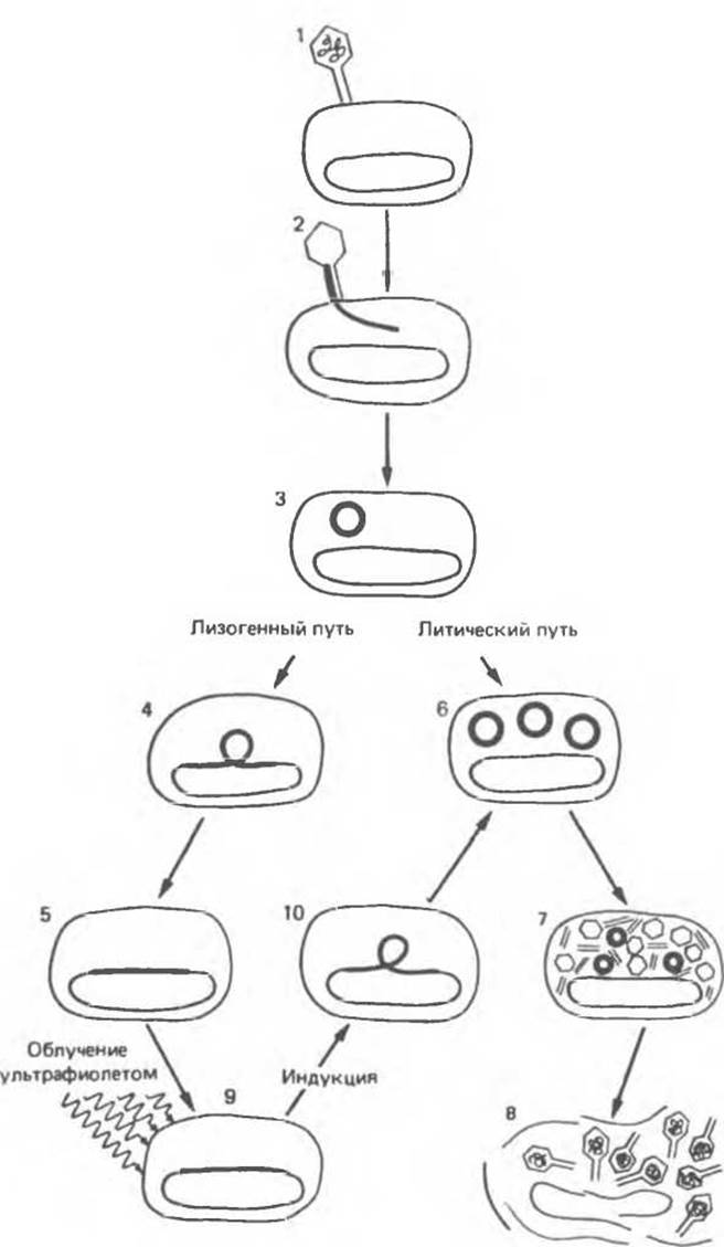
Fig. 41.5. Infection of E. coli by phage λ begins with the adsorption of the phage particle to the bacterial cell surface (1). The next stage is the injection of the phage DNA (dark line) into the cell (2, 3). Subsequent events proceed along one of two possible pathways. In the lysogenic pathway, the phage DNA integrates into the bacterial chromosome (4, 5). In this case, the phage DNA replicates as an integral part of the chromosome—passively during cell division. Cells carrying the integrated ("dormant") virus are termed lysogenic, and the integrated phage itself is called a prophage. In the alternative Lytic Pathway of infection, the phage DNA replicates independently (6) and directs the synthesis of phage Proteins (7). About 100 new phage particles are formed. Phage propagation ultimately leads to lysis of the host cell (8). The prophage can be induced by various factors, such as ultraviolet irradiation (9). The inducing agent switches the operation of two alternative sets of gene pathways. Specifically, the phage DNA is excised from the host chromosome (10), and the lytic cycle begins. (Reproduced, with permission, from Ptashne M., Johnson A. D., Pabo С. О A genetic switch in a bacterial virus. Sсi. Am. [Nov.] 1982, 247, 128.)
Integrated into the host cell genome, the phage λ DNA persists in a "latent" state (as a prophage) until it is activated by exposing the lysogenic cell to various DNA-damaging agents. In response to such Treatment, the prophage is "induced"—triggering the transcription and translation of phage genes required for the excision of phage DNA from the host chromosome, its Replication, packaging into the protein capsid, and cell lysis. This development is triggered by a switch-like mechanism corresponding to option C in Fig. 41.1. This means that once prophage induction occurs, reversal is impossible: the process proceeds inexorably to cell lysis and the release of new phage particles. The switching of development pathways from lysogenic (the prophage state) to lytic (virulent phage) has been thoroughly elucidated at the Molecular and genetic levels and is presented hereafter as a paradigm.
The switching of the phage development pathway involves an 80-base-pair DNA region known as the "right operator" (OR) (Fig. 41.6A). The right operator is flanked on the left by the structural gene for the lambda phage repressor and on the right by the structural gene for another regulatory protein called cro. The only phage gene expressed while the phage DNA resides within the host chromosome (i.e., in the prophage state) is the repressor gene. During lytic development, the repressor gene is not expressed, whereas the cro gene, along with many other phage genes, is actively expressed. Thus, when the repressor gene is ON, the cro gene is OFF, and conversely, when the cro gene is ON, the repressor gene is OFF. As we will see below, these two genes regulate one another, ultimately determining the choice between the lytic and lysogenic developmental pathways of phage λ.
The operator region consists of three tandemly arranged, discrete, similar but not identical 17-base-pair sequence segments (Fig. 41.6B). Each of these three segments, OR1, OR2, and OR3, can bind the repressor or the cro protein, predominantly through contacts between the protein molecule and the minor groove of the DNA double helix. The DNA region between the repressor gene and cro also contains two promoter sequences that direct RNA polymerase binding in a specific orientation. One promoter directs transcription to the right, thereby transcribing cro and other distal genes. The other promoter directs transcription to the left, i.e., in the direction of repressor gene transcription (Fig. 41.6C).

Fig. 41.6. Schematic representation of the right operator (OR) of phage λ (a series of diagrams with successively increasing structural detail). The operator region is a stretch of phage DNA approximately 80 bp in length. A. The operator region is flanked by the lambda repressor gene (left) and the cro regulatory protein gene (right). B. The operator region consists of three functional segments OR1, OR2, OR3, each 17 base pairs long. All three segments are recognized by both the repressor and the cro protein. These segments overlap with the sequences of two promoters—that is, the RNA polymerase binding sites necessary to initiate mRNA synthesis (wavy line), which subsequently directs the Synthesis of the corresponding protein. C. Nucleotide sequence of the OR1 segment. (Reproduced with permission from Ptashne M., Johnson A. D, Pabo C.O. A genetic switch in a bacterial virus. Sci. Am. [Nov.] 1982, 247, 128.)
The repressor gene product, the repressor protein consisting of 236 Amino Acids, is organized into a two-domain structure, in which the N-terminal domain binds to the DNA of the operator region, and the C-terminal domain is responsible for binding to another repressor molecule to form a dimer. The dimeric repressor binds to the operator DNA more tightly than the monomer (Fig. 41.7A–C).
The product of the cro gene, the Cro protein, consists of 66 Amino Acids and features a single-domain structure, yet it binds much more tightly to the operator in its dimeric form (Fig. 47.7, D). Evidently, the single domain of the Cro protein is responsible for both DNA binding and dimerization.
In a lysogenic bacterium harboring phage λ in the prophage state, the λ repressor binds preferentially to OR1, and through cooperative interactions, facilitates the binding of a second dimeric repressor molecule to the OR2 region (Fig. 41.8). Of the three operator sites, OR3 exhibits the lowest affinity for the repressor. The binding of the repressor to OR1 leads to two main effects. First, RNA polymerase is prevented from binding to the rightward promoter, and consequently, the crо gene is not expressed. Second, as mentioned above, the repressor dimer bound to OR1 enhances the binding of another dimer to OR2. Repressor binding to OR2 exerts an important additional effect: it increases the efficiency of RNA polymerase binding to the leftward promoter, which overlaps with OR2, thereby upregulating expression of the repressor gene. This enhancement appears to be mediated by protein–Protein Interactions between the repressor bound to OR2 and the RNA polymerase bound to the promoter. Thus, the λ repressor acts simultaneously as a negative regulator, preventing transcription of the crо gene, and a positive regulator, stimulating transcription of its own gene. This dual role of the repressor ensures the Stability of the prophage state: it not only suppresses the expression of lytic genes but also enhances its own expression. Both factors contribute to maintaining the lysogenic status of the cell (i.e., the existence of the bacteriophage as a prophage). When the repressor concentration reaches very high levels, it becomes able to bind to OR3, which in turn reduces the efficiency of repressor gene transcription from the left promoter. As a result, the repressor concentration drops to levels that trigger the dissociation of the repressor-OR3 complex.
When a DNA-damaging signal, such as ultraviolet irradiation, affects a lysogenic bacterial cell, the resulting single-stranded DNA fragments activate a specific bacterial protease encoded by the recA gene (Fig. 41.8). The activated RecA protease cleaves the region of the repressor molecule that connects its N- and C-terminal domains. This Cleavage leads to the dissociation of the repressor dimer, followed by the disruption of its complexes with OR2 and OR1. The consequences of removing the repressor from OR1 and OR2 are readily predictable. RNA polymerase immediately gains access to the rightward promoter and initiates transcription of the crо gene. Furthermore, the stimulatory effect of the repressor–OR2 complex on leftward transcription is lost (Fig. 41.8).
The Cro protein, produced by translation of the newly formed transcript, also binds to the operator region as a dimer, but the order of operator site preference for Cro is the reverse of that for the repressor protein. Specifically, Cro binds most tightly to OR3, with no cooperative effect whatsoever regarding the binding of a second dimer to OR2. As its concentration increases, Cro begins to bind to OR2 and subsequently to OR1.
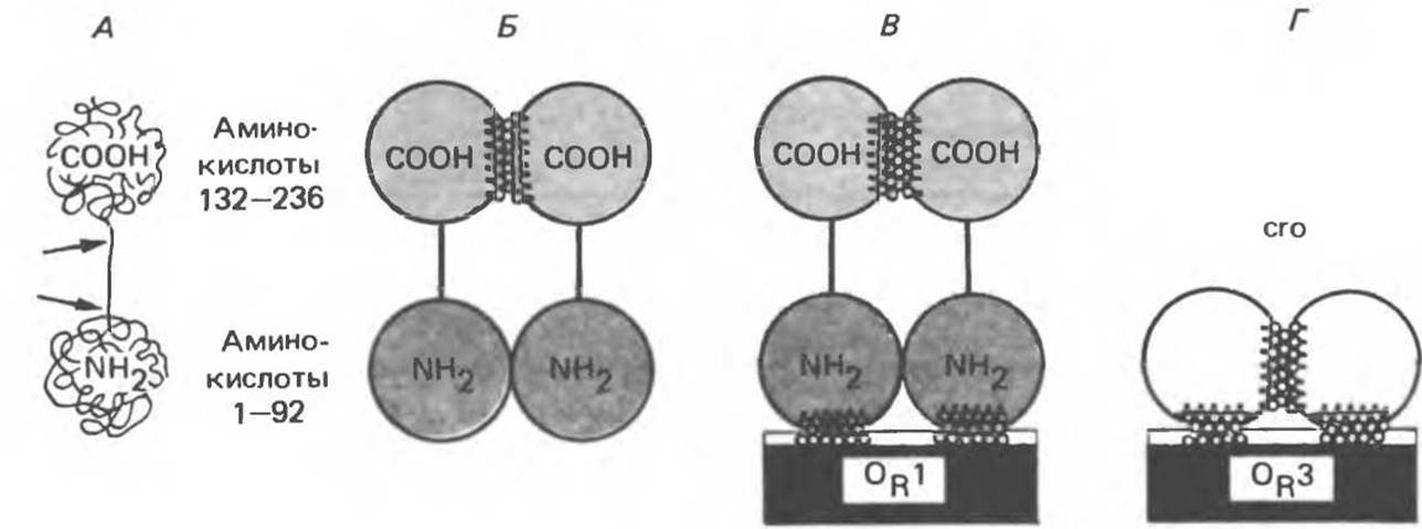
Fig. 41.7. The lambda repressor protein is a 236-amino-acid polypeptide. The polypeptide folds into a dumbbell-shaped structure containing two distinct domains: an N-terminal and a C-terminal domain. These two domains are connected by a protease-sensitive segment of the polypeptide chain (A). Individual repressor molecules (monomers) associate into dimers (B). The dimer can reversibly dissociate back into monomers. Monomers are held together within the dimer primarily through C-terminal domain interactions (shaded contact region). Repressor dimers can reversibly bind to the operator region, exhibiting the highest affinity for OR1 site (B). Contacts with DNA (shaded area) are mediated primarily by the N-terminal domains. The Cro protein (D) is a single-domain protein containing dimerization sites; in its dimeric form, this protein binds to the operator, preferentially at the OR3 site. (Reproduced with permission from Ptashne M., Johnson A. D., Pabo C. O. A genetic switch in bacterial virus Sci. Am [Nov.] 1982, 247, 128.)
The binding of the Cro protein to OR3 immediately shuts down leftward transcription and thus prevents further expression of the repressor gene. This triggers a complete switch: the cro gene is expressed, while the repressor gene is turned off. This event is irreversible and is followed by the expression of the remaining phage genes, initiating the normal lytic development cycle of phage λ. When the concentration of the Cro protein becomes sufficiently high, it binds to the OR1 site, thereby lowering the expression level of its own gene, which is essential for the execution of the late Stages of the lytic cycle.

Fig. 41.8. Four stages of the λ phage life cycle and the pathway-switching scheme. The lysogenic pathway (virus in the prophage state) is chosen when a dimeric repressor binds to OR1. Repressor binding to OR1 facilitates the binding of another repressor molecule to OR2. In the prophage state (top), repressor dimers bound to OR1 and OR2 prevent RNA polymerase from binding to the right promoter, thereby blocking Cro Protein Synthesis (negative control). Simultaneously, this stimulates polymerase binding to the left promoter (positive control), leading to more active transcription of the repressor gene (repressor mRNA is shown as a wavy line) and, consequently, more efficient production of the repressor protein required to maintain the lysogenic state. The prophage can be induced when UV-activated RecA protease cleaves the repressor monomers. The equilibrium between free monomer, dimer, and operator-bound dimer molecules is disturbed, and dimers leave the operator site. Nothing further facilitates RNA polymerase binding to the left promoter, and repressor synthesis ceases. During induction, all operator sites are vacated, polymerase binds to the right promoter, and Cro protein production begins. Early in the lytic cycle, a single Cro protein dimer binds to the OR3 site, for which it has a high affinity. Polymerase can no longer bind to the left promoter, but the right promoter remains accessible. Polymerase continues to bind to it, driving the transcription of the crо gene and other early lytic genes. The lytic pathway is thus established. (Reproduced with permission from Ptashne M., Johnson A. D., Pabo C. O. A genetic switch in bacterial virus. Sci. Am. [Nov.] 1982, 247, 128.)
The three-dimensional structures of both the Cro protein and the repressor protein have been resolved using X-ray crystallography. Models of their DNA-binding interactions have been proposed and tested, and the associated molecular and genetic events thoroughly analyzed. To this day, phage λ remains the most thoroughly studied model for the molecular mechanisms regulating gene expression.
Transcriptional Attenuation
Bacterial operons responsible for Amino acid Biosynthesis frequently employ an additional control system for gene expression based on premature transcription termination. This process, termed attenuation, operates independently of the promoter-operator regulatory system. Attenuation is utilized to regulate expression in response to various physiological cues. The attenuation-mediated regulatory mechanism involves Translation initiation, ribosomal stalling, and the shifting of alternative RNA secondary structures, one of which forms a transcription terminator, while the other prevents terminator formation. In E. coli, attenuation regulates the Tryptophan, phenylalanine, Histidine, Threonine, leucine, isoleucine, and valine operons.
Let us examine the attenuation of the tryptophan (Trp) operon, as it is the most fully characterized system. The structure of the Trp operon is illustrated in Fig. 41.9. Its promoter-operator regulatory system is analogous to that described above for the Lac operon. Repression of the Trp operon results in a 70-fold decrease in transcription levels; however, mutants lacking a functional repression system still retain the ability to respond to tryptophan starvation with an 8–10-fold increase in Trp mRNA synthesis. Analysis of other E. coli mutants revealed that attenuation is linked more closely to the efficiency of translating tryptophan codons than to a direct effect of fluctuations in free tryptophan concentration within the medium. It was soon discovered that premature transcription termination can occur within the TrpL region of the operon (Fig. 41.9), preventing the transcription of the distal genes (TrpEDCBA). This premature termination occurs when the translation of tryptophan codons in TrpL proceeds at a normal rate. As a result of this termination (attenuation), a so-called leader transcript of 140 NUCLEOTIDES is produced instead of the extended polycistronic transcript of the entire operon required to synthesize all Enzymes of the tryptophan biosynthetic pathway. Genetic Engineering techniques enabled the isolation of Mutations within the attenuator region and the analysis of their nucleotide sequences. The combination of genetic and recombinant DNA approaches has helped reconstruct the following dynamic picture of attenuation.
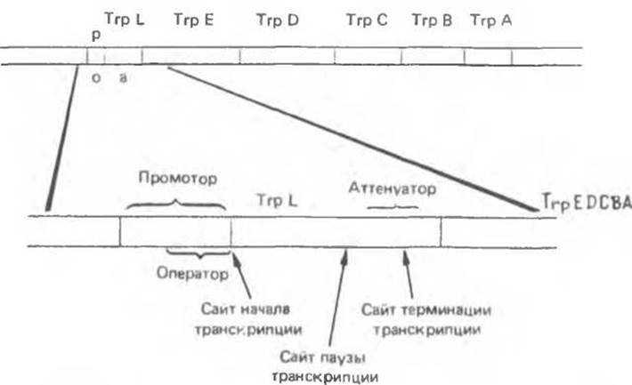
Fig. 41.9. Regulatory region and structural GENES OF THE E. coli Trp operon. Transcription initiation is controlled by the promoter-operator. Transcription termination is controlled by the attenuator located within the 162-nucleotide leader sequence TrpL. All polymerase molecules transcribing the operon pause temporarily at the attenuation site before proceeding further. (Reproduced with permission from Yanofsky C. Attenuation in control of expression of bacterial operons. Nature 1981, 289, 751.)
At the Trp promoter region, RNA polymerase, free from repressor control, initiates transcription of the operon and proceeds to the 90th nucleotide (Fig. 41.10), where it pauses temporarily. During this pause, a ribosome attaches to the newly formed 5' end of the leader transcript near the AUG start codon (positions 27–29), and translation of a 14-amino-acid leader peptide ensues. Starting at position 54, two tandem tryptophan codons are located sequentially in the transcript, requiring the presence of tRNATrp for translation to continue. It is worth noting that tryptophan is a relatively rare amino acid. Even rarer is the occurrence of two consecutive tryptophan residues within Polypeptides; thus, translating the leader peptide serves as a way to "test" the level of tRNATrp, which in turn reflects the intracellular tryptophan concentration. When RNA polymerase resumes transcription past the pause site (at position 90), the ribosome translating the leader peptide reaches the stop codon (at position 70) if tRNATrp is abundant. When tRNATrp is scarce, the ribosome stalls earlier—at the site containing the two tandem tryptophan codons. THE POSITION OF the ribosome on the leader transcript dictates the choice between two alternative secondary structures formed by the RNA transcript.
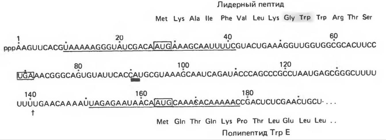
Fig. 41.10. Nucleotide sequence of the 5' end of Trp mRNA. The non-terminated transcript is shown. Transcription termination at the attenuator yields a 140-nucleotide transcript, whose 3' end is indicated by an arrow. The 3' end of the 90-nucleotide transcript generated during pausing at the transcription pause site is marked with a bold bar. Two ribosome binding sites centered on AUG triplets are underlined. Translation start (AUG) and stop (UGA) codons are boxed. The predicted Amino acid sequences of the leader peptide and the beginning of the TrpE protein are shown. (Reproduced with permission from Yanofsky C. Attenuation in the control of expression of bacterial operons. Nature 1981, 289, 751.)
The nucleotide sequence of the leader transcript is such that hairpin structures can form between regions designated as 1 and 2, and 3 and 4 (Fig. 41.11), in accordance with complementary base-pairing rules (A:U, G:C). The hairpin formed between regions 3 and 4 acts as a transcription termination signal. In this case, transcription terminates around the 140th nucleotide, yielding a prematurely terminated transcript 140 nucleotides long.
Regions 2 and 3 of the transcript can also pair to form a hairpin. This generates an alternative Secondary structure that prevents the Formation of the 3:4 hairpin terminator signal. When the ribosome stalls temporarily at the two Trp codons (Fig. 41.11, B), region 1 is "shielded," while regions 2 and 3 form a hairpin structure. This precludes premature termination, allowing RNA polymerase to continue transcription past the 140th nucleotide and produce the polycistronic mRNA encoding the enzymes of tryptophan biosynthesis.
If the cell contains an adequate supply of Trp–tRNATrp, the ribosome traverses the Trp codons in the leader sequence and reaches the translation stop signal (UGA) in region 2, thereby masking both regions 1 and 2. Under these conditions, segments 3 and 4 can form a hairpin—the premature transcription termination signal—beyond which RNA polymerase cannot transcribe the operon. Instead of a polycistronic mRNA encoding the tryptophan operon enzymes, a prematurely terminated 140-nucleotide transcript is produced. The formation of mutually exclusive secondary structures (between regions 2 and 3, or 3 and 4) serves as the intracellular signal informing RNA polymerase whether the cell can successfully translate tryptophan codons.
Figure 41.12 shows The amino acid sequences of the leader Peptides, predicted from their corresponding nucleotide sequences, for several other operons in E. coli and Salmonella typhimurium. The figure clearly demonstrates that the leader sequence is significantly enriched in precisely that amino acid whose biosynthetic enzymes are encoded by the respective operon.
Recently, the attenuation phenomenon has also been described in mammalian cells. The underlying mechanism remains unknown, but it must clearly differ significantly from the bacterial system, given that transcription and translation in eukaryotes take place in separate intracellular compartments.

Fig. 41.11. Model of attenuation for the E. coli Trp operon. In the presence of excess tryptophan, a ribosome translates the leader RNA, resulting in the synthesis of the complete leader peptide. The ribosome masks regions 1 and 2 of the mRNA chain, thereby preventing the formation of the 1:2 and 2:3 hairpins. Under these conditions, only the 3:4 hairpin forms freely, and RNA polymerase (not shown) transcribes the leader peptide until transcription is halted. During tryptophan starvation, charged tRNATrp becomes limiting, causing the ribosome to stall at the tandem tryptophan codons of the leader peptide. Here, the ribosome masks only region 1, allowing the 2:3 hairpin to form, which precludes the formation of the 3:4 terminator hairpin. Consequently, RNA polymerase continues transcription into the structural gene region. When the leader transcript is not translated, the 1:2 hairpin can form immediately after the synthesis of the respective segments during transcription, which in turn favors the formation of the 3:4 terminator hairpin. (Reproduced with permission from Oxender D., Zurawski G., Yanofsky C. Attenuation of the Escherichia coli tryptophan operon: Role of RNA secondary structure involving tryptophan codon region. Proc. Natl. Acad. Sci. USA 1979, 76, 5524.)
Last update: 06/08/2026
Editorial and Educational Adaptation: This material has been compiled based on the primary/original source text. The project team performed an editorial review, corrected technical inaccuracies, structured sections, and adapted the content for an educational format.
What was processed:
- elimination of formatting defects (OCR errors, structural breaks, corrupted characters);
- editorial organization of content;
- standardization of terminology in accordance with academic sources;
- verification of factual statements against the original source text.
All mentions of the author, publication year, and origin of the primary text have been preserved in accordance with the source.