Human Biochemistry, Volume 1 - Murray R. 1993
Bioenergetics and Carbohydrate and Lipid Metabolism
Metabolism of Acylglycerols and Sphingolipids
Metabolism of Acylglycerols
Catabolism OF TRIACYLGLYCEROLS
The catabolism of triacylglycerols begins with their Hydrolysis to Fatty acids and glycerol, catalyzed by lipase; this process occurs predominantly in adipose tissue. The released fatty acids enter the Blood Plasma, where they bind to serum albumin. Free Fatty acids are then transported into Tissues, where they are either oxidized or re-esterified. The tissues of many Organs (Liver, Heart, Kidneys, Muscles, Lungs, Testes, Brain) as well as adipose tissue are capable of oxidizing long-chain fatty acids. However, The entry of these acids into brain Cells is restricted. As for The Fate of glycerol, it depends on whether the tissue contains the necessary activating enzyme, glycerokinase (Fig. 25.1). Significant amounts of this enzyme are found in the liver, kidneys, intestines, brown adipose tissue, and Mammary Glands during Lactation.
Biosynthesis of Acylglycerols
Although reactions reverse to The breakdown of triacylglycerols can be carried out under laboratory conditions, in living organisms The biosynthesis of acylglycerols proceeds via a different pathway. Prior to The formation of acylglycerols, both glycerol and fatty acids must be activated with the participation of ATP. Glycerokinase catalyzes the phosphorylation of glycerol, resulting in the formation of sn-glycerol-3-phosphate. In the absence of this enzyme or when its activity is low, as is the case in muscles and adipose tissue, the bulk of glycerol-3-phosphate must be generated from an intermediate of The Glycolytic Pathway, dihydroxyacetone phosphate, the reduction of which to glycerol-3-phosphate using NADH is catalyzed by glycerol-3-phosphate dehydrogenase (Fig. 25.1).
Class="center">
Fig. 25.1. Biosynthesis of triacylglycerols and Phospholipids. 1 — monoacylglycerol pathway; 2 — glycerophosphate pathway; 3 — dihydroxyacetone phosphate pathway. Phosphoethanolamine diacylglycerol transferase is absent in the liver.
A. Triacylglycerol. Fatty acids are activated through interaction with CoA to form acyl-CoA; this reaction is catalyzed by acyl-CoA synthetase and proceeds with the involvement of ATP. Two molecules of acyl-CoA react with glycerol-3-phosphate, yielding 1,2-diacylglycerol phosphate (phosphatidate). This process takes place in two stages. First, glycerol-3-phosphate acyltransferase catalyzes the formation of lysophosphatidate, and then 1-acylglycero-3-phosphate acyltransferase (lysophosphatidate acyltransferase) catalyzes the formation of phosphatidate. Next, phosphatidate is hydrolyzed by phosphatidate phosphohydrolase to 1,2-diacylglycerol. In the intestinal mucosa, an alternative pathway operates for the formation of 1,2-diacylglycerol from monoacylglycerol, a reaction catalyzed by monoacylglycerol acyltransferase. Subsequently, 1,2-diacylglycerol is acylated by a third molecule of acyl-CoA and converted into triacylglycerol (this reaction is catalyzed by diacylglycerol acyltransferase). Most of the Enzymes of this pathway are located in The Endoplasmic reticulum, with only a few, such as glycerol-3-phosphate acyltransferase, residing in the Cell/35.html">Mitochondria. Phosphatidate phosphohydrolase activity is found predominantly in the supernatant fraction, with a portion associated with membranes.
Dihydroxyacetone phosphate can be acylated and converted into lysophosphatidate via reduction involving NADPH. There is still no consensus regarding the physiological significance of this pathway. It likely plays an important role in Peroxisomes, where it participates in the biosynthesis of Ether-linked Lipids.
B. Phosphoglycerols. These phospholipids are formed either from phosphatidate (e.g., phosphatidylinositol) or from 1,2-diacylglycerol (e.g., phosphatidylcholine and phosphatidylethanolamine). In the synthesis of phosphatidylinositol, cytidine triphosphate (CTP) reacts with phosphatidate to yield cytidine diphosphate diacylglycerol (CDP-diacylglycerol), which then reacts with Inositol in the presence of the enzyme CDP-diacylglycerol inositol transferase, producing phosphatidylinositol (Fig. 25.1). Sequential phosphorylation of phosphatidylinositol leads to the formation first of phosphatidylinositol 4-phosphate and then of phosphatidylinositol 4,5-bisphosphate. The latter is hydrolyzed to yield diacylglycerol and inositol trisphosphate (a process triggered by Hormones, particularly vasopressin, which elevate the concentration of Ca2+). These two products act as second messengers in hormone action (Fig. 44.5).
During the biosynthesis of phosphatidylcholine and phosphatidylethanolamine (lecithin and cephalin), Choline and ethanolamine must first be converted into their active forms. In The First stage of the process, reaction with ATP yields the corresponding monophosphate, which subsequently reacts with CTP to form either cytidine diphosphocholine (CDP-choline) or cytidine diphosphoethanolamine (CDP-ethanolamine). In this active form, choline (or ethanolamine) enters into a reaction with 1,2-diacylglycerol; the phosphorylated base (phosphocholine or phosphoethanolamine) is transferred to diacylglycerol, forming either phosphatidylcholine or phosphatidylethanolamine. The regulatory enzyme in The pathway of phosphatidylcholine formation appears to be cytidylyltransferase.
Phosphatidylserine is synthesized via the direct interaction of phosphatidylethanolamine and Serine. Phosphatidylserine can undergo decarboxylation to yield phosphatidylethanolamine. In the liver, there is an alternative pathway whereby phosphatidylcholine is synthesized from phosphatidylethanolamine through the successive methylation of the ethanolamine residue utilizing S-adenosylmethionine as the methyl group donor.
Mitochondria contain the phospholipid cardiolipin (diphosphatidylglycerol). It is formed from phosphatidylglycerol, which in turn is synthesized from CDP-diacylglycerol (Fig. 25.1) and glycerol-3-phosphate, as shown in the scheme presented in Fig. 25.2.

Fig. 25.2. Biosynthesis of cardiolipin.
Pulmonary surfactant is a secretion with high surface-active properties that prevents the collapse of pulmonary alveoli. These surfactant properties are attributed mainly to the presence of the phospholipid dipalmitoylphosphatidylcholine, which is synthesized in the lungs of a full-term fetus just prior to birth. A deficiency of this compound in the lungs of premature infants is the cause of respiratory distress syndrome.
C. Ether-linked Glycerophospholipids and plasmalogens. The precursors of plasmalogens are diacylglycerols containing an alkenyl residue at position 1 (or 2) that forms an aldehyde-type ether bond. The precursor of the glycerol moiety is dihydroxyacetone phosphate (Fig. 25.3); by reacting with acyl-CoA, it is converted into 1-acyldihydroxyacetone phosphate. Next, the acyl group is replaced by the alkoxy group of a long-chain alcohol to form 1-alkyldihydroxyacetone phosphate containing an ether bond; in the presence of NADPH, the latter is converted into 1-alkylglycerol-3-phosphate. Subsequent acylation at position 2 leads to the formation of 1-alkyl-2-acylglycero-3-phosphate (an analog of phosphatidate, Fig. 25.1), which is hydrolyzed to 1-alkyl-2-acylglycerol (Fig. 25.3). Plasmalogens are formed by the dehydrogenation of the corresponding 3-phosphoethanolamine derivative (Fig. 25.3). In mitochondria, the majority of phospholipids are represented by plasmalogens. Platelet-activating factor (PAF) is synthesized from the corresponding 3-phosphocholine derivative and has been identified as 1-alkyl-2-acetyl-sn-glycero-3-phosphocholine. It is produced in most Blood Cells as well as in A number of tissues and, at concentrations on the order of 10-11 mol L-1, induces platelet aggregation. PAF also causes a drop in blood pressure.

Fig. 25.3. Biosynthesis of ether lipids, plasmalogens, and platelet-activating factor (PAF).
Degradation and Turnover of Glycerophospholipids
Many complex molecules, such as Proteins, undergo complete degradation in tissues. Therefore, a turnover time can be determined for them. Phospholipids also actively degrade, but in this case, the turnover time varies for each part of the molecule. For example, the turnover time of the phosphate group differs from that of the 1-acyl group; this is due to the presence of enzymes that catalyze the partial hydrolysis of phospholipids, which can be followed by their resynthesis (Fig. 25.4). Phospholipase A2 catalyzes the hydrolysis of the ester bond at position 2 of glycerophospholipids, yielding a free Fatty acid and a lysophospholipid, which in turn is re-acylated by acyl-CoA with the participation of an acyltransferase. Alternatively, a lysophospholipid (e.g., lysolecithin) is attacked by lysophospholipase (phospholipase B), resulting in the Cleavage of the remaining 1-acyl group and the Formation of the corresponding glycerophosphoryl base. The latter can subsequently be cleaved by a hydrolase into glycerol-3-phosphate and the base. Phospholipase A1 attacks the ester bond of phospholipids at position 1 (Fig. 25.5), whereas phospholipase C attacks at position 3 (in the latter case yielding 1,2-diacylglycerol and a phosphoryl base). Phospholipase C is one of the major Bacterial toxins. Phospholipase D, found primarily in plants, catalyzes the removal of the nitrogenous base from the phospholipid.

Fig. 25.4. METABOLISM of phosphatidylcholine (lecithin).
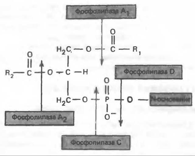
Fig. 25.5. Ester bonds of the phospholipid substrate hydrolyzed by phospholipases.
Lysolecithin can be synthesized via an alternative pathway involving lecithin-Cholesterol acyltransferase (LCAT). This enzyme, found in blood plasma and produced in the liver, catalyzes The transfer of a fatty acid residue from position 2 of a lecithin molecule to cholesterol, forming a cholesteryl ester. It is believed that the bulk of cholesteryl esters—components of plasma Lipoproteins—is synthesized through the action of LCAT. The consequences of LCAT deficiency are discussed on p. 281.

In phospholipids, long-chain saturated fatty acids are predominantly located at the sn-1 position, whereas polyunsaturated fatty acids (such as prostaglandin precursors) are most frequently found at the sn-2 position. The incorporation of fatty acids into the lecithin molecule occurs during de novo phospholipid synthesis, via transacylation between cholesterol ester and lysolecithin, and through the direct acylation of lysolecithin by acyl-CoA. This mechanism allows for the continuous turnover of fatty acids, with the incorporation of Essential Fatty Acids into phospholipid molecules being of particular importance.
Last update: 06/08/2026
Editorial and Educational Adaptation: This material has been compiled based on the primary/original source text. The project team performed an editorial review, corrected technical inaccuracies, structured sections, and adapted the content for an educational format.
What was processed:
- elimination of formatting defects (OCR errors, structural breaks, corrupted characters);
- editorial organization of content;
- standardization of terminology in accordance with academic sources;
- verification of factual statements against the original source text.
All mentions of the author, publication year, and origin of the primary text have been preserved in accordance with the source.