Chemistry and Biology of Proteins - F. Haurowitz 1953
Conjugated Proteins
Nucleoproteins
Although Nucleoproteins are one of the most vital classes of complex Proteins, they remain relatively little studied. Their profound biological significance is underscored by the fact that they are present in all living Cells and are also Key Components of Viruses and Bacteria [246]. Nucleoproteins consist of a protein moiety and a prosthetic group, which is a nucleic acid. Since many researchers view Selection/9.html">Nucleic Acids AS templates or matrices for Protein Synthesis in vivo, a closer examination of The Structure of these compounds is essential.
Complete Hydrolysis of nucleic acids yields equivalent amounts of phosphoric acid, a pentose, and purine or pyrimidine bases. Enzymatic hydrolysis using gastric or pancreatic juices breaks down nucleic acids into mononucleotides. Mononucleotides contain 1 molecule of phosphoric acid, 1 molecule of pentose, and 1 molecule of a purine or pyrimidine base [247, 248]. Further Cleavage of mononucleotides can be brought about by Enzymes from the intestinal juice or the Liver [249—251]. To state the products of complete nucleic acid degradation more precisely: such hydrolysis yields (1) phosphoric acid, (2) ribose or deoxyribose, (3) the purine bases adenine and guanine, and the pyrimidine bases thymine, cytosine, and uracil [252, 253].
The general structure of mononucleotides can be represented as follows: base — pentose — phosphoric acid. The pentosides that make up mononucleotides are known as nucleosides. The carbohydrate moiety of a nucleoside is linked to the nitrogen of a purine base at position 9 or to the nitrogen of a pyrimidine base at position 3. Phosphoric acid is attached via an ester bond to either the third or the fifth hydroxyl group of the pentose.
The structure of a typical nucleoside and mononucleotide is illustrated by the following formulas:
Class="center">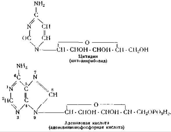
In nucleic acids, mononucleotides are linked together by phosphoric acid residues, forming chains of the following structure:
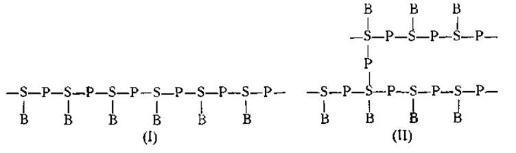
In the formulas above, S denotes the carbohydrate (ribose or deoxyribose), P represents phosphoric acid, and B stands for a purine or pyrimidine base [250].
Many questions concerning the arrangement of nucleotide chains within nucleic acid molecules remain unresolved. It is still unclear whether these chains are unbranched (formula I) or branched (formula II), whether they contain smaller subunits such as tetra- or pentanucleotides, and, finally, whether purine and pyrimidine bases alternate regularly along the nucleotide chains or are distributed at random [254, 255].
Nucleic acid preparations were initially obtained by alkali extraction followed by acid precipitation. However, exposure to acids and alkalis alters nucleic acid macromolecules; at pH <5.6 and > 10.9 [256], their solutions lose their characteristic high viscosity, presumably due to macromolecular disaggregation. The disaggregation of highly viscous polymeric nucleic acids is also triggered by two enzymes: Ribonuclease and deoxyribonuclease [257, 258]. Both enzymes were isolated in crystalline form from the Pancreas by Kunitz (see Ch. XII).
Preparations of natural macromolecular nucleic acids are typically obtained from nucleoproteins extracted from Tissues using salt solutions or Water. Careful dialysis of these nucleoproteins or vigorous shaking with chloroform [259] causes them to dissociate into protein and polymeric nucleic acid. The protein can subsequently be removed by Denaturation or salting out with sodium chloride [260]. According to various researchers [261—264], the Molecular Weight of native thymonucleic acid macromolecules ranges from 820,000 to 3,700,000. The length of the thread-like molecule of this acid is approximately 5,000 Å, and its cross-sectional diameter is 18—20 Å [262—264].
Depending on the type of pentose they contain, nucleic acids are divided into two categories: Ribonucleic Acids (RNA) and Deoxyribonucleic Acids (DNA). There is currently no Evidence for the existence of nucleic acids containing both types of pentose simultaneously [256]. Ribonucleic acid is identical to the "Yeast nucleic acid" of earlier authors, whereas deoxyribonucleic acid corresponds to "thymonucleic acid" isolated from the Thymus gland. These older designations have proven obsolete, as it was discovered that yeast also contains small amounts of deoxyribonucleic acid. The RNA/DNA ratio in yeast ranges from 30 to 50 [265]. The nuclei of animal and plant cells contain predominantly DNA, whereas the Cytoplasm of these cells is rich in RNA [266, 267] (see Ch. XVII).
RNA is degraded by Treatment with 1 N sodium hydroxide solutions, whereas DNA is more resistant and withstands this Procedure. This difference is exploited for the Quantitative determination of both nucleic acids when they occur together in solution [268]. After RNA is destroyed by sodium hydroxide, DNA can be precipitated by adding acidic protein solutions [268].
In tissues, both Types of Nucleic acids can be differentiated using the Feulgen reaction [269]. Tissue sections or a Cell suspension are treated with a 1 N Hydrochloric acid solution and subsequently with the so-called aldehyde reagent, which is fuchsin-sulfurous acid. Upon treatment with 1 N hydrochloric acid, only deoxyribose forms an aldehyde, most likely ω-oxylevulinic aldehyde (СН2ОН ∙ СО ∙ СН2∙ СН2∙ СНО) [270]. This aldehyde yields a red color with the fuchsin reagent. It is also responsible for the diphenylamine reaction proposed by Dische [271]. A positive reaction with both of these Reagents provides conclusive evidence that DNA is indeed present in the tissue.
Another important method used to differentiate RNA and DNA is the treatment of tissues with solutions of specific enzymes: ribonuclease and deoxyribonuclease. Each of these enzymes cleaves only its respective nucleic acid. By treating tissue sections with basic Dyes before and after the action of these enzymes, one can also determine the Intracellular Localization of nucleic acids [272].
Purine and pyrimidine bases intensely absorb ultraviolet light, with an absorption maximum near 2,600 Å, i.e., in a spectral region where proteins show no appreciable absorption. This makes it possible to use a quartz Microscope and ultraviolet micrography to detect nucleic acids [273]. By combining this method with The Use of specific enzymes, both types of nucleic acids can be differentiated. If tissue RNA is pre-digested with ribonuclease, any remaining Absorption in the ultraviolet region will indicate the presence of DNA. This same approach can also be used to determine the exact Location of DNA within the tissue.
As noted above, it remains unknown whether the nucleotide chains composing the molecules of both types of nucleic acids are straight or branched. The species Specificity of nucleic acids is more easily explained by the presence of branched chains. However, the existence of such branched chains has not yet been experimentally confirmed in any way [274].
Until recently, it was assumed that the macromolecule of nucleic acids is a polymer of tetra- and pentanucleotides. This view was based on the fact that nucleic acid isolated from yeast contains approximately 4 moles of phosphoric acid per each mole of adenine, guanine, cytosine, and uracil. In RNA isolated from the pancreas, there are approximately 5 moles of phosphoric acid per each mole of the aforementioned bases [275].
However, quantitative determinations using partition Chromatography yielded different figures, which are summarized in Table 15 [254, 276].
Table 15 Composition of Nucleic acids
|
Nucleic acids |
Purine and pyrimidine bases, moles per |
1 mole of phosphoric acid |
|||
|
adenine |
guanine |
cytosine |
uracil |
thymine |
|
|
From yeast |
0.260 |
0.255 |
0.245 |
0.0825 |
— |
|
From pancreas |
0.166 |
0.402 |
0.205 |
0.046 |
— |
|
From thymus |
0.260 |
0.205 |
0.172 |
— |
0.242 |
|
From pig liver |
0.310 |
0.17–0.18 |
0.200 |
0.13 |
— |
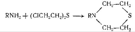
As can be seen from the figures in the table, the obtained data do not support the hypothesis that nucleic acids are polymers of tetra- or pentanucleotides. Only a few of these values are close to the expected figures (0.250 for a tetranucleotide and 0.200 for a pentanucleotide), while the majority deviate quite significantly. In this regard, the hypothesis was advanced that macromolecular nucleic acids are "statistical tetranucleotides" in which purine and pyrimidine bases are arranged more or less randomly [256]. Enzymatic hydrolysis revealed that pyrimidine bases are cleaved much faster than purine ones. This led to the Conclusion that purine bases are located primarily in the interior of the nucleic acid molecule [277–279]. It was also established that adenine and cytosine entering into the composition of nucleic acids are capable of undergoing deamination and, consequently, contain free amino groups [280]. These groups can also react with mustard gas to form thiazane rings [281]:
The Current state of our knowledge does not allow us to definitively resolve the question of whether nucleic acids possess species specificity. Meanwhile, the Assessment of the Biological Role of nucleic acids depends entirely on the resolution of this question. If it turned out that each animal species and each organ had its own specific nucleic acids, one could consider that nucleic acids play a decisive role in the transmission of hereditary traits. In the absence of species specificity, The Significance of nucleic acids in this regard would have to be considered secondary.
Due to their high phosphoric acid content, nucleic acids exhibit a pronounced acidic character. Like many other macromolecular anions, they react with proteins to form salt-like compounds that are insoluble within the pH range bounded by the isoelectric points of the proteins and nucleic acids. Thus, for example, egg albumin and other proteins are precipitated by nucleic acids under slightly acidic reaction conditions [282, 283]. There is no doubt that proteins bind to nucleic acids via electrovalent linkages. Like Other Compounds of the same type, nucleoproteins dissolve in concentrated solutions of neutral salts [283].
One of the primary difficulties encountered in the chemistry of nucleoproteins stems from the fact that we are still unable to answer whether nucleoproteins pre-exist in tissue cells as salt-like compounds of Nucleic Acids and Proteins, or as compounds formed through Other types of linkages, or whether they are formed only during the isolation process when favorable conditions are created for proteins to combine with nucleic acids via salt linkages [244]. Most nucleoproteins contain protamines or Histones as their protein component. Both types of these proteins are characterized by distinctly pronounced alkaline properties (see Ch. VIII, p. 197), enabling them to readily form salt-like bonds with nucleic acids. Nucleoprotamines and nucleohistones differ from other proteins in that the former lack Tyrosine, whereas the latter lack Tryptophan [284]. Apparently, cells contain either nucleoprotamines or nucleohistones exclusively, rather than mixtures of both types [284].
Table 16 Tryptophan and tyrosine content in proteins, histones, and protamines
|
Compound |
Tryptophan |
Tyrosine |
|
Proteins |
+ |
+ |
|
Histones |
0 |
+ |
|
Protamines |
0 |
0 |
Nucleoprotamines have been isolated from fish sperm, while nucleohistones have been obtained from the thymus and other mammalian tissues. The nucleic acid content in these nucleoproteins ranges from 31 to 66% [245]. Nucleoproteins isolated from plants or bacteria contain neither protamines nor histones; their composition includes only true proteins [285]. Similar true nucleoproteins have been found in animal tissues (see Ch. XVII).
Upon cellular extraction with a 1-percent sodium chloride solution, only proteins pass into the extract, whereas nucleoproteins remain insoluble. DNA-containing nucleoproteins can be extracted by repeated treatment with 6–11-percent sodium chloride solutions. These nucleoproteins precipitate from such solutions upon dilution with water [286]. Nucleoprotein solutions are highly viscous and exhibit strong Flow Birefringence. Based on this, it has been suggested that nucleoprotein molecules possess a thread-like shape. Their axial ratio varies from 40 : 1 to 60 : 1 [287].
A nucleoprotein containing DNA and histone was obtained by extracting the thymus gland with a sodium chloride solution. It constitutes a significant portion of the euchromatin in the nuclei of cells from this gland [287], whereas nuclear heterochromatin contains a nucleoprotein incorporating RNA.
The question of whether Chromatin contains histone nucleinate or some other specific compound of histone with nucleic acid remains finally unresolved. Extraction of the thymus gland with water, followed by high-speed centrifugation of the extract, yielded a nucleoprotein insoluble in isotonic salt solutions [288]. This nucleoprotein was designated as genoprotein T. It is hypothesized that both the salt-soluble and insoluble nucleoproteins are pre-existing in the gland cells.
As noted above, the greatest obstacle in resolving various questions concerning nucleoprotein chemistry is our inability to distinguish between nucleoproteins pre-existing in cells and those formed during the isolation procedure. The possibility of forming such "synthetic" protein nucleinates is evidenced by the fact that the thermal coagulation of Proteins can be prevented by adding small amounts of nucleic acids to the protein solution [289]. This is further supported by the disappearance of nucleic acid birefringence upon The addition of proteins to their solutions [290]. It has not yet been established whether solely electrovalent bonds participate in The formation of protein nucleinates. Since proteins can combine with polar compounds that do not dissociate into ions, such as sugars (see p. 232), there is no reason to rule out the potential involvement of dipole bonds alongside electrovalent bonds in the formation of protein nucleinates. The same applies to "natural" nucleoproteins. The combination of nucleic acids with proteins in these nucleoproteins may likewise proceed via electrovalent bonds, dipole bonds, or dipole-ion interactions [291].
A certain differentiation between natural nucleoproteins and artifacts arising during isolation can be achieved using calcium salts. Native nucleoproteins extracted from streptococci are not precipitated by calcium salts. However, if these nucleoproteins are pre-treated with acids, the subsequent addition of calcium salts induces precipitation [292]. These findings are interpreted as indicating either that the phosphoric acid incorporated into native nucleoproteins is inaccessible to Calcium Ions, or that soluble calcium compounds are formed under the action of calcium salts. Nucleoproteins isolated from the liver precipitate upon the addition of 0.03–0.06-percent calcium chloride solutions. Nevertheless, it remains uncertain whether the formation of a precipitate in this case indicates the creation of insoluble calcium compounds or merely the coagulation of nucleoprotein granules under METABOLISM/18.html">The Influence of calcium ions [293].
As mentioned earlier, many proteins, even crystalline ones, have proven to be non-uniform compounds rather than mixtures of different proteins. Therefore, it is hardly surprising that nucleoproteins have also turned out to be mixtures in many cases. The molecular weight of individual components of these mixtures in liver cells varies from 1,600,000 to 2,300,000 [287]. X-Ray Structural Analysis of nucleoproteins indicates the presence of short periods of 3.3 Å [294], i.e., periodicities identical to those discovered in Peptides.
Last update: 06/08/2026
Editorial and Educational Adaptation: This material has been compiled based on the primary/original source text. The project team performed an editorial review, corrected technical inaccuracies, structured sections, and adapted the content for an educational format.
What was processed:
- elimination of formatting defects (OCR errors, structural breaks, corrupted characters);
- editorial organization of content;
- standardization of terminology in accordance with academic sources;
- verification of factual statements against the original source text.
All mentions of the author, publication year, and origin of the primary text have been preserved in accordance with the source.