INVERTEBRATE ZOOLOGY IN THREE VOLUMES - BOOK 3 - H.I. Shcherbak - 1997
DEUTEROSTOMIA
PHYLUM ECHINODERMATA
SUBPHYLUM ECHINOZOA
This subphylum combines two classes of echinoderms: Holothuroidea and Echinoidea.
CLASS HOLOTHUROIDEA, OR SEA CUCUMBERS (HOLOTHUROIDEA)
Unlike other echinoderms, holothurians are less demanding regarding Water salinity; some apodous holothurians can live even in considerably freshened waters of mangrove swamps. A total of 1,100 species are known; eight of these have been recorded in the Black Sea, with the most widespread species being Stereoderma kirschbergi.
Holothurians are relatively large animals with an average size of 10–40 cm, although there are species with a body length exceeding 2 m. The majority of holothurians are brown, dirty white, or gray, but some species exhibit bright colorations of various hues.
Unlike other echinoderms, the body of holothurians is elongated along the axis from the oral to the aboral pole, and most of them resemble thick worms. However, there are nearly cylindrical, fusiform, spherical, or otherwise shaped species.
In holothurians, one distinguishes the anterior end, which is immediately recognizable by a ring of tentacles around the Mouth, and the opposite posterior end, where the anus is located (Fig. 181). Unlike other echinoderms, the oral-aboral body axis of holothurians is oriented not perpendicularly, but parallel to the substrate, and the animals lie as if on their side. The side facing the substrate is conventionally called the ventral side, and the opposite one is the dorsal side. In many holothurians, the ventral side is flattened to a certain extent, while the dorsal side is convex.
Class="center">
Fig. 181. Cucumaria frondosa:
1 — anus; 2 — tube feet of the bivium; 3 — tentacle ampullae; 4 — tentacles; 5 — tube feet of the trivium
The body of holothurians is covered with a non-ciliated epithelium. The integument of most species is soft due to a significant reduction of the Skeleton, which is represented only by microscopic calcareous bodies of various shapes (Fig. 182) scattered throughout the cutis and its various outgrowths. In addition to this peculiar external skeleton, holothurians also possess an internal skeleton — a circumoral calcareous ring comprising five large radial and many small interradial ossicles. It serves as an attachment site for various Muscles and protects the circumoral nerve ring.
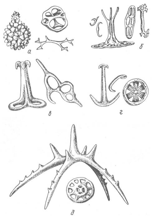
Fig. 182. Skeletal elements of the Skin in holothurians:
a — dendrochirotid, b — aspidochirotid, c — dolioliform, d — apodous, e — pelagothuriid holothurians
Due to skeletal reduction, the musculature in holothurians is highly developed. A continuous layer of circular musculature lies beneath the skin, and beneath it lies the longitudinal musculature consisting of five bands. At the anterior end of the body, these bands depart from the body wall and attach at an angle to the margin of the mouth opening. Contraction of these muscles causes the retraction of the anterior end of the body along with the tentacles. A series of muscles attached at one end to the calcareous ring around the Pharynx and at the other to the anterior end of the body brings about the reverse movement. Contraction of the circular musculature causes the holothurian's body to elongate, whereas contraction of the longitudinal musculature causes it to shorten sharply.
Beneath the Muscle layers lies a ciliated peritoneal epithelium that lines the spacious body cavity containing the Internal Organs. In holothurians, the body cavity performs the mechanical function of supporting the soft body walls, which lack a skeleton.
The ambulacral system of holothurians begins with a small madreporite located in one of the interradii of the bivium behind the anterior end of the body; in most holothurians, it does not reach the body surface and opens into the coelom. A stone canal extends from the madreporite; sometimes There are many such canals. The stone canal or canals empty, as in all other echinoderms, into the ring canal located directly behind the circumoral skeletal ring. Five radial canals extend from it; in holothurians, these first run forward, sending branches into the circumoral tentacles (which are modified tube feet), and then turn backward, positioned between the circular and longitudinal muscles (see Fig. 181). Lateral branches extend from the radial canals to the tube feet, from which elongated ampullae project into the body cavity, protruding from beneath the bands of longitudinal musculature (Fig. 183). The tube feet are arranged on the body surface in meridional rows: three of them (the trivium) along the ventral side, and two (the bivium) along the dorsal side. The tube feet of the trivium bear suckers and participate in locomotion. The feet of the bivium often lose their suckers, become thinner, and perform only a sensory function. In apodous holothurians, the radial canals of the ambulacral system are entirely absent. These holothurians move solely through the action of body muscles.
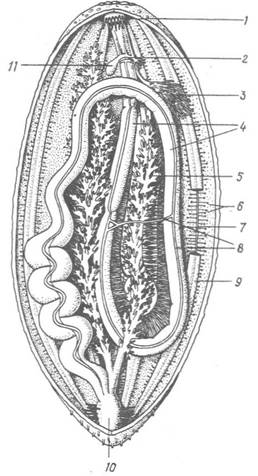
Fig. 183. Diagram of the internal anatomy of a holothurian:
1 — retracted tentacles; 2 — ring canal; 3 — gonadal tubules; 4 — intestine; 5 — respiratory trees (water Lungs); 6 — ampullae of tube feet; 7 — radial canal of the ambulacral system; 8 — Blood Vessels; 9 — longitudinal muscles; 10 — cloaca; 11 — Polian vesicle
Typically, a single thin-walled, often large sac with easily distensible walls extends from the ring canal between the radial canals. This is the so-called Polian vesicle, which serves as a reservoir for the ambulacral fluid. Occasionally, there may be many Polian vesicles — up to 20 or more.
The Digestive System begins with the mouth opening, which can be closed by means of specialized circular muscles. The mouth is surrounded by tentacles, which, as mentioned above, are modified tube feet. The number of tentacles ranges from 8 to 30, and their Structure varies greatly among representatives of different orders (Fig. 184). They play a significant role in obtaining food, both through active capture and by gathering it from the substrate surface or burrowing into it. In addition, they participate in locomotion, serve as organs of Touch, and sometimes assist in Respiration.

Fig. 184. Diagram of The structure of holothurian oral tentacles:
a — branched; b — peltate; c — hand-shaped; d — pinnate; e — digitate
The mouth leads into a pharynx that passes through the calcareous ring and continues into a relatively narrow Esophagus, which sometimes terminates in an enlargement. The esophagus opens into a long midgut that extends toward the posterior end of the body (descending limb), forms a loop in the posterior third and turns forward (ascending limb), and then turns back again toward the anus, in front of which the intestine widens to form a cloaca (see Fig. 183). Throughout its length, the alimentary canal is suspended from the body wall by mesenterial strands, while the cloaca is additionally supported by well-developed muscles. In a few holothurians, the intestine has a different structure.
In some aspidochirote holothurians, the ducts of the so-called Cuvierian organs empty into the cloaca. These are glandular, tubular structures that, when the holothurian is disturbed, are expelled through the cloaca and transform into long, white, sticky threads that entangle the stimulus-causing object or Organism.
The perhemal system in holothurians consists solely of radial canals (Fig. 185).

Fig. 185. Cross-section of the body wall of Cucumaria frondosa:
1 — transverse Muscles of the interradial body regions; 2 — longitudinal muscles of the radial body regions; 3 — peritoneal epithelium; 4 — hyponeural Nervous system; 5 — radial blood lacuna; 6 — radial perhemal canal; 7 — radial ambulacral canal; 8 — ectoneural nervous system; 9 — Connective Tissue within the body wall
The Circulatory system is better developed than in other echinoderms. In addition to numerous lacunae in the connective tissue, they possess a circumoral lacunar ring, five pairs of radial lacunae located between the radial ambulacral canals and nerves, and a well-developed network of blood vessels within the intestinal walls (Fig. 186), among which the ventral and dorsal vessels are the most prominent. The ventral vessel closely adheres to the intestine, accompanying it along its entire length and giving off fine branching twigs that encircle the gut wall. The dorsal vessel is connected to the intestine by a mesentery wrapped in numerous branches of this vessel. Transverse vessels frequently cross the body cavity freely, surrounded by coelomic epithelium, connecting the vessels of different intestinal loops. In some holothurian groups, the partitions of the dorsal vessel form a complex network known as the "rete mirabile" (admirable net). Occasionally, the rete mirabile envelops the left respiratory tree (see below), allowing oxygen to pass directly from it into the blood rather than the coelomic fluid.

Fig. 186. CIRCULATORY SYSTEM OF Cucumaria frondosa associated with the intestine:
1 — anterior descending intestinal loop; 2 — ventral and 3 — dorsal blood vessels; 4 — crop; 5 — mesentery; 6 — esophageal blood vessel; 7 — esophagus; 8 — ascending intestinal loop; 9 — anastomoses between the ascending and descending PARTS OF THE ventral vessel; 10 — posterior descending intestinal loop; 11 — rete mirabile
Respiration in many holothurians occurs through the skin and ambulacral tentacles, whereas representatives of three orders (Dendrochirota, Aspidochirota, and Molpadonia) possess specialized respiratory organs known as water lungs (see Fig. 183). These consist of two long, frequently highly branched, and sometimes brightly colored trunks lying on either side of the intestine within the body cavity, occupying a significant portion of it. They are connected to the body wall and intestinal loops by muscular and connective tissue strands. Posteriorly, the trunks of both respiratory trees unite and open into the cloaca via a common duct. The inner walls of the lungs are lined with ectodermal ciliated epithelium, followed by connective tissue, a layer of well-developed muscles, and another layer of connective tissue covered by coelomic epithelium. Through rhythmic contractions and relaxations of the muscles, water is alternately drawn through the cloaca into the lungs, filling their finest branches, and expelled. Dissolved oxygen then diffuses through the thin walls of the lungs into the coelomic fluid and is distributed throughout the body.
In addition to respiration, the water lungs also perform an excretory function, as amoeboid Cells (celomocytes) laden with metabolic wastes are discharged from the coelomic fluid through their walls and subsequently expelled via the cloaca. Specialized excretory organs are absent; only apodous holothurians possess structures somewhat analogous to them. In these holothurians, ciliated funnels are arranged along the intestinal mesentery, capturing waste-laden celomocytes, which then coalesce into distinct corpuscles before being discharged back into the coelomic fluid.
The Nervous System of holothurians is submerged beneath the skin. The ectoneural nervous system consists of a nerve ring situated within and protected by the calcareous circumoral ring, along with five thick radial nerve cords. The latter run parallel to the radial ambulacral canals and hyponeural nerve cords, almost fusing with them. A ring component of the hyponeural system and an entirely apical nervous system are absent in holothurians. Lateral nerve fibers branch off from the circumoral ectoneural nerve ring to supply each tentacle surrounding the mouth, as well as the oral membrane and pharynx. All ambulacral tube feet are innervated by the radial cords, as are numerous sensory cells in the skin, which are most densely concentrated at the anterior and posterior ends of the body. Equilibrium organs, or statocysts (Fig. 187), found in certain deep-sea forms, are also associated with the radial nerves. They take the form of small vesicles containing floating statoliths. In a few holothurians, photoreceptor cells are located at the Base of the tentacles, enabling the animal to detect light levels.

Fig. 187. Statocysts of holothurians (cross-section):
1 — statocyst; 2 — statoliths; 3 — statocyst nerve; 4 — radial canal of the ambulacral system; 5 — radial nerve
Most holothurians are gonochoric (1.e., dioecious); unlike other echinoderms, they lack a genital strand and possess only a single gonad consisting of a variable number of long, blind-ending tubules upon whose outer walls Germ Cells are formed (see Fig. 183). All tubules unite into a single genital duct that opens in the dorsal interradius near the anterior end of the body, occasionally at the base or even at the tip of one of the tentacles.
A certain number of holothurian species are hermaphroditic, with the gonad producing both eggs and spermatozoa—alternately in some species and simultaneously in others. In both cases, male Gametes are typically shed before female ones. The eggs are dispersed freely in the water and do not form egg masses or clusters. Fertilization in most species occurs externally in the water.
The Development of holothurians can proceed via two pathways. In species with yolk-poor eggs, the egg develops into a ciliated blastula or gastrula that swims freely for several weeks, undergoing profound structural changes during this period. Initially, it develops into a bilaterally symmetrical dipleurula, after which a sinuous ciliated band forms; at this larval stage, the larva is called an auricularia. It swims freely in the water, actively feeding on small planktonic organisms, which enables rapid growth and transition to the next stage—the doliolaria. The latter is easily recognized by its barrel-shaped body and wreaths of cilia encircling it. The doliolaria swims initially, then settles to the bottom; it does not feed, relying instead on nutrient reserves accumulated during the auricularia stage. Subsequently, the rudiments of the circumoral tentacles and ambulacral tube feet appear, the ciliated bands disappear, and this stage, known as a pentactula, already resembles a young juvenile (Fig. 188).

Fig. 188. Larvae of holothurians:
a — auricularia of Synapta vittata; b, c — doliolaria and pentactula of Cucumaria planci; 1 — ciliated band; 2 — mouth; 3 — Stomach; 4 — anus; 5 — preoral lobe; 6 — circumoral tentacles; 7 — ambulacral tube feet
In holothurians with yolk-rich eggs, the egg directly hatches into a so-called pseudodoliolaria, which differs from a true doliolaria in THE POSITION OF its mouth; it soon develops into a pentactula. Species in which the egg hatches directly into a pentactula are also known.
A number of holothurian species, particularly inhabitants of Arctic waters, exhibit parental care. In the simplest cases, eggs and larvae develop On the surface of the mother's body, protected by various dermal outgrowths; in others, body depressions appear, such as brood chambers extending into the coelomic cavity, or development takes place within the Ovary or body cavity. How fertilization occurs in these instances remains unclear, but development proceeds via a different pathway.
Holothurians are characterized by a high capacity for regeneration. It is known that under severe stress, holothurians are capable of autotomy of a number of organs—the cloacal wall ruptures, through which the animal expels either just the intestine, the left respiratory tree, or the Gonads, or even all internal organs. Despite this, the animal does not die, and within a short period regenerates all lost parts. Some holothurians can divide crosswise into anterior and posterior halves, after which each part regenerates the missing half, meaning asexual reproduction takes place.
As already mentioned, holothurians are bottom-dwelling animals; typically, they move slowly along the seabed using tube feet, tentacles, or muscular body contractions, more rarely burrowing into the sediment and even more rarely swimming throughout their entire lives. When disturbed, holothurians retract the anterior part of the body along with their tentacles, expel water from the cloaca, and curl up into a dense lump.
The economic importance of holothurians stems from the fact that about 40 of their species, collectively known as trepang or sea cucumbers, are edible. Trepang fisheries are especially developed off the coasts of Japan, China, Indonesia, and the Philippines, and they are also harvested along the shores of Africa, the Americas, and Australia. Trepangs are sold dried, boiled, salted, or canned. In Eastern medicine, trepangs are referred to as "marine ginseng." Their flesh is rich in Proteins and valuable mineral salts.
Holothurians provide shelter to many invertebrates (Protozoans, worms, Mollusks, crustaceans) and Fishes that live on The surface of their bodies, in their intestines, Polian vesicles, respiratory trees, circulatory system, etc.
The Class Holothuroidea is divided into six orders.

Order Dendrochirota. This is a fairly large order of holothurians, all representatives of which possess relatively long, heavily branched tentacles (see Fig. 184) and exhibit more pronounced pentamerous radial Symmetry than other holothurians.
The body shape of individual species varies greatly and is well adapted to their living conditions. Most dendrochirotids inhabit coastal waters and intermediate depths (down to 1000 m), with only isolated species living at great depths.
Well-known Representatives of the family Cucumariidae are the so-called sea cucumbers. They possess a more or less cylindrical or spindle-shaped body, sometimes strongly curved, with 10 tentacles. Widespread along the shores of the Sea of Japan, the Sea of Okhotsk, and the Yellow Sea is the Japanese sea cucumber (Cucumaria japonica, Fig. 189, a), which is the target of intensive commercial fisheries. It features a flattened ventral side, on which long tube feet with powerful suckers are arranged along the radii, used for locomotion. The dorsal tube feet, meanwhile, have transformed into sensory papillae. Belonging to the family Cucumariidae is the previously mentioned species *Stereoderma kirschbergi*, which lives in the Black Sea near the coast of Crimea. While most sea cucumbers lead a sluggish lifestyle lying on the seabed, members of the family Sclerodactylidae often burrow into the sediment in such a way that only the anterior end with the mouth and the posterior end with the anus protrude above the surface.

Fig. 189. Dendrochirotid holothurians: a — Cucumaria japonica; b — Psolus phantopus
Members of the family Psolidae possess an interesting structure. Their body is flattened, and part of the ventral side has transformed into a flat creeping sole covered with soft skin, while the convex upper portion is covered with scale-like skeletal plates (Fig. 189, b).
Order Dactylochirotida. This order includes holothurians that have 8–30 digitiform tentacles and a body shape quite unusual for holothurians. For instance, in members of the family Rhopalodinidae, the body is club- or flask-shaped due to strong reduction of the dorsal side and expansion of the ventral side. These animals possess 10 radii and 10 interradiuses: five descend along the "neck" of the flask-like body and five ascend upward. The mouth and anus are located almost side by side at the elongated end of the body, with the genital opening situated between them. In members of the family Ypsilothuridae, such as *Ypsilothuria bitentaculata*, the body is nearly spherical, covered with large transparent plates that feature openings for the tube feet (Fig. 190).

Fig. 190. Dactylochirotid holothurians:
a — structural diagram of Rhopalodina lageniformes; b — Ypsilothuria bitentaculata; 1, 2 — anal and oral openings
Order Aspidochirota. This is the most species-rich order of holothurians, whose representatives possess short, peltate tentacles that cannot be retracted inside the body due to the absence of appropriate muscles. Externally, many of them resemble large worms. This order includes the majority of commercially harvested species—trepangs (Fig. 191, a, b).

Fig. 191. Aspidochirotid holothurians:
a, b — trepangs from the Indian Ocean and the Sea of Japan, respectively; c — deep-sea species Euphronides tanneri
The largest family in this order, the Holothuriidae (true sea cucumbers), consists of warm-water inhabitants that feed on organic detritus from the sediment. Among true sea cucumbers, there are poisonous species; for example, the toxin of the black sea cucumber (*Ludwigothuria atra*) is used by Pacific islanders to stun fish. However, humans are not susceptible to its poison, and this holothurian species is used for food.
An interesting swimming adaptation is found in members of the family Synallactidae, which inhabit greater depths. For instance, *Euphronides tanneri* features a well-developed marginal brim and a distinctly separated anterior body region (Fig. 191, c). This holothurian is capable of swimming for extended periods, rising a considerable distance off the bottom. It obtains its food both from the water Column and from the seabed.
Order Elasipoda. This order comprises both deep-sea benthic animals and true pelagic forms that spend their entire lives swimming in the water column. As in members of the previous order, elasipods have short peltate or otherwise shaped tentacles that do not retract into the body. In deep-sea forms, large ventral tube feet are located along the sides of the body; on the dorsal side, they are reduced or modified into complex outgrowths (Fig. 192, a, b).

Fig. 192. Side-swimming sea cucumbers:
a — Scotoplanes murrayi; b — Elpidina curilensis; c — Pelagothuria ludwigi
Sea cucumbers of the family Pelagothuriidae (Fig. 192, c) exhibit a somewhat different form, leading a pelagic lifestyle. Externally, they resemble small jellyfish. These fragile, pink or purple creatures have a gelatinous body devoid of any skeletal elements, which significantly reduces their weight. Surrounding the mouth, which is encircled by 12–16 short tentacles, a swimming disc is formed by the expansion of the body wall, with long appendages located along its margins. Each of these appendages contains a canal that communicates with the ambulacral canal of the tentacles.
Order Molpadida (Molpadonia). The body of representatives of this order is barrel- or spindle-shaped, with the posterior end always extended into a long or short tail (Fig. 193); ambulacral feet are reduced, and tentacles are greatly shortened (see Fig. 184), adapted for burrowing through sediment. In the dermis, aside from typical skeletal plates of various shapes, there are round red or brown bodies containing phosphorus. Barrel-shaped sea cucumbers inhabit muddy sediments, burying themselves in them while leaving the tail with the terminal anus exposed above the surface. Through this opening, oxygen-rich water enters the respiratory trees.

Fig. 193. Barrel-shaped sea cucumbers:
a — Molpadia musculus; b — Trichostoma arcticum
Order Apodida (Apoda). The body of apodous sea cucumbers is worm-like, evenly rounded at the posterior end. The skin is transparent, but typically rough or even warty due to accumulations of skeletal ossicles. Ambulacral feet, as well as radial canals, are absent. The mouth is surrounded by tentacles of various structures, which communicate directly with the ring ambulacral canal.
Apodous sea cucumbers respire through their thin integument, lacking respiratory trees. As already mentioned, unlike other sea cucumbers, apodous forms possess special excretory organs — ciliated funnels. Many species burrow into sediment or hide under stones; tropical species live among corals. It is precisely the apodous sea cucumbers that are less demanding regarding water salinity, with some species inhabiting the heavily brackish waters of mangrove swamps.
Four species of these sea cucumbers inhabit the Black Sea. One of them, *Oestergzenia thomsoni*, has the ability at the slightest disturbance to fragment into separate pieces; the tentacle-bearing end burrows into the sediment and regenerates the lost parts, while the other pieces perish. Similar behavior is characteristic of some other species of the family Synaptidae, which includes both the largest sea cucumber species, *Synapta maculata*, reaching 2 m 10 cm in length (with a diameter of 5 cm), and the smallest, *Leptosynapta minuta*, measuring just 0.5 cm long.
An interesting lifestyle is observed in *Synaptula hydriformes* (Fig. 194), which lives among Algae or coral branches, clinging to them with its tentacles. These sea cucumbers feed exclusively on algae. They brood their young within the body cavity.
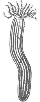
Fig. 194. Apodous sea cucumbers: Synaptula hydriformes
Last update: 13/08/2026
Editorial and Educational Adaptation: This material has been compiled based on the primary/original source text. The project team performed an editorial review, corrected technical inaccuracies, structured sections, and adapted the content for an educational format.
What was processed:
- elimination of formatting defects (OCR errors, structural breaks, corrupted characters);
- editorial organization of content;
- standardization of terminology in accordance with academic sources;
- verification of factual statements against the original source text.
All mentions of the author, publication year, and origin of the primary text have been preserved in accordance with the source.