INVERTEBRATE ZOOLOGY IN THREE VOLUMES - BOOK 3 - H.I. Shcherbak - 1997
DEUTEROSTOMIA
PHYLUM ECHINODERMATA
SUBPHYLUM CRINOZOA, OR STALKED ECHINODERMS
CLASS CRINOIDEA (SEA LILIES)
Class="center">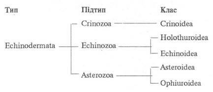
About 4,000 fossil species and more than 620 extant species are known. They are distributed primarily in the tropical Regions of the Indian and Pacific Oceans, although some occur in Antarctic and Arctic waters. The size of sea lilies ranges from a few centimeters to 2.5 m. The class name refers to the resemblance of these animals to flowers, which is further emphasized by their bright coloration. Some crinoids remain attached to the substrate throughout their lives or may detach for short periods to move a small distance. Others lead a free-swimming or crawling lifestyle, always keeping their oral surface directed upward; they periodically stop and attach to the substrate (Fig. 171).

Fig. 171. Heliometra glacialis:
1 — pinnules; 2 — rays, or arms; 3 — cirri
The body of sea lilies consists of a small, more or less cup-shaped trunk from which five long rays, or arms, extend. These arms fork at their base, increasing the number to ten, but typically the arms branch repeatedly, and their number can reach up to 200.
A single-layered epithelial covering is present only in juveniles; in adults, all external skeletal plates are exposed.
Connective Tissue lies beneath the epithelium in the non-skeletal areas.
On the flat oral side of the trunk, large skeletal plates are present only in some juveniles and a few species throughout their lives; however, as a rule, the Skeleton of the oral side is underdeveloped and represented by small calcareous ossicles.
The aboral cup-shaped side is covered by a solid armor formed by a lower central plate (often hemispherical in shape) and two to three whorls of five skeletal plates each, some occupying radial and others interradial positions. In some sea lilies, the aboral skeleton is significantly reduced, resulting in a nearly disk-shaped body. In certain crinoids, the function of the aboral trunk skeleton is performed by the arm ossicles (see below).
In most sessile species, a stalk extends from the aboral side of the body, consisting of cylindrical or disk-shaped calcareous ossicles connected by Muscle fibers, which provides flexibility to the stalk. The length of the stalk in extant species is 75–90 cm, whereas in fossil forms it reached up to 21 m. The uppermost ossicle of the stalk typically fuses with the central plate of the aboral trunk side, forming the Base of the calyx.
Sea lilies attach to the substrate in various ways. In some species, attachment is mediated by the terminal ossicle of the stalk, which expands into a disk, hook, or similar Structure; others possess numerous thin outgrowths, or ROOT-like holdfasts; while still others have movable cirri, which may also occur on other stalk ossicles.
Cirri are also present in stalkless sea lilies. They are located on the central aboral skeletal plate, which features special sockets for their attachment (Fig. 172). Nerves pass to the cirri through openings at the bottom of these sockets. Using numerous cirri (numbering up to 100) with terminal claws, stalkless crinoids temporarily anchor themselves to the bottom.

Fig. 172. STRUCTURE OF THE lower part of a sea lily:
1 — radial plate; 2, 3 — First and Second arm ossicles; 4 — arms; 5 — cirri; 6 — central plate; 7 — sockets for cirral attachment
The arms of sea lilies possess a well-developed skeleton consisting of individual cylindrical ossicles, or vertebrae, also known as brachials, as well as a small soft part containing certain Internal Organs (Fig. 173). The first ossicle of each arm fuses with the plates of the aboral trunk skeleton, while the second articulates with it via two articular sockets, with several more ossicles closely adjoining it. Beyond this lies the freely movable portion of the arm.

Fig. 173. Cross-section through the arm of Heliometra glacialis:
1 — brachial trunk of the apical Nervous system; 2 — bulk of the ossicle; 3 — interbrachial Muscles; 4 — oral coelomic sinus; 5 — radial canal of the ambulacral system; 6 — lateral branch of the ambulacral system; 7 — ambulacral groove; 8 — radial perhaemal canal; 9 — ectoneural nervous system; 10 — genital sinus; 11 — aboral coelomic sinus
The arm ossicles are connected by elastic ligaments and muscle fibers, which ensures their exceptional flexibility and mobility. However, in some joints, The connection between ossicles lacks muscles, and it is precisely along the boundaries of such ossicles that autotomy (spontaneous shedding of a body part) very frequently occurs. In the center of each arm vertebra, There is a canal through which the brachial trunk of the apical nervous system passes. Almost every vertebra bears lateral branches on either the right or left side—the pinnules—which share the same Anatomical Structure as the arms. On the inner side of the vertebrae and their ramifications, including the pinnules, there is a variably developed groove traversed by a radial ambulacral furrow lined with ciliated epithelium.
Using their arms, free-living crinoids can crawl or swim quite dexterously. In addition, the arms form a perfect trapping apparatus that supplies the crinoids with food.
Musculature is poorly developed. Muscles are present only in the arms (between the arm ossicles, in the pinnules, and in the ambulacral tentacles), as well as between the ossicles of the stalk and cirri.
The body cavity of adult animals is largely filled with a network of connective-tissue trabeculae lined with coelomic epithelium, making the coelom spongy. Free coelomic spaces persist near the oral side of the body, extending into the arms, and in the lower part of the calyx near the central plate, where the coelomic cavity is divided by partitions into five chambers, hence referred to as the five-chambered organ (Fig. 174). The coelom extends from the calyx into the stalk and cirri. In the central part of the trunk lies the axial coelomic sinus, which houses the spongy portion of the so-called axial organ, originating as a solid cord within the five-chambered organ.

Fig. 174. Horizontal section through the calyx of a sea lily:
1 — radial nerve cord; 2 — central nerve capsule; 3 — partition of the five-chambered sinus; 4 — its chamber; 5 — muscles; 6 — axial organ; 7, 8 — central and radial plates of the aboral skeleton, respectively
The ambulacral system consists of a circumoral ring and five radial canals that branch in accordance with the branching of the arms, including the pinnules. Lateral canal branches depart from these canals and enter two rows of ambulacral tentacles located on the oral side of the rays and pinnules. The tentacles lack suckers and function as respiratory and tactile organs. Stone canals—ranging from five to 150 in individual species—extend from the ring canal. They open freely into the body cavity with their distal ends. The entire oral side of the crinoid body functionally corresponds to the madreporite, being perforated by numerous so-called Water pores that connect the coelom with the surrounding water. The movement of the ambulacral tentacles is driven by muscular action and the pressure of the coelomic fluid.
The Digestive System begins with the Mouth, which occupies a central position on the oral disc (Fig. 175) in some species, whereas in others it is shifted slightly to the side. The mouth leads into the Esophagus, which continues into a long endodermal intestine lined with ciliated epithelium; it initially runs toward the aboral bottom of the body, then forms from one (in species with a centrally positioned mouth) to four loops, and turns back toward the oral side, where it opens via a short hindgut through the anus situated on a small prominence located in one of the interradii.

Fig. 175. Oral disc of Heliometra glacialis:
1 — mouth surrounded by papillae; 2 — ambulacral grooves; 3 — anal cone; 4 — first pair of pinnules; 5 — arms; 6 — vestigial plates of the oral skeleton; 7 — water pores
Crinoids feed on numerous microscopic animal and plant organisms, as well as detritus particles. Through the dense network of tentacles on the ambulacral grooves of the arms, food is filtered from the water and directed along the grooves toward the mouth by a water current generated by rapid rotational Movements of the tentacles and the beating of cilia. In some laboratory-studied species, notably the North Atlantic species Antedon petasus, food particles were observed to be coated with mucus secreted by mucous Cells.
The perihemal system is underdeveloped; it is represented by five slender radial canals extending along the
arms beneath the radial canals of the ambulacral system. Both at the base of the arms and at their tips, these canals end blindly.
The Circulatory system consists of a circumoral plexus of lacunae that transition into the lacunae of the intestinal walls and the axial coelomic sinus. Radial lacunae, located within the walls of the genital cord of the arms and pinnules (see below), also extend from the circumoral ring.
Respiration in crinoids occurs through areas of the Skin lacking a skeleton and via the ambulacral tentacles.
The Nervous System of crinoids is characterized by a strong development of its apical part, which consists of an aggregation of Nerve Cells (the central nerve capsule) located inside the five-chambered organ, and five radial nerves piercing all the arm ossicles and pinnules. This system controls the vital movements of the crinoid arms.
The ectoneural part of the nervous system consists of a circumoesophageal ring and five radial nerve cords running along the ambulacral grooves of the arms. The hyponeural nervous system, located deeper than the ectoneural one, has a similar structure. These systems are less developed than the apical one. It is believed that they are associated only with sensory cells, which are abundant in the cutaneous epithelium and function as chemo- and photoreceptors.
All crinoids are dioecious, but they exhibit no Sexual Dimorphism. Gonads develop within the arms. A genital stolon originates from the oral end of the aforementioned axial organ, from which five genital cords extend into the arms, branching in accordance with the arm branching. The terminal branches enter the pinnules, where they transform into closed sacs in which Germ Cells develop. The release of male Gametes from the pinnules to the exterior occurs through specialized pores, whereas female gametes are released via ruptures in the pinnule walls. In many species, sperm is first shed into the water, which stimulates female individuals to spawn eggs.
Fertilization is mostly external. When fertilization is internal, spermatozoa actively penetrate the body of the female. The fertilized egg develops into a sac-like larva—a dolicholaria—which lacks both mouth and anus, bearing an apical tuft of cilia and five ciliated bands (Fig. 176, a); it leads a planktonic existence for two to three days, after which it settles to the bottom and attaches by its anterior end to a substrate or some hard object, and occasionally to the body of its parents.
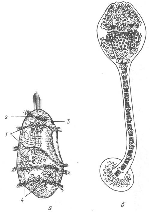
Fig. 176. Larvae of the sea lily Antedon rosacea — dolicholaria (a) and pentacrinoid (b):
1 — continuous ciliary bands; 2 — semi-ring-like anterior ciliary band; 3 — attachment pit; 4 — skeletal plates
The body of the larva begins to elongate and divide into a stalk and a calyx-shaped trunk, at the apex of which the mouth breaks through; the ciliary rings and apical tuft degenerate. Skeletal plates are laid down in the mesoderm of the calyx and stalk. This stage is called the pentacrinoid stage due to its similarity to stalked crinoids of the genus Pentacrinus. Outgrowths appear along the edges of the calyx, which transform into arms (Fig. 176, b).
The further development of stalked and stalkless forms proceeds differently. In the former, new and new stem ossicles are formed, and cirri and additional branches develop, with which the stem firmly attaches to the substrate. In the latter, after several months of attached life, the calyx spontaneously breaks off, and the crinoid shifts to a free-living mode of life.
All modern sea lilies belong to the subclass Articulata, which includes four orders.

The first three orders, comprising only about 80 species, unite stalked sea lilies, while the fourth and most numerous order comprises stalkless ones.
Order Isocrinida. This order includes species with a long, rope-like stem bearing whorls of cirri throughout its length, with five in each ring (Fig. 177). The arms are relatively long and branched compared to the body. The skeleton of the aboral part of the body also includes brachial plates of the arms. On the oral side, it is represented by numerous small ossicles, whose Condensation into tegminal plates along the food grooves almost completely covers the latter.
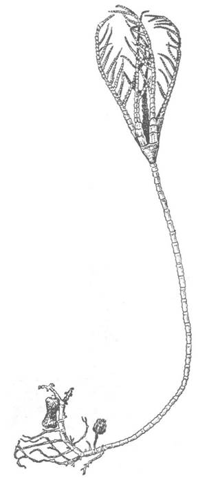
Fig. 177. Order Isocrinida: Metacrinus nobilis
Most species of this order belong to the genus Metacrinus, for instance, the Pacific species M. suborbus with a body length up to 19 cm and a stem up to 1.5 m long and 0.8 cm thick, living at a depth of 200–300 m, or M. nobilis, which has an almost white stem and a yellowish or red-orange crown, found in the seas of the Indo-Malayan region at a depth of about 250 m.
Order Millericrinida. Representatives of this order differ from isocrinids by their smaller size and the absence of cirri on the stem, which may only be present at its base to serve for attachment to the substrate. One of the most famous species of this order is Rhizocrinus lofotensis (Fig. 178), which is widely distributed in the Atlantic at depths from 140 to 4000 m. It is a small species reaching 8–10 cm in total length including the stem. It possesses a five-rayed, and occasionally four- or seven-rayed, calyx. Its rays do not branch, but are covered with sturdy pinnules. The North Atlantic species Bathycrinus carpenterii is significantly larger in size; the length of its stem is 27 cm, and the arms are 3 cm; the stem ends in several coarse rootlets by which the animal attaches to the substrate.

Fig. 178. Order Millericrinida: Rhizocrinus lofotensis
Order Cyrtocrinida. Representatives of this order differ significantly from other stalked crinoids, primarily in the shape of the body and arms. It is believed that their structure is closest to ancestral forms. Thus, in the deep-water species Hyocrinus bethelianus, living at depths of 3000–5000 m in the southern Pacific Ocean and reaching 15 cm in length, the body calyx (Fig. 179) is relatively quite high compared to other stalked sea lilies. The skeletal plates of the aboral side are not fused together and are not bound by brachial plates. The arms originate from the edge of the calyx. On the oral side, there are large calcareous plates, and small tegminal plates surround the food grooves. Cirri are never present on the stem. An even more original structure is possessed by Holopus rangii, found at shallow depths (200–300 m) in the Caribbean Sea. It has a short (about 4 cm high) robust calyx whose skeletal plates are completely fused, the stem is underdeveloped, and the animal attaches to a hard substrate by the bottom of its calyx. The mouth is located in the center of the oral side and is surrounded by five stout arms that fork dichotomously above the first basal ossicle. All ten arms of the animal are of unequal length: they are longer on one side than on the other. The pinnules on the arms, unlike those of other crinoids, are turned inward and form an almost continuous tube along each arm, through which water currents carry planktonic organisms to the mouth.

Fig. 179. Order Cyrtocrinida: Hyocrinus bethelianus (a) and Holopus rangii (b):
1 — anus; 2 — mouth; 3 — arms; 4 — first arm branching; 5 — first arm ossicle; 6 — radial plate; 7 — basal plate; 8 — stem
Order Comatulida. This order unites all sea lilies that lead a free-living existence in the adult stage. It includes more than 550 species, representing the vast majority of extant (currently living) sea lilies. During development, the body skeleton in many comatulids is significantly reduced, and brachial plates replace it to some extent, forming the walls of a small calyx bearing relatively long branched arms with long pinnules. On the aboral side of the calyx, many comatulids possess numerous highly mobile cirri, with which the animal attaches to the substrate.
The cirri vary in structure depending on the type of sediment inhabited by particular species. If the bottom is muddy, the cirri are long, thin, and straight, supporting the animal On the surface like stilts; whereas if it is rocky, the cirri are short and hook-shaped,
allowing the animal to firmly grasp stones, coral skeletons, etc. Some species lie on the bottom all the time, but most sea lilies periodically detach from their support and swim for some time. Swimming is intermittent: having covered a small distance, the animal settles to the bottom and attaches to it, then swims again, and so on. Comatulids move by means of their arms; multi-rayed individuals use alternating sections of all arms during swimming, whereas few-rayed ones use different arms. Similarly, during swimming, the animal catches onto some object with the forward-reaching part of its arms using pinnules that can also secrete a sticky substance. Then these arms contract simultaneously, while the free arms push off from the substrate.
Almost all comatulids are brightly colored with a wide range of hues—monochrome, variegated, or spotted.
Feeding in comatulids takes place only in a quiescent state.
The largest family of the order is Antedonidae, which numbers over 130 species. Antedonids are found from the littoral zone (the intertidal area of the seabed exposed during low tide) to a depth of 6000 m and quite frequently in temperate latitudes. They are mostly ten-rayed. Representatives of the genus Antedon inhabiting European coastal waters, particularly Antedon bifida (Fig. 180), have been thoroughly studied. This is a sea lily with variable coloration: ranging from intense purple to pink and yellow, and sometimes variegated. It lives at depths from 5 to 450 m, attaching itself with strong curved cirri to the rhizomes and stems of Algae. It has thin, flexible rays 12.5 cm long. They are very fragile, and it is rare to find a specimen that possesses all ten undamaged arms; as a rule, some of them are in The process of regeneration.

Fig. 180. Order Comatulida:
a — Antedon bifida; b — Comatula purpurea
The regenerative capacity of antedons, like that of many other comatulids, is so great that they restore not only broken rays but even half of the body if cut in half.
Extremely striking members of the quite numerous family Comasteridae (comprising up to 100 species). Most of these are multi-armed species with long arms (up to 20–25 cm) that inhabit the coastal waters of the tropics and feature a bright, variegated coloration that makes them look even more like flowers. Their mouth is shifted toward the margin, while the anus occupies a central position. Another distinctive feature is the presence of so-called oral pinnules, consisting of numerous laterally compressed segments with tiny Teeth on their upper side, giving the tips of the pinnules a serrated appearance. It is believed that the animal uses these to capture small organisms and transfer them to the food grooves. Among comasterids, there are species with varying arm lengths. One can distinguish anterior (feeding) arms and shorter posterior ones, where gametes are developed.
Antarctic feather stars that exhibit parental care are of particular interest. For instance, in feather stars of the genus Phrixometra, embryos develop in brood chambers located within the genital pinnules of females. In some species, such as Phrixometra longipinna, the juveniles emerge from the capsules at the pentacrinoid stage, whereas in others (Phnutrix), they emerge only after the adult form is fully developed.
Last update: 13/08/2026
Editorial and Educational Adaptation: This material has been compiled based on the primary/original source text. The project team performed an editorial review, corrected technical inaccuracies, structured sections, and adapted the content for an educational format.
What was processed:
- elimination of formatting defects (OCR errors, structural breaks, corrupted characters);
- editorial organization of content;
- standardization of terminology in accordance with academic sources;
- verification of factual statements against the original source text.
All mentions of the author, publication year, and origin of the primary text have been preserved in accordance with the source.