INVERTEBRATE ZOOLOGY IN THREE VOLUMES - BOOK 3 - H. Y. Shcherbak - 1997
DEUTEROSTOMIA
PHYLUM ECHINODERMATA
SUBPHYLUM ECHINOZOA
CLASS ECHINOIDEA
Sea urchins, much like sea lilies, inhabit exclusively marine waters with a salinity of about 35 % and are highly sensitive to even the slightest decrease in salinity. These benthic, sluggish animals range in body size from one to two up to thirty centimeters in diameter. Many exhibit bright, sometimes mottled coloration. Currently, about 900 extant and more than 2,500 extinct species are known.
The body shape is mostly rounded or spherical, although egg-shaped, disk-shaped, or Heart-shaped species also occur. The vast majority of sea urchins face the substrate with a slightly flattened side, in the center of which the Mouth is located. This side is termed the oral surface, whereas the opposite side, bearing the anus, is referred to as the aboral surface.
The entire body of sea urchins—with the exception of two small, soft, leathery areas around the mouth (the peristome) and the anus (the periproct)—is covered by a continuous shell (test) formed of calcareous skeletal plates immovably joined at their edges. An exception is found in members of the family Echinothuriiidae, in which individual skeletal plates are separated by areas of Skin, rendering the plates mobile. When such an urchin is removed from the Water, its spherical body deflates and becomes disk-shaped.
The test consists of 20 rows of plates arranged in ten pairs of bands: five ambulacral (radial) and five interambulacral (interradial) bands extending from the peristome to the periproct along the meridians (Fig. 195). The ambulacral bands consist of two rows of small plates (several dozen in each row) featuring pores through which the ambulacral tube feet emerge. Each such band terminates at the aboral pole in a small ocular plate bearing a tiny eyespot. The interambulacral bands likewise consist of two rows of plates, but these are significantly larger and lack pores. These bands terminate in genital plates bearing a gonopore. One of these plates also Functions as the madreporite (see below).
Class="center">
Fig. 195. Diagram of the sea urchin test Structure:
a — general view; b — part of the test from the aboral side; 1 — tubercles for spine attachment; 2 — ambulacral plates with pores for tube feet; 3 — madreporite; 4 — spines; 5 — interambulacral plates; 6 — ocular plate; 7 — genital pore; 8 — genital plate; 9 — anus; 10 — periproct
The outer surface of the skeletal plates bears numerous hemispherical tubercles, at the base of each of which lies an articulating boss that connects via a socket to a cylindrical calcareous rod—the spine. The articulation between the spine and the tubercle is enclosed in a capsule containing Muscle fibers that move the spine. Spines vary in length (sometimes being two to three times longer than the urchin's body diameter) and shape (Fig. 196, a, b). They can be smooth, spiny, ringed, or even branched, ranging from very robust to nearly Hair-like. In many urchins, the spines are arranged in more or less regular meridional rows and are used for locomotion.

Fig. 196. Sea urchin spines:
a — spine attachment diagram; b — types of spines; c, d, e, f — pedicellariae: ophiocephalous, globiferous, triphyllous, and tridentate, respectively;
1 — test plate; 2 — articular HEAD; 3 — Muscles; 4 — nerve ring; 5 — articular capsule; 6 — epithelium; 7 — spine
Alongside regular spines, the skeletal plates bear modified spines known as pedicellariae. These consist of a movable stalk with a skeletal support axis and three movable terminal jaws that, driven by specialized muscles, can open and close to function as tiny pincers. Pedicellariae vary in shape (Fig. 196, c–f) and function. The primary role of most is sanitary. The elevated position of the anus, through which undigested food residues are expelled, combined with the uneven body surface, makes the urchin prone to fouling. Any foreign particle landing on the body surface is immediately caught by a pedicellaria, which passes it to neighboring pedicellariae until the particle reaches the tube feet, which push it away into the surrounding water. Pedicellariae are also involved in food capture and defense. Globiferous pedicellariae, for instance, possess venom glands located within the stalk and the expanded head. The secretions of these glands contain a potent toxin dangerous even to humans.
In most sea urchins (with the exception of heart urchins), In addition to the external Skeleton, There is a fairly large internal Skeletal structure—the masticatory apparatus known as Aristotle's lantern (Fig. 197). It consists of skeletal elements, connecting ligaments, and muscles that actuate them. Aristotle's lantern is shaped like a five-sided pyramid with its apex directed toward the mouth. The Main Components of the lantern are five pyramids, or jaws, inside each of which freely moves a single continuously growing tooth. Each tooth is slightly curved along its entire length, hooks at the aboral end, and is pointed at the oral end, projecting outward from the mouth. The surface of the Teeth is covered with enamel.

Fig. 197. Aristotle's lantern:
a — general view; b — pyramid with tooth; 1 — tooth; 2 — interpyramidal muscles; 3 — pyramids; 4 — aboral tooth hook; 5 — oral end of the tooth; 6 — internal groove of the pyramid; 7 — ridges for muscle attachment
The body of sea urchins is externally covered by a single-layered ciliated epithelium that also extends onto the spines (only their tips remain bare). The general body cavity (coelom) is quite spacious and filled with coelomic fluid.
The ambulacral system begins with the madreporite, which slightly protrudes above the surface of the aboral pole. Besides the gonopores, it is pierced by numerous tiny pores leading into a stone canal, which begins with an expansion (ampulla), then sharply narrows and extends downward to a small ring canal lying atop Aristotle's lantern. In the interradii, small spongy outgrowths with a system of lacunae extend from the ring canal; previously considered Polian vesicles, these are structurally closer to the Tiedemann's bodies of sea stars (see p. 269), along with radial canals. The latter descend along the lantern to the oral pole and then ascend along the meridians of the sphere all the way to the periproct, running centrally between the two rows of ambulacral plates in each radius.
Numerous lateral branches extend from the radial canals to the ambulacral tube feet (Fig. 198).

Fig. 198. Dissected region of the radial ambulacral canal:
1 — radial ambulacral canal; 2 — lateral canals; 3 — ampulla; 4 — paired tubuled canals; 5, 6 — ambulacral podium and its sucker
The lateral canals are short and, almost immediately after branching off, expand into ampullae that are strongly flattened in the oral-aboral direction. Two small tubules extend from each ampulla, penetrating the walls of the radial skeletal plate (hence each podium corresponds to two pores) and, fusing together, open into the cavity of the ambulacral podium. The podium resembles a long, narrow tube capable of substantial extension and contraction due to the action of strong muscles in its walls. At its tip, the podium expands into a sucker in most sea urchins. Calcareous spicules are scattered throughout the walls of the podia; in addition, skeletal structures are present in the suckers (Fig. 199).

Fig. 199. Skeletal elements of the ambulacral podium:
1 — sucker; 2 — sucker plates; 3 — sucker ring; 4 — spicules
Most ambulacral podia serve for locomotion and Respiration. A small proportion of longer and thicker podia, located around the peristome and at the aboral pole, perform a sensory function.
The Digestive System of sea urchins begins with the mouth opening surrounded by the five teeth of Aristotle's lantern. It leads into the Pharynx located inside the lantern, which transitions into the Esophagus that emerges from the lantern and ascends
almost to the aboral pole, where it expands abruptly into the midgut. The latter forms two loops within the body cavity and terminates in a short hindgut that opens at the aboral pole via the anus. The midgut has a scalloped appearance and is attached to the body wall by a mesentery (Fig. 200).

Fig. 200. General view of a dissected Strongylocentrotus droebachinensis:
a, b — oral and aboral halves, respectively: 1 — spines; 2 — siphonal groove (siphon); 3 — Aristotle's lantern; 4 — esophagus; 5, 6 — hindgut and midgut; 7 — genital duct; 8 — gonad; 9 — axial complex; 10 — ambulacral podia; 11 — interradial plates of the test; 12 — intestinal mesentery; 13 — ampullae of the ambulacral podia
A thin tube known as the siphon is associated with the intestine. It originates at the boundary between the esophagus and the midgut, runs parallel to the intestine, and re-enters it near the beginning of the second loop. Water swallowed with food passes through the siphon. It is believed that the siphon performs a respiratory function.
In irregular sea urchins, concomitant with the reduction in body volume, a shortening of all sections of the digestive tract is observed.
Regular sea urchins feed primarily on plant material, notably Algae, which they scrape from rock surfaces using their teeth, although many species are omnivorous. They also consume hydrozoan polyps, Annelids, Sponges, ascidians, and various detritus and dead organic remains.
Irregular sea urchins are predominantly detritivores. Their intestines are invariably filled with sand along with diatoms, foraminiferans, and fragments of sponges, worms, Mollusks, and Cnidarians.
A unique feature of sea urchin feeding, perhaps unparalleled in the animal kingdom, is the formation within their digestive tract of discrete, regularly shaped food pellets coated with a stable, non-digestible mucous membrane. This coating plays a crucial role in protecting the intestinal walls from sharp particles that may be present in the food. The defecation process also presents certain advantages: the waste material is eliminated in the form of compact, slippery pellets that easily pass through the anus.
Sea urchins lack specialized excretory Organs. Some excretory products are deposited in the skin and certain Tissues, but the majority are eliminated externally by amoeboid Cells contained within the coelomic fluid, Circulatory system, and periproctal/perhemal systems. Amoeboid cells laden with waste products exit the body through the integument, primarily via the thinnest-walled dermal branchiae (skin gills). The population of amoeboid cells is replenished in the axial organ (see p. 258).
The perhemal system lacks a ring canal, possessing only five radial canals.
The CIRCULATORY SYSTEM OF sea urchins is well developed. Parallel to the ambulacral ring lies the oral ring Blood lacuna, from which five radial lacunae extend; all of these are situated as slits between the radial ambulacral and perhemal canals (Fig. 201). In addition, small lateral branches extend from the ring lacuna into the Polian vesicles. At the opposite pole lies the anal ring lacuna, from which lacunae branch off within the interradii to the Gonads.

Fig. 201. Cross-section through a decalcified radius of an adult sea urchin:
1 — radial ambulacral canal; 2 — radial blood lacuna; 3 — radial perhemal canal; 4 — radial band-shaped nerve cord; 5 — epineural canal; 6 — peritoneal epithelium; 7 — decalcified body of the skeletal plate; 8 — boundary between skeletal plates
Both ring lacunae are interconnected by the so-called axial organ, which has a rather complex structure. It is formed by a network of blood lacunae embedded in Connective Tissue located between two axial coelomic sinuses; the pulsation of the right sinus (known as the Pericardium) drives the flow of fluid through the circulatory system. Furthermore, external and internal blood lacunae extend along the intestine. Lateral branches originating from them form a plexus on the intestinal surface. Both lacunae merge into a single vessel that runs along the esophagus to the circumoral ring. Nutrients are continuously absorbed from the intestinal walls into the intestinal circulatory system, after which they are distributed throughout the body via the axial organ and radial lacunae; thus, The primary function of the circulatory system is nutrient transport.
Gas Exchange in sea urchins is associated with the coelomic fluid, which, among other components, contains cells with respiratory pigments (such as echinochrome and spinochrome). Particularly intensive gas exchange takes place in the so-called peristomial gills, which are outgrowths of a specialized region of the oral coelom surrounding the esophagus. The walls of these gills are covered with a thin layer of skin, allowing dissolved oxygen in the water to easily diffuse through them into the coelomic fluid. A certain role in gas exchange is also played by the ambulacral system and the siphon associated with the intestine.
In The Nervous system of sea urchins, the ectoneural division is the best developed; its oral ring lies parallel to the ambulacral ring canal on Aristotle's lantern. Five radial nerves depart from it, extending deep beneath the skin and lying at the floor of the epineural canals beneath the radial perihemal and ambulacral canals (see Fig. 201). The hyponeural division of the nervous system is either underdeveloped (represented only by five clusters of Nerve Cells located close to the ring nerve of the ectoneural division) or entirely absent in heart urchins. The apical division is represented by a small ring nerve and five interradial nerve cords that innervate the gonads.
In addition to numerous sensory cells scattered across the entire body surface, as in all Echinoderms, sea urchins possess five primitive ocelli mentioned previously, as well as small spherical or club-shaped modified spines known as sphaeridia at their tips, which presumably function as organs of balance (Fig. 202).
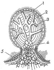
Fig. 202. Sphaeridium of a sea urchin (cross-section):
1 — ciliated epithelium; 2 — calcareous body of the sphaeridium; 3 — canal system; 4 — stalk of the sphaeridium articulated with a test tubercle; 5 — sensory cells
Sea urchins are dioecious animals; Hermaphroditism occurs very rarely. In young individuals, the genital cord surrounds the hindgut; it has a pentagonal shape and, as it grows, gives rise to five gonads located interradially. Genital ducts extend from the glands (sac-like Ovaries or Testes), leading to large interradial plates at the aboral pole and opening through them via genital pores.
Concluding the Overview of the internal anatomy of sea urchins, mention should be made of the so-called axial complex, which is also characteristic of Representatives of the next two classes: Asteroidea and Ophiuroidea. It is a spatial association comprising the axial organ, the stone canal with the madreporite, two separate coelomic regions—the left and right axial sinuses—and the genital sinus (the region of the coelom around the hindgut containing the genital cord). The axial complex will be examined in more detail within the Class Asteroidea.
Sea urchins are quite prolific: the Ovary of a single individual can simultaneously develop 10–60 million eggs, and spawning occurs several times throughout the year. Fertilization is external and typically takes place within the first day after gamete release. Embryonic development culminates in The formation of a flagellated blastula; in some species, the animal pole bears a tuft of longer, motionless flagella. The blastula sheds its membranes and becomes a free-swimming larva. Upon completion of Gastrulation, structural Changes in the larva lead to the Formation of the echinopluteus (Fig. 203)—a bilaterally symmetrical larva characteristic of sea urchins, distinguished by four to six pairs of lateral extensions called arms and containing a rather complex larval skeleton. The echinopluteus leads a pelagic lifestyle, swimming in a spiral while actively feeding on small planktonic organisms.

Fig. 203. Larvae of sea urchins:
a — blastula of Echinocyamus pusillus; b, c — echinoplutei of Clypeaster humilis and Lovenia elongata, respectively
The transition to the radially symmetrical adult is quite rapid (taking up to an hour) and is accompanied by the formation of the juvenile urchin on the side of the larva, seemingly budding off from the echinopluteus, the greater part of which degenerates. During metamorphosis, as arm reduction begins, the animal's body becomes more compact and heavy, settling to the bottom; the newly formed urchin immediately starts crawling.
In some sea urchins possessing large yolk-rich eggs, development is direct.
Despite their robust test and toxic pedicellariae, sea urchins are readily preyed upon by many other animals: sea stars, crustaceans, gastropods, birds, sea otters, and arctic foxes. Observations indicate that certain birds pick up sea urchins and drop them from a height onto rocks to peck out the soft parts.
The roe of certain sea urchin species is highly nutritious and consumed by humans raw, salted, or boiled. It is also canned for long-term preservation.
The Class Echinoidea comprises two subclasses and nine orders.
Last update: 13/08/2026
Editorial and Educational Adaptation: This material has been compiled based on the primary/original source text. The project team performed an editorial review, corrected technical inaccuracies, structured sections, and adapted the content for an educational format.
What was processed:
- elimination of formatting defects (OCR errors, structural breaks, corrupted characters);
- editorial organization of content;
- standardization of terminology in accordance with academic sources;
- verification of factual statements against the original source text.
All mentions of the author, publication year, and origin of the primary text have been preserved in accordance with the source.