INVERTEBRATE ZOOLOGY IN THREE BOOKS - BOOK 3 - H.Y. Shcherbak - 1997
PHYLUM BRYOZOA
CLASS PHYLACTOLAEMATA
Class="center">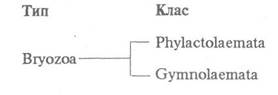
Phylactolaemates are a small group (about 50 species) of exclusively freshwater bryozoans distributed worldwide. They form monomorphic colonies of various structures (Fig. 134) that are generally sessile, with only a few capable of slow movement, such as the very common Cristatella mucedo. An exception is represented by two species of the genus Monobryozoon, which lead a solitary lifestyle.

Fig. 134. Phylactolaemate bryozoans:
а — Fredericella sultana; б — Austrarella indica; в — general view of the Cristatella mucedo colony; г — its cross-section; 1 — polypides; 2 — budding zone; 3 — statoblast
Phylactolaemate colonies are characterized by a high degree of cystid fusion. The partitions, or septa, between them, consisting of two layers of perigoneal epithelium, are often incomplete or entirely absent; if present, they are perforated by numerous pores.
The epidermis of the colony's outer wall, covered with a thin chitinous cuticle, often contains numerous glandular Cells that secrete a gelatinous substance covering the entire colony in a continuous layer, except for the polypides. At the boundary between the anterior, protrusible part of the polypide and the cystid, the epidermis forms a fold that closes the aperture when the polypide retracts. Beneath the epidermis lies a thin layer of circular and longitudinal Muscles lined with peritoneal epithelium. Contractions of the body-wall musculature of the colony's lower wall, together with the gelatinous secretions, facilitate the movement of those colonies that do not attach to the substrate.
At the anterior end of each polypide, There is a horseshoe-shaped body outgrowth—the lophophore, which bears numerous (20—80) tentacles lined on their inner side with ciliated epithelium. Near their base, the tentacles are joined together for about 1/3 of their length by an intertentacular membrane.
The common cavity of the colony, as already mentioned in the Description of the phylum, directly opens into the cavity of each zooid. Near the lophophore, there is a septum that separates the anterior compartment of the coelom from the main coelom; this anterior compartment forms a ring canal and extends into the lophophore and tentacles. There is a pore in the septum through which both coelomic regions communicate with each other.
Among the specialized muscles, the largest is the polypide retractor Muscle, which extends from the lower wall of the colony to the anterior end of the polypide, where it continues into the muscles running along the lower side of the lophophore. Upon its contraction, the polypide, along with the tentacles, is retracted into the cystid; their protrusion occurs under the pressure of the coelomic fluid, which increases due to the contraction of the colony's body walls. The retractor muscle relaxes during this process.
The Digestive System (Fig. 135) begins with the Mouth, which is located at the Base of the lophophore between the tentacles and is covered from above by a small hollow outgrowth—the epistome (hence the name of the class). The mouth leads into an epithelium-lined Pharynx, which passes into the Esophagus, separated from The Stomach by a circular muscle lying in a circular fold of the peritoneal epithelium. The stomach begins with a descending cardiac portion that transitions into a cecum, from which a narrow, so-called Cytology/practical/109.html">Pyloric part of the midgut is directed toward the anterior end of the body. The digestive tract ends with a small ectodermal hindgut opening via the anus. There is a layer of circular muscles in the intestinal walls. Food (microscopic diatoms, radiolarians, rotifers, detritus particles) entering the pharynx rotates due to Ciliary movement; its further movement is driven by peristaltic contractions of the intestinal walls. Digestion takes place in the stomach and is exclusively extracellular.

Fig. 135. Diagram of The Structure of a phylactolaemate zooid (cross-section):
1 — intertentacular membrane; 2 — coelomoduct; 3 — ganglion; 4 — muscles running along the lophophore; 5 — anus; 6 — pyloric part of the stomach; 7, 8, 9 — circular muscles, cecum, and stomach retractor; 10 — funiculus; 11 — forming statoblast; 12 — young bud; 13 — septum; 14 — peritoneal epithelium; 15 — Muscles of the colony wall; 16 — cardiac part of the stomach; 17 — trunk coelom; 18 — esophagus; 19 — pharynx; 20 — Diaphragm; 21 — lophophoral coelom; 22 — mouth; 23 — epistome; 24 — tentacles
Externally, the entire gut is covered by peritoneal epithelium. From the bottom of the stomach cecum, an extension of this epithelium arises in the form of a thin mesentery, or funiculus, which extends backward through the body cavity, connecting with the peritoneal epithelium of the zooid's body wall.
The excretory system of phylactolaemates is represented by two ciliated canals (coelomoducts) located in the lophophoral coelom between the mouth and the anus. Each canal begins in the trunk coelom with a funnel-shaped expansion and then passes into the lophophoral coelom, where both canals merge into a single excretory duct. Through the funnels, amoebocytes filled with excretory products enter the coelomoducts from the coelomic fluid. There is no permanent excretory pore, so the amoebocytes filling the excretory duct are periodically discharged to the outside through a temporary pore. In addition to the coelomoducts, amoebocytes carrying excretory products are eliminated from the body through the tentacular epithelium and the intestinal wall, and then expelled outward through the anus along with undigested food particles.
Circulatory and respiratory systems are absent. Gas exchange occurs through the tentacles and pores that penetrate the upper wall; nutrient transport takes place via the coelomic fluid. The periodic retraction and protrusion of the polypide, along with the peristaltic Movements of the gut, drive the coelomic fluid to all PARTS OF THE zooid and the entire colony.
The central part of the zooid's Nervous system is the cerebral ganglion; from it arise two powerful lophophoral nerve cords, which give off nerves to the tentacles. Two nerves enter each tentacle, along with fine Branches of the radial cords, forming a nerve plexus in each tentacle. A similar plexus is present in the intertentacular membrane (Fig. 136). The cerebral ganglion also gives rise to a cord that forms a ring around the esophagus, nerves innervating the epistome, and cords extending to the posterior end of the polypide, innervating all Internal Organs. A nerve plexus lies within the colony walls, consisting of numerous Nerve Cells interconnected by their processes.
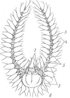
Fig. 136. Diagram of the structure of a part of the gymnolaemate nervous system:
1 — cerebral ganglion; 2 — epistomal nerves; 3 — lophophoral nerve cord; 4 — tentacular nerves; 5 — radial nerve cords; 6 — circumesophageal nerve ring
The formation of Gonads is closely associated with the peritoneal epithelium. In most species, Testes develop within the funiculus, or occasionally in the walls of the septa separating the cavities of individual zooids; Ovaries develop on the dorsal wall of the colony.
Fertilization is internal, and self-fertilization also occurs. The entire development up to larval formation takes place within the maternal Organism inside the so-called ooecia, which are outpocketings of the ventral wall of the cystid.
Cleavage is holoblastic, partially equal, but irregular. After the diploblastic embryo is formed, a specialized ectodermal outgrowth—the Placenta—develops, through which it receives nutrients from the maternal organism. The fully formed larva ruptures the wall of the ooecium and enters the body cavity of the zooid. The pathways of larval release remain unstudied; this may occur through the apertures of degenerating polypides.
Most of the larva is covered with ciliated epithelium, while the non-ciliated portion serves as the primordium of the ectoderm of the future colony wall. Above this primordium, the polypides of the future colony develop inside the larva (Fig. 137). For example, There are two in Plumatella and four in Cristatella. The free-swimming larva has a short lifespan—from a few minutes to two days—after which it attaches to the substrate by the end covered with larval ciliated epithelium. This is followed by the rapid proliferation of the definitive ectoderm, which causes the larval ciliated epithelium to end up inside the larval cavity, where it is subsequently resorbed and phagocytosed. Pores rupture in the upper wall of the larva, through which the polypides evaginate. The entire process of larval metamorphosis into a young colony takes only a few minutes.

Fig. 137. Larva (a) and young colony (b) of Cristatella mucedo:
1 — mesodermal Cell layer; 2 — larval ciliated ectoderm; 3 — internal cavity; 4 — fully formed polypide; 5 — young buds; 6 — ectodermal epithelium with numerous gelatinous inclusions; 7 — remnants of larval epithelium
Subsequent colony growth is associated with asexual reproduction via internal budding. The bud develops from specialized undifferentiated cells adjacent to the inner wall of the colony. The Cells of the outer epidermis overlying the bud do not participate in this process. A fully formed polypide develops from the bud, after which an aperture forms in the colony wall above it, allowing the polypide to communicate with the external environment. Buds form at the margins of the colony, where the surface area of its wall increases concurrently. Meanwhile, the funiculus extending from the stomach of the young polypide attaches near its mouth, and growth of the colony wall begins between them, gradually pushing the funiculus further away from the polypide. The next bud arises on the newly formed colony wall, and the cycle repeats (Fig. 138).

Fig. 138. Asexual reproduction of phylactolaemates:
a — diagram of budding; b, c — spinoblasts of Lophopodella carteri and Cristatella mucedo, respectively; 1—6 — sequence of bud appearance; 7 — float (annulus); 8 — chitinous hooks
A peculiar form of internal budding in phylactolaemate bryozoans is the formation of so-called statoblasts, which possess specialized adaptations for surviving adverse environmental conditions—such as low temperatures or the drying out of Water bodies, during which the parent colony perishes. Statoblasts are formed from specialized undifferentiated cells that initially localize in the outer epidermis and then actively migrate into the funiculus, as well as from the cells of the funiculus itself. A fully formed statoblast contains a group of mesodermal cells surrounded by a bilayered epithelial envelope, which secretes a thin but strong, complexly structured chitinous shell on its outer surface.
Several varieties of statoblasts are recognized. Piptoblasts are oval or bean-shaped. After the disintegration of the colony, they drop onto the substrate, and under favorable conditions, a new colony develops from them. Floatoblasts possess a specialized chitinous cellular ring (annulus) filled with air, which allows them to float passively in the water Column and be dispersed by currents to new locations. Spinoblasts have the most complex structure; they feature chitinous hooks on their outer chitinous shell or float, which they use to cling to moving objects or animals, such as the feathers or legs of birds, thereby dispersing over long distances.
A single zooid develops within each statoblast, emerging after the chitinous shell of the statoblast splits open, which initiates a rapid process of conventional budding that leads to the formation of a colony.
It is interesting to note that in some species, such as Cristatella mucedo, transverse fission of the colony produces two daughter colonies, which then regenerate the missing parts.
Phylactolaemate bryozoans inhabit both running and standing waters; they are found in large rivers, streams, deep lakes (such as Lake Baikal), ponds, ditches, and so on. Their colonies settle on various substrates, including muddy bottoms, and on the underside of leaves and stems of aquatic plants; they encrust stones, submerged objects, and hydraulic structures. Some species, particularly from the genus Plumatella, are quite tolerant to pollution. Freshwater bryozoans play a certain role in water self-purification, but on the other hand, as a major component of biofouling on vessels and various hydraulic structures, they can cause significant damage. In water supply systems, including in Ukraine, bryozoans such as Plumatella fungosa pose a particular threat: after the colonies die, their fragments and statoblasts clog the water distribution network.
Last update: 13/08/2026
Editorial and Educational Adaptation: This material has been compiled based on the primary/original source text. The project team performed an editorial review, corrected technical inaccuracies, structured sections, and adapted the content for an educational format.
What was processed:
- elimination of formatting defects (OCR errors, structural breaks, corrupted characters);
- editorial organization of content;
- standardization of terminology in accordance with academic sources;
- verification of factual statements against the original source text.
All mentions of the author, publication year, and origin of the primary text have been preserved in accordance with the source.