GENERAL MICROBIOLOGY - T.P. Pyrog - 2004
5. STRUCTURE OF THE MICROBIAL CELL
5.3. INTRACELLULAR STRUCTURES
Ribosomes. Ribosomes are the sites of Protein Synthesis. On electron micrographs, they appear as particles scattered throughout the Cytoplasm. Bacterial ribosomes measure approximately 16 x 18 nm. Roughly 80-85% of all bacterial RNA is contained within ribosomes. They consist of protein (35-40%) and RNA (60-65%). Because intact (whole, undamaged) bacterial ribosomes sediment at a rate of about 70 Svedberg units (S) during ultracentrifugation, they are referred to as 70S ribosomes. Eukaryotic cytoplasmic ribosomes are larger, at 80S. Ribosomes similar in size to bacterial ones can also be isolated from eukaryotic Cell/35.html">Mitochondria (for example, from Yeast mitochondria).
A bacterial 70S ribosome consists of two subunits: a 30S and a 50S subunit (Fig. 5.9). A bacterial cell contains anywhere from 5,000 to 50,000 ribosomes. Their number increases with the growth rate of The Cell. During active protein synthesis, regular chains of ribosomes can be observed on electron micrographs. These are ribosomes strung together like beads along Messenger RNA chains, known as polyribosomes or Polysomes.
Class="center">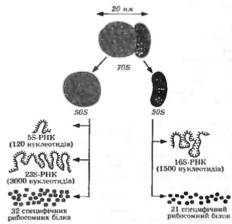
Fig. 59. Schematic diagram of a bacterial ribosome Structure
The difference between bacterial (70S) and eukaryotic (80S) ribosomes is of critical importance for combating infectious diseases. Certain Antibiotics partially or completely inhibit protein synthesis occurring on 70S ribosomes while leaving the Functions of 80S ribosomes unaffected.
Vacuoles. Gas vacuoles (aerosomes) are characteristic of aquatic Bacteria, particularly Purple and green sulfur bacteria, mud-dwelling species, and certain soil bacteria. Gas vacuoles consist of gas vesicles arranged in parallel rows to form a honeycomb structure. Gas vesicles are hollow cylinders enclosed by a single-layered protein membrane. In this protein membrane, hydrophobic Amino Acids face the interior of the cylinder, while hydrophilic ones face outward. This arrangement of amino acids prevents Water from entering the vesicle. The cylinders are filled with a gas whose composition is similar to that of the surrounding environment. Aerosomes act as buoyancy regulators for bacteria. They enable non-motile bacteria to move vertically in bodies of water and soil capillaries, thereby positioning themselves optimally relative to light sources, dissolved oxygen concentrations, and nutrients. In other words, gas vacuoles serve an adaptive function in bacteria.
As for Eukaryotic Cells, they contain a large vacuole enclosed by a single-layered membrane. Its functions have not been definitively established. It has been shown to contain hydrolytic Enzymes, polyphosphates, Lipids, low-molecular-weight cellular intermediates, and Metal Ions. It is likely that the vacuole serves as a storage reservoir for nutrients and hydrolytic enzymes.
Carboxysomes. These structures have been found in the cells of cyanobacteria, certain purple bacteria, and nitrifying bacteria. They appear as four- or six-sided inclusions up to 500 nm in diameter, enclosed by a single-layered protein membrane, and containing enzymes involved in carbon dioxide fixation via The Calvin Cycle.
Magnetosomes. Found in the cells of bacteria exhibiting magnetotaxis. These are membrane-enclosed Fe3O4 particles that vary in shape, number, and distribution within the cell.
Reserve Materials. Under certain environmental conditions, many microorganisms deposit substances that can be considered reserves—Polysaccharides, fats, polyphosphates, and sulfur. These substances accumulate when the growth medium contains specific precursor compounds, yet Microbial growth is restricted or entirely prevented due to a shortage of certain nutritional components or the presence of inhibitors. Reserve materials are held within the cell in an osmotically inert, water-insoluble form. Under growth-favorable conditions, when these reserves are needed, they are re-channeled into METABOLISM. Reserve Polysaccharides, neutral fats, and poly-β-hydroxybutyric acid can serve as sources of carbon and energy. Consequently, in the absence of external Energy Sources, they can prolong cell survival, and in spore-forming species, they can provide the conditions necessary for spore formation even when exogenous substrates are lacking. Polyphosphates can act as a reserve source of phosphorus, and sulfur as a potential electron donor.
In some microorganisms, starch (blue coloration) or Glycogen (brown coloration) can be identified using a color reaction with Lugol's iodine solution. Unlike Cell wall polysaccharides, reserve polysaccharides are formed from α-glucose, with glucose molecules linked by 1,4-α-bonds. Due to these α-bonds, polyglucose chains are not elongated but instead form a helical twist. Granulose, a starch-like compound, is a specific reserve substance found in bacteria of the genus Clostridium. Glycogen (animal starch) resembles amylopectin, but its chains are even more branched (due to The formation of 1,6-bonds). Glycogen is more common in bacteria than starch and has also been found in Yeasts and Fungi.
Granules and droplets of fat are frequently found in microbial cells. They can be stained with lipophilic Dyes—Sudan III and Sudan Black—making them visible under a Microscope. Unstained, they can be observed microscopically due to their strong light-refracting properties.
The granules of many bacteria are composed of poly-β-hydroxybutyric acid. This is a polyester containing about 60 residues of β-hydroxybutyrate. The proportion of this substance in the dry biomass can reach up to 80%. Poly-β-hydroxybutyric acid is synthesized by aerobic bacteria, as well as cyanobacteria and anaerobic phototrophic bacteria. The formation of poly-β-hydroxybutyric acid is observed in aerobes under conditions of oxygen limitation.
Neutral fats (triglycerides) are deposited in particularly large amounts within the vacuoles of yeasts and fungi. Waxes (esters of Fatty acids and long-chain alcohols) have been found in mycobacteria (which may contain up to 40% waxes), Nocardia, and actinomycetes. The content of reserve fats is determined by the COMPOSITION OF THE nutrient medium (a high C/N ratio), and these fats can be extracted directly from the cells.
Fungi, yeasts, many bacteria, and green Algae are capable of accumulating phosphoric acid in the form of polyphosphate granules. Such granules were first described in the bacterium Spirillum volutans, which is why they are frequently called volutin granules (volutin).
In many bacteria that oxidize sulfide to sulfate, sulfur is temporarily stored as highly light-refracting globules. The amount of sulfur that can accumulate depends on the hydrogen sulfide content in the environment: in the absence of hydrogen sulfide, sulfur is oxidized to sulfate. For aerobic sulfur bacteria, sulfur serves as an energy source, whereas for anaerobic phototrophic purple bacteria, it acts as an electron donor.
Nucleoid. The question of whether bacteria possess a Nucleus was a matter of debate for many decades. The small size of the cells and the limitations of research techniques made it difficult to detect nuclear structures in bacteria, although there was no doubt about the presence of a hereditary apparatus, since reproducing cells of one bacterial species yielded offspring of the same species. Modern electron-microscopic and genetic studies have established that bacteria possess structures analogous to the nuclei of eukaryotic cells, yet differing in several key aspects:
1) bacterial nuclei lack a nuclear envelope (membrane), meaning the DNA is in direct contact with the cytoplasm;
2) there is no division into Chromosomes; instead, a single DNA thread acts as an analog of the eukaryotic chromosome and is referred to as the bacterial chromosome (multiple copies of it may be present in the cell);
3) Meiosis and mitosis are absent.
Consequently, the nuclear apparatus of bacteria is called the bacterial nucleus, or nucleoid. It has been established that the bacterial chromosome takes the form of a closed ring. This is a giant DNA molecule with a Molecular Weight of 109 daltons. Its length varies among different bacteria: from 0.25 mm in Mycoplasmas to up to 3 mm in cyanobacteria. Prokaryotic Cells may contain multiple nucleoids and multiple copies of the chromosome (e.g., from 2 to 9 chromosomes distributed across several nucleoids in Bacillus subtilis, and about 40 chromosomes in a single nucleoid in Azotobacter vinelandii).
Bacterial chromosomes are highly ordered structures of two types: membrane-associated and free, with sedimentation coefficients of 3200-7000S and 1600-2000S, respectively. The DNA within these structures exists in a supercoiled state, forming 20-140 loops connected to a dense central region composed of RNA, which is responsible for maintaining their compact shape (Fig. 5.10). The bacterial chromosome is always tethered to the membrane (the number of attachment sites can reach 20 or more) either directly via specific Membrane Proteins or through membrane-bound ribosomes.

Fig. 5.10. Model of nucleoid Organization (a) and structural compaction of the Escherichia coli chromosome (b):
1 — chromosome loop; 2 — RNA connecting the loops; ori — origin of Replication on the bacterial chromosome; r — ribosomes; om — outer membrane; m — murein; cm — cytoplasmic membrane
Unlike eukaryotes, prokaryotes do not exhibit significant Changes in the state of the nucleoid during the Cell Cycle, except for its Condensation prior to spore formation. In bacterial cells, DNA Synthesis occurs continuously, and DNA accounts for approximately 1–3% of the cell's dry weight.
Last update: 13/08/2026
Editorial and Educational Adaptation: This material has been compiled based on the primary/original source text. The project team performed an editorial review, corrected technical inaccuracies, structured sections, and adapted the content for an educational format.
What was processed:
- elimination of formatting defects (OCR errors, structural breaks, corrupted characters);
- editorial organization of content;
- standardization of terminology in accordance with academic sources;
- verification of factual statements against the original source text.
All mentions of the author, publication year, and origin of the primary text have been preserved in accordance with the source.