ECOLOGICAL MICROBIOLOGY - M. I. Chernyavskaya - 2016
TOPIC 3. AUTOECOLOGY. THE INFLUENCE OF CHEMICAL ENVIRONMENTAL FACTORS ON MICROBIAL ACTIVITY
Osmotic pressure largely depends on Water activity (aw), i.e., The amount of dissolved substances it contains. When the salt concentration in the medium is lower than inside The Cell (hypotonic solutions), water tends to enter the cell along the concentration gradient of dissolved solutes, causing osmotic pressure to rise, which may lead to lysis. Thanks to The Cell wall, this condition poses no danger to most Bacteria. Conversely, a high salt concentration in the medium (hypertonic solutions) causes cellular dehydration (plasmolysis) and growth inhibition. The degree of growth inhibition depends on the type of medium and The properties of the microorganism.
According to their Salt Tolerance, microorganisms are classified into the following groups:
✵ freshwater (non-halophilic) microorganisms, including inhabitants of ultra-fresh waters — they develop in media with a salt content of less than 0.01% and are typically sensitive to a 3.0% NaCl concentration;
✵ marine microorganisms — generally grow within a narrow range of salt concentrations (2.5 – 5.0% NaCl); the optimal salinity is about 3.5%. Typical marine bacteria include Photobacterium, Vibrio, and Alteromonas;
✵ halotolerant microorganisms — usually withstand higher salt concentrations (up to 13.0% NaCl) and frequently inhabit environments with fluctuating salinity, such as soil;
✵ mildly halophilic microorganisms — grow at 1.5 – 5.0% NaCl;
✵ moderate halophiles — grow within a salinity range of approximately 5.0 – 15.0% NaCl;
✵ extreme halophiles — develop at NaCl concentrations ranging from 12.0 – 15.0% up to saturated salt solutions.
A distinct group is formed by haloalkaliphiles, which grow at high soda concentrations and combine the properties of both halo- and alkaliphiles. Typical habitats of these microorganisms are highly mineralized soda lakes.
The primary mechanism of microbial adaptation to high environmental osmotic pressure is the synthesis of osmoprotectants (osmolytes) — low-molecular-weight Organic compounds that are neutral toward cellular metabolites and whose concentration in the Cytoplasm balances the external osmotic pressure. Osmoprotectants include Amino Acids and their derivatives (Glycine betaine, Proline, glutamate), sugars (sucrose, trehalose), alcohols (glycerol, mannitol), and heteroglycosides. The composition of osmolytes depends on the NaCl concentration in the medium and varies among different microorganisms, sometimes accumulating in significant amounts. For example, Dunaliella viridis accumulates up to 30% glycerol of its total cell mass. Osmolytes retain water exceptionally well and are therefore used in the cosmetic industry as ingredients in moisturizing creams (e.g., ectoine from halophilic anoxygenic phototrophs). Adaptation to elevated salinity in extremely halophilic archaea (order Halobacteriales) is based on the accumulation of K+ ions. The intracellular potassium ion concentration can be up to 1,000 times higher than that of the surrounding environment, meaning that halobacterial Enzymes function in a saline solution. A similar osmoadaptation strategy has been discovered in certain eubacteria — Salinibacter ruber and members of the order Haloanaerobiales. The Proteins of halophiles are more "acidic" (rich in aspartate and glutamate), forming hydrophobic interactions that lead to a more compact globule packing. A "protein shield" mechanism (S-layers) operates on the cell surface, where the COOH groups of Amino acids are exposed outward to retain Na+ ions. These same groups form a "hydrated" cell shell through the electrostatic orientation of water dipoles. Halophiles actively transport ions out of the cell, thereby maintaining a specific "osmostasis".
For terrestrial organisms, adaptation to dryness and exposure to air is of great importance. Water stress and the threat of desiccation occur on the surfaces of cliffs, rocks, trees, various structures, and in soils, particularly desert soils. The primary defense mechanism against drying out is The formation of mucous capsules and survival structures (spores, conidia, cysts). High resistance to desiccation is exhibited by certain mycobacteria with a high lipid content in their cell walls.
Environmental acidity is a crucial factor determining the survival of prokaryotes. Hydrogen ion concentration has both a direct effect on the cell—affecting its electrical charge, membrane state, and the occurrence of redox reactions—and an indirect effect by determining the ionic state of metals and acids, as well as their availability and toxicity.
The pH values of various natural waters and solutions supporting Microbial growth span almost the entire theoretically possible pH range — from 1 – 2 in acidic springs to 10 in soda lakes. Based on their pH response, microorganisms are divided into several physiological groups:
✵ neutrophils — microorganisms for which the optimal pH is close to neutral (6.0 – 8.0), while growth is possible within a pH range of 4.0 – 9.0 (e.g., Escherichia coli, Bacillus megaterium, Enterococcus faecalis, etc.). Most natural habitats (freshwater lakes and rivers, many soils, and the internal environment of PLANT AND ANIMAL organisms) have a nearly neutral reaction; seawater is slightly alkaline;
✵ acidophiles — microorganisms whose pH optimum is shifted toward acidic values (typically below 4.0), with growth possible in the pH range of 0 – 5.5. Acidophilic microorganisms are subdivided into obligate and facultative forms capable of growing at neutral pH. Examples of acidophiles include lactic acid and acetic acid bacteria;
✵ alkaliphiles — microorganisms that grow within a pH range of 8.5 – 11.5, which are also divided into facultative (capable of growing in neutral media) and obligate groups. Examples of alkaliphiles include urea-degrading bacteria and cyanobacteria.
Despite extreme environmental pH levels, the intracellular pH remains relatively stable and is maintained at approximately ~7.5 in both alkalo- and acidophilic microorganisms. The constancy of intracellular pH (pH Homeostasis) is facilitated by the low permeability of the cytoplasmic membrane to H+ protons. However, because protons slowly diffuse down their concentration gradient, the cell employs energy-dependent export mechanisms. Under slight acidification, bacteria utilize Na+ and K+ proton antiporters. When experiencing a sharp drop in pH, the synthesis of specialized chaperones ("acid Shock" proteins) is triggered, which prevent acid Denaturation of cytoplasmic proteins and restore the conformation of denatured proteins.
During their metabolic activity, microorganisms can alter environmental pH by producing acidic or alkaline products. For instance,
E. coli responds to increased environmental acidity by synthesizing amino acid Decarboxylases. The resulting amines alkalize the medium. Conversely, an increase in environmental pH stimulates the synthesis of amino acid deaminases, leading to medium acidification. A striking example of bacterial pH regulation is the two-phase acetone-butanol Fermentation process in Clostridium acetobutylicum. The first phase is the acid phase, during which butyric and acetic acids are produced via glucose fermentation. The second phase is the acetone-butanol phase, where as the medium becomes increasingly acidic (pH below 5.0), the synthesis of enzymes leading to the production and accumulation of neutral products—primarily n-butanol, acetone, and ethanol—is induced.
Microorganisms harness energy from redox reactions, making the oxidation-reduction potential of the environment (Eh) one of the most important factors influencing their growth. The Eh value characterizes the reduction state of the medium and determines the thermodynamic feasibility of redox reactions. Microbial activity leads to Changes in the Redox Potential of the environment. The primary reductants in nature are H2 and H2S, frequently produced by bacteria, while molecular oxygen serves as the main oxidant. The redox potential, which ranges from -500 to +800 mV, can indicate the presence of oxygen in the medium: the more negative the value, the more reduced the environment (anaerobic conditions).
Molecular oxygen acts on microorganisms not only as a factor determining environmental redox conditions and the feasibility of Chemical Reactions, but also as a crucial catabolic substrate and electron acceptor for aerobic microorganisms. Oxygen concentrations in natural habitats vary widely. Atmospheric air contains 21% oxygen. The solubility of O2 in water is low (about 8 mg/L at 20 °C) and decreases significantly with rising Temperature and salinity. In the presence of readily oxidizable substrates, primarily organic compounds, oxygen is rapidly consumed by aerobes and, under conditions of limited atmospheric exchange, becomes a limiting factor. In many natural habitats (sludge, cyanobacterial mats), sharp O2 gradients develop over fractions of a millimeter, culminating in its complete depletion. A similar drop in oxygen concentration is observed in soil, where aerobic and anaerobic microzones coexist within a single soil particle.
Based on their oxygen requirements, microorganisms are divided into two groups: aerobes, which grow only in the presence of oxygen, and anaerobes, which can grow without it (Table 4). Both aerobic and anaerobic forms are found among bacteria and Protozoa. Fungi are aerobes, although certain species, typically Yeast-like anamorphs, are facultative anaerobes. There is also a small number of obligately anaerobic species.
Class="center">Table 4. Groups of microorganisms according to their relationship with molecular oxygen
Group of microorganisms |
Characteristics |
Example |
Obligate aerobes |
Require molecular oxygen; type of METABOLISM is aerobic Respiration |
Micrococcus luteus |
Facultative anaerobes |
Do not require molecular oxygen but grow better in its presence; type of metabolism is aerobic or Anaerobic respiration, fermentation |
Escherichia coli |
Microaerophiles |
Require molecular oxygen, but at concentrations lower than atmospheric; type of metabolism is aerobic respiration |
Spirillum volutans, Beggiatoa spp. |
Aerotolerant anaerobes |
Can grow in the presence of small amounts of oxygen; type of metabolism is fermentation |
Streptococcos pyogenes, Lactococcus lactis |
Obligate anaerobes |
Molecular oxygen inhibits their growth or leads to death; type of metabolism is fermentation or anaerobic respiration |
Heliobacteria, methanogenic bacteria |
The Toxic Effect of O2 on microorganisms involves the inactivation of oxygen-sensitive proteins (e.g., Nitrogenase) as well as the generation of highly reactive oxygen species. The formation of reactive oxygen species occurs with the participation of several enzymes:
1) О2 + 4e- —> 2O2- (oxide anion) — cytochrome oxidase, laccase, tyrosinase;
2) О2 + 2e- —> О2-2 (Н2О2) — flavin enzymes (glucose oxidase, amino acid oxidase, xanthine oxidase);
3) О2 + 1e- —> О2- (superoxide radical);
4) О2- + Н2О2 + Н+ —> Н2О + О2 + ОН (hydroxyl radical) — aldehyde oxidase, NADH oxidase.
Hydrogen peroxide and the superoxide radical, which are formed most frequently, are removed by specialized enzymes.
Catalase decomposes peroxide via the reaction 2Н2О2 —> 2Н2О + О2, and peroxidase via the reaction RH2 + Н2О2 —> R + 2Н2О, where R is the oxidized substrate of the peroxidase reaction.
The superoxide radical is removed by superoxide dismutase (SOD):
2О2- + 2Н+ —> Н2О2 + О2.
Singlet oxygen may form in the presence of light:
Р + hv —> Р*, Р* + О2 —> О2, where P is a sensitizing pigment.
Carotenoids perform the function of "quenching" singlet oxygen within the cell.
Aerobes and facultative anaerobes possess SOD, catalase, and peroxidase in their Cells, whereas obligate anaerobes lack these enzymes. However, catalase and SOD have been found in certain methanogens and Clostridia, playing a crucial role in their survival under fluctuating anaerobic conditions. Hydrogen-using sulfate reducers are capable of actively combating oxygen by carrying out sulfate respiration. It should be noted that aerobes also contain oxygen-sensitive enzymes (such as nitrogenase). In such cases, cells employ specific defense mechanisms (e.g., ROOT nodules in Rhizobium, capsules in Azotobacter, heterocysts lacking Photosystem II in cyanobacteria). Despite the toxicity of oxygen to obligate anaerobes, they can persist in permanently aerobic habitats by living in communities with aerobic and facultative anaerobic microorganisms that consume O2. Examples of such communities include cyanobacterial mats, where the upper layer is aerobic and oxygenic, and the lower layer is anaerobic; and the oral microbiota, where the obligately anaerobic bacterium Porphyromonas gingivalis inhabits dental calculus deposits.
1. What effects do hypoosmotic and hyperosmotic environmental conditions have on microorganisms?
2. Into what physiological groups are microorganisms divided based on their attitude to environmental salinity?
3. Name the Mechanisms of microbial adaptation to life under conditions of elevated environmental osmotic pressure.
4. Into what groups are microorganisms divided based on their attitude to environmental acidity? Give examples of representatives for each group.
5. Name the main mechanisms for maintaining intracellular pH homeostasis in a bacterial cell.
6. What groups of microorganisms are distinguished in relation to oxygen?
7. What causes the toxic effect of oxygen on microbial cells?
8. List the mechanisms protecting microorganisms against highly reactive oxygen species.
Laboratory Work 5. Effect of Osmotic Pressure and Medium Acidity on Microbial Growth
Objective: to study the growth of microorganisms from various taxonomic groups on nutrient media with differing acidity and salinity (osmotic pressure).
Materials and equipment: PDB with various pH values (3.0, 5.0, 7.0, 8.0, 9.0, 10.0), PDA supplemented with 0.5, 5, 10, 15% NaСI, automatic micropipette (2 — 20 µl), sterile serological pipettes (1 — 2 ml), pipette tips (10 — 200 µl), microbiological loop, spectrophotometer or photoelectrocolorimeter, spirit lamp, incubator.
1. Effect of osmotic pressure on microbial growth.
1) Streak the test bacterial cultures onto Petri dishes containing PDA supplemented with 0.5, 5, 10, and 15% NaСl.
2) Incubate the dishes at 28 — 37 °С for 48 — 72 h.
3) Evaluate the results visually by comparing bacterial growth on plates with different NaСl concentrations. Record the findings in the table (Table 5).
4) Draw a Conclusion regarding the salinity tolerance of the studied microbial strains.
Table 5. Growth of the studied microorganisms at various NaCl concentrations
Microorganism |
NaСl concentration, % |
|||
0,5 |
5 |
10 |
15 |
|
Note: "+++" — abundant growth, "++" — moderate growth, "+" — weak growth, "—" — no growth.
2. Microbial growth at various medium pH values.
1) The day before the laboratory session, inoculate the test bacterial cultures into PDB and incubate at the optimal temperature for 24 — 48 h.
2) Add 10 µl of each grown culture to six test tubes containing 2 ml of PDB with different pH values (3.0, 5.0, 7.0, 8.0, 9.0, 10.0). Ensure that the initial cell concentration is identical across all tubes.
3) Incubate the tubes containing the test cultures at the optimal temperature for 24 — 48 h.
4) Quantify the results using a spectrophotometer or photoelectrocolorimeter. Compare the optical density of the culture with that of the PDB at the corresponding pH value (blank control). Measure the absorbance at a wavelength of 600 nm (OD600).
5) Plot a graph based on the obtained measurements (Fig. 5).
Fig. 5. Biomass accumulation of bacterial cultures as a function of nutrient medium pH (after 24 h of cultivation)
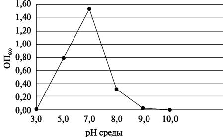
6) Determine the optimal pH value for the growth of the studied microorganisms.
Last update: 12/08/2026
Editorial and Educational Adaptation: This material has been compiled based on the primary/original source text. The project team performed an editorial review, corrected technical inaccuracies, structured sections, and adapted the content for an educational format.
What was processed:
- elimination of formatting defects (OCR errors, structural breaks, corrupted characters);
- editorial organization of content;
- standardization of terminology in accordance with academic sources;
- verification of factual statements against the original source text.
All mentions of the author, publication year, and origin of the primary text have been preserved in accordance with the source.