Diagnosis and treatment of patients with recurrent gastroduodenal hemorrhage - Shaprynskyi V.O. 2009
Morphological features of the gastric and duodenal mucosa in patients with peptic ulcer disease complicated by acute recurrent gastrointestinal hemorrhage
Histopathological studies have provided insights into the pathological processes occurring in the ulcer base that directly lead to bleeding. Tissues from gastric and duodenal ulcer craters were examined, establishing that acute bleeding occurs against the Background of exacerbated inflammation of the surrounding mucosa and intensified necrotic processes within the ulcer crater affecting the walls of adjacent vessels. However, certain morphological features of bleeding gastroduodenal ulcers complicated by acute recurrent bleeding remain insufficiently studied. This raises the question of a potential morphological substrate specific to ulcers complicated by recurrent GIB, which our study aimed to investigate.
Microscopic examination of gastric ulcer margins revealed the following Changes in the superficial epithelium: flattening, nuclear pyknosis, disorganized nuclear arrangement, fragmentation, cytoplasmic vacuolation, and, in some areas of the foveolar epithelium, mucoid occupying almost the entire Cytoplasm and displacing the nuclei to the basal region, subepithelial edema, leukodiapedesis, and Cell desquamation. Pronounced signs of regenerative processes, such as increased mitotic activity, hyperchromatic nuclei, cytoplasmic basophilia, and displacement
of nuclei in the apical direction, and replacement of the destructively altered epithelium with new cellular elements were observed in only 2% of patients.
Approaching the ulcer defect, degenerative changes in the epithelium intensified, presenting as distinct zones in the ulcer area (Fig. 3.1): Exudation zone. The superficial and foveolar epithelium is necrotic and represented by a homogeneous, structureless mass mixed with polymorphonuclear leukocytes and a small number of lymphoid elements.
Fibrinoid necrosis zone, which encompassed most of the lamina propria of the mucosa. At its periphery, significant polymorphonuclear infiltration was noted, with a predominance of leukocytic elements in gastric ulcers and lymphoid Cells in duodenal ulcers. The contours of individual glands were preserved in places, but The Cell boundaries were blurred. The cytoplasm of glandular cells was homogeneous and basophilic. Most of the nuclei were hyperchromatic, and in some areas pyknotic and fragmented.
The fibrinoid necrosis zone could be divided into two layers: superficial and compact. The Superficial layer of the fibrinoid necrosis zone was located between the weakly expressed exudate zone and the compact zone. It had a loose, clumpy Structure and was focally infiltrated by macrophages and leukocytes. Fragmented Collagen fibers could occasionally be seen here. The clumps composing this layer showed a weakly positive PAS reaction (Fig. 3.2) and did not stain with Alcian blue.
The structural characteristics of these ulcer zones varied across different sections. The superficial necrotic layer was mostly of significant thickness, though in some areas, particularly in duodenal ulcers, it was weakly expressed. In certain regions, it appeared as fresh coagulative oxyphilic necrosis with 'shadows' of dead cells and fibers, without leukocytic infiltration. The compact layer of fibrinoid necrosis was well-developed, often presenting with numerous hemorrhages (Fig. 3.3) and destroyed capillary walls.
Class="center">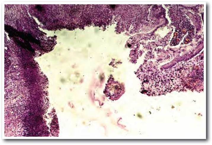
Fig. 3.1. Chronic Gastric Ulcer complicated by ARGIB. Undermined margin of the ulcer defect. Fibrinoid necrosis zone of the gastric ulcer base. Hematoxylin-eosin staining x 100.
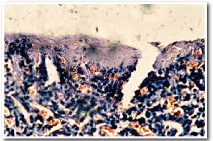
Fig. 3.2. Chronic gastric ulcer complicated by ARGIB: PAS-positive reaction of the superficial layer of the fibrinoid necrosis zone of the gastric ulcer base. PAS stain x 400.
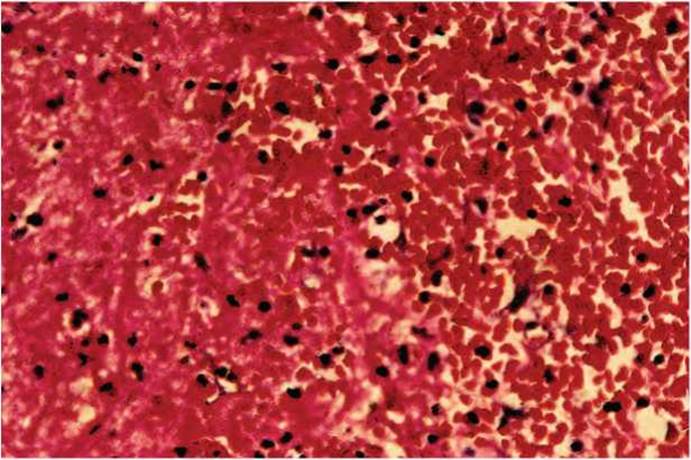
Fig. 3.3. Chronic gastric ulcer complicated by ARGIB: numerous hemorrhages in the fibrinoid necrosis zone of the gastric ulcer base. Hematoxylin-eosin staining x 400.
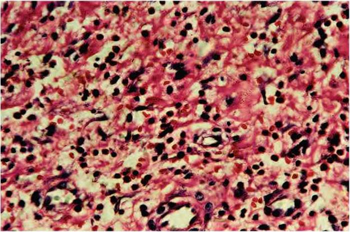
Fig. 3.4. Chronic gastric ulcer complicated by ARGIB: predominantly leukocytic infiltration of the granulation tissue of the gastric ulcer base. Hematoxylin-eosin staining x 400.
Horizontally oriented capillaries were filled with erythrocytes, often exhibiting sludge phenomenon and stasis. The endothelium of some vessels showed dystrophic changes and desquamation, while in others, angioblast proliferation predominated. Approaching the scar zone, a decrease in the number of leukocytes and macrophages and an increase in fibroblasts were observed. Lymphoid infiltration remained at the same level in some areas (Fig. 3.4).
A characteristic feature of duodenal ulcers in 60% of cases was the absence or poor development of granulation tissue.
The next zone was characterized by the proliferation of Connective Tissue of varying degrees of maturity. In some areas, it extended beyond the ulcer defect and penetrated into the submucosal and subserosal layers, as well as between the fibers of the muscularis propria, causing sclerosis and fiber fragmentation. Compared to The structure of ulcers without recurrence, an unstable scar zone predominated here, containing destroyed fibroblasts and loose, destructively altered collagen fibers (Fig. 3.5).
Almost throughout the entire section, the Base of the ulcer defect was located at the level of the submucosal layer. In some areas, destruction of the gastric muscularis propria was noted, with its replacement by fine-fibrillar connective tissue. In these regions, the proliferation of a significant number of large fibroblasts with highly basophilic cytoplasm could occasionally be observed.
In the lamina propria of the mucosa, the most common pathological manifestation was infiltration, which varied in character. Thus, in cases of gastric ulcer disease, leukocytic, lymphocytic, and plasmacytic infiltration was observed, whereas in duodenal ulcers, lymphocytic infiltration predominated, located mainly in the superficial PARTS OF THE mucosa. In gastric ulcers, the infiltration extended to the serous layer, in some places dissecting the Muscle fibers (Fig. 3.6).
The basement membrane of the lamina propria of the gastric and duodenal mucosa was loose in places. The submucosa was edematous, showing extensive areas of Hemorrhage, with the walls of small Arteries exhibiting plasmatic infiltration, and some showing signs of fibrinoid necrosis.
The muscularis mucosae itself was hypertrophied; the thickened muscle bundles varied in appearance and orientation. They penetrated the mucosal stroma, separating glandular complexes from one another, with individual fibers reaching the gastric pits and positioning themselves between them. The secretory glands appeared as if embedded in the muscular layer.
In the scar zone, the muscular layer was divided into separate bundles by thick layers of coarse-fibrillar connective tissue, which in some places completely replaced muscle fibers and were clearly visible with van Gieson's picrofuchsin staining (Fig. 3.7).
Collagen fibers presented as loose, net-like structures with parallel and intertwined bundles, tangles, and large tendon-like formations. Compactly arranged thickened collagen fibers in the scar zone were eosinophilic, orthochromatic when stained with toluidine blue, moderately PAS-positive, and hyalinized in places. Among the scar tissue, small-caliber Blood Vessels were identified, sometimes surrounded by circularly arranged collagen fibers forming a massive sleeve. The vessels were highly tortuous and appeared as closely spaced transverse, tangential, and longitudinal sections relative to the gastric surface.

Fig. 3.5. Chronic duodenal ulcer complicated by acute gastrointestinal bleeding: mild macrophage infiltration and randomly arranged fibroblasts of the granulation tissue at the base of the duodenal ulcer. Hematoxylin and eosin stain, ×400.
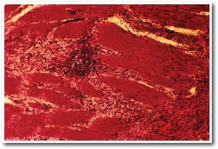
Fig. 3.6. Chronic gastric ulcer complicated by acute gastrointestinal bleeding: lymphoid aggregates disrupting and dissecting the fibers of the muscular layer in gastric ulcer disease. Hematoxylin and eosin stain, ×100.
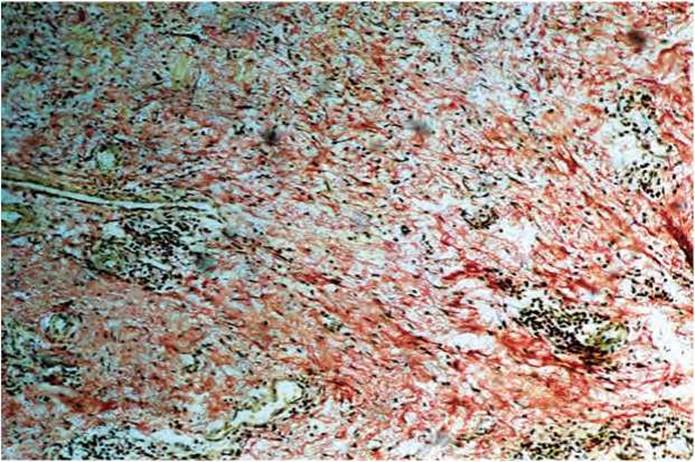
Fig. 3.7. Chronic duodenal ulcer complicated by acute gastrointestinal bleeding: replacement of the muscular layer with fibrous tissue. Van Gieson's stain, ×100.
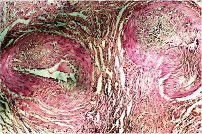
Fig. 3.8. Chronic duodenal ulcer complicated by acute gastrointestinal bleeding: narrowing of the arterial lumen in the cicatricial zone of the duodenal ulcer base by more than 2/3 due to intimal hyperplasia. Hematoxylin and eosin stain, ×100.
Elastic fibers in these areas of the muscular layer were unevenly distributed. In some places they were completely absent, while in others they formed dense plexuses with hyperchromia and uneven thickening (hyperelastosis). In the areas adjacent to the ulcer base, fragmentation of elastic fibers was observed.
One of the most Characteristic Features of the vascular bed of The Stomach and duodenum in PEPTIC ULCER DISEASE complicated by acute gastrointestinal bleeding was the presence of sclerotically altered arteries.
This morphological pattern resulted from a significant change in the angioarchitecture of the periulcerous zone: hemodynamic structural remodeling of vessels of various calibers and Functions (vessels of distribution, resistance, adaptation, and exchange) was observed. Musculoelastic and muscular arteries lost their circular lumen, elastic membranes formed deep folds, and musculoelastic elements of the tunica media underwent reorientation and tended to be randomly arranged. With distance from the ulcer defect, the vascular lumen narrowed, and proliferation of the subendothelial layer was noted. Intimal proliferation sometimes completely exceeded the thickness of the arterial muscular coat (Fig. 3.8) and was accompanied by the spread of fibrous elements in the muscular coat, adventitia, and intima of the vessels (Fig. 3.9).
A large amount of acid glycosaminoglycans is formed in the ground substance, and myoelastofibrosis occurs with The formation of a new lumen at the height of the folds. Neoangiogenesis was also observed: the vessels had swollen endothelium, and The Development of vasa vasorum was enhanced, which penetrated the entire thickness of the vessel wall, often extending beyond it as sinusoids in the perivascular areas. The adventitia and external elastic membrane hyperplased and coarsened. As the ulcer defect was approached, especially in the zone of fibrinoid necrosis, arterioles and arteries were frequently encountered, the walls of which were entirely involved in the zone of fibrinoid necrosis, resulting in their complete destruction. In such vessels, destructive changes of the internal elastic membrane—its duplication, fragmentation, and lysis—were highly pronounced, indicating the early onset of changes specifically in these regions.
Sclerotically degenerated walls of large arteries with complete lumen obliteration were also quite frequently detected (Fig. 3.10). Both circular and focal sclerosis were observed in the arteries, sometimes with pronounced perivascular edema. Fairly large arteries with collapsed walls (Fig. 3.11) and narrowed lumens were also encountered.
Changes in the Veins located in the areas adjacent to the ulcer largely resembled the changes in the veins within the ulcer itself, although they were less pronounced. The vein wall was thickened due to focal or diffuse proliferation of connective tissue. In these veins, elastic fibers broke down into separate fragments scattered throughout all layers of the wall, with only small particles remaining in some places. Another portion of the veins had very thin walls, with fragmented elastic fibers, or their complete absence in other segments. Veins of varying thickness across all segments were also found, due to both sclerotic thickening and plasmatic infiltration of the walls. In small groups of veins, the irregular shape was caused by spastic contraction of the walls. However, in most cases (80%), veins were in an atonic state. The walls of these veins were thickened, loosened, locally deformed, with evidence of erythrocyte sludge in the lumen. Flask-like dilations of the veins were also observed in the venous bed. The endothelium of most of these veins was closely adherent to the basement membrane, occasionally focally absent, and erythrocyte sludging and adhesion to the walls were observed at the sites of endothelial defects.
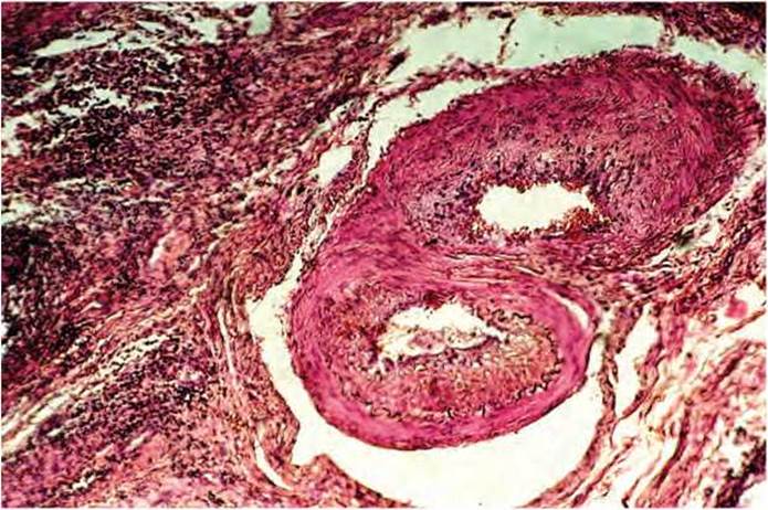
Fig. 3.9. Chronic gastric ulcer complicated by acute gastrointestinal bleeding: narrowing of the arterial lumen in the cicatricial zone of the gastric ulcer base by more than 2/3 due to intimal hyperplasia and proliferation of fibrous elements in the wall. Hematoxylin and eosin stain, ×100.
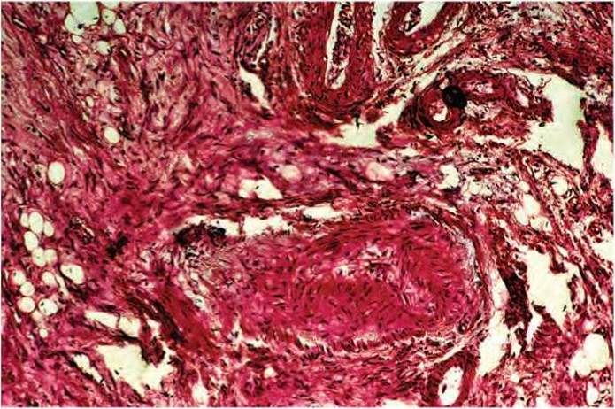
Fig. 3.10. Chronic gastric ulcer complicated by acute gastrointestinal bleeding: sclerotically altered arterial wall in the cicatricial zone of the gastric ulcer base with lumen obliteration. Hematoxylin and eosin stain, ×100.
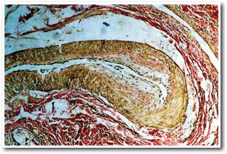
Fig. 3.11. Chronic duodenal ulcer complicated by acute gastrointestinal bleeding: collapse of the arterial wall. Van Gieson's stain, ×100.

Fig. 3.12. Chronic gastric ulcer complicated by acute gastrointestinal bleeding: dilated VEINS OF THE gastric muscular layer with fibrin thrombi in the lumens. Hematoxylin and eosin stain, ×100.
In the walls of small arteries, morphological changes predominated in the form of thinning, flattening, straightening, and destruction of the internal elastic membrane against the background of severe fibrosis of the intima and media, which was accompanied by impaired endothelial adhesive properties and the formation of small mural thrombi and platelet aggregates. Arteriolohyalinosis was detected significantly more often in these same areas. Changes in predominantly small vessels were more diffuse in nature. They manifested as congestive hyperemia with stasis, perivascular edema, loosening and homogeneous Swelling of the vascular wall, and paretic dilation of the lumens.
Notably, vascular dilation with impaired blood filling and rheological properties of Blood Cells was not limited to subepithelial vascular plexuses but was also found in the muscular and serous plexuses. Fibrin thrombi that were just beginning to form were found in the lumens of individual veins (Fig. 3.12).
Thus, it can be concluded that destructive-necrotic processes occur in the zone of the bleeding ulcer, which also extend to the vessel wall. Morphological signs of chronic tissue Hypoxia in the form of sclerosis, obliteration of microcirculatory bed areas, and signs of acute local Circulatory Disorders such as sludge, thrombosis, edema, hemorrhages, and especially areas of tissue necrosis, indicate a link between hypoxia and the occurrence of such a severe complication as bleeding.
The mucous membrane at the margins of the duodenal ulcer has the appearance of villous structures and is covered with flattened columnar epithelium with focal infiltration, predominantly by lymphocytes and plasma cells (Fig. 3.13).
Along with focal infiltration of the lamina propria, there was significant thinning of the layer, a decrease in the height of the duodenal crypts, dystrophic changes in the epithelium, and hyperplasia of Brunner's glands.
In individual villi, severe congestion, intensive infiltration with plasma and lymphoid cells, vacuolation and desquamation of the surface epithelium (sometimes complete, exposing the villous stroma) were observed. Circulatory disorders occurred mainly in the form of congestive hyperemia and hemorrhages, primarily of the diapedetic type.
Tissue hypoxia caused by circulatory disorders may be accompanied by the development of dystrophic, necrobiotic, and necrotic changes in the intestinal crypts. Some epithelial cells swell and vacuolate, the nuclei of some of them undergo lytic changes, and the continuity of the brush border of the epithelial layer is disrupted. Desquamation of the affected epithelium is accompanied by exposure of the stroma with secondary development of stasis and thrombosis in superficially located blood vessels.
Cystic changes in the crypts are often observed. The cysts are usually filled with mucus, sometimes mixed with desquamated epithelial cells and leukocytes. The crypt epithelium shows atrophic changes.
It should be emphasized that in 80% of patients with duodenal ulcers, mucosal atrophy was accompanied by hyperplasia of Brunner's glands. In some areas, the loss of demarcation between the glands and their fusion was detected. The displacement of A large number of Brunner's glands from the submucosal layer into the lamina propria of the mucosa was accompanied by remodeling of the Glandular Epithelium, similar to the epithelium of the colonic crypts. Hemorrhages into the lamina propria of the mucosa, its edema, and erythrocyte stasis in the capillaries were also observed.
The base of the ulcer defect, almost throughout its entire extent, was represented by a thin layer of fibrinoid necrosis. In 86% of patients with duodenal ulcer disease, the zone of exudation was absent. Beneath the necrotic layer, there was granulation tissue with a typical arrangement of its capillaries (mostly perpendicular to the ulcer surface) and severely swollen endothelial cells. Underneath the granulation tissue, within the submucosal layer, lies a layer of connective tissue, mostly delicate and in some areas coarse-fibered, occasionally of significant thickness, which is twice or more the thickness of the muscular coat. The Blood vessels of the submucosal connective tissue layer are unevenly congested.
Accumulations of hemosiderophages and small hemorrhages of various ages, predominantly of the diapedetic type, are observed.
In the muscular coat preserved beneath the ulcer defect, a significant thickening of the connective tissue septa is revealed. Their delicate Fibrous connective tissue is infiltrated with plasma and lymphoid cells, as well as polymorphonuclear leukocytes. In the myenteric nerve ganglia, chromatolysis and pyknosis of the nuclei of individual ganglion cells are observed. The nerve trunks are corkscrew-like and tortuous in many places, and the perineurium is thickened. The blood vessels in the area of the ulcer lesion have a tortuous course, showing moderately pronounced changes of the productive endarteritis and endophlebitis type. The serous coat is thickened, loose, and congested.
Pronounced morphological changes are observed not only in the area of the ulcers but also in the periulcerous zone, manifesting as the development of scar tissue with severe thickening and sclerosis of the submucosal layer, as well as hypertrophy and myofibrosis of the mucosa.
In the periulcerous zone, the mucosa is thinned, with significant atrophic changes in the glandular apparatus and intensive infiltration by plasma cells, lymphocytes, and leukocytes, which extends to the muscularis mucosae of the stomach, splitting it into separate bundles in many places. Lymphoid follicles are found in large numbers in the mucosa.
In some areas, the mucosa is severely thinned; the gastric pits are short and occasionally dilated. The surface of these mucosal areas is lined with cuboidal and columnar epithelium with a weakly pronounced PAS reaction in the cytoplasm. In the inter-foveolar stroma, There is a rather intensive cellular infiltration with a predominance of plasma cells. Deeper down, almost the entire thickness of the mucosa consists of confluent lymphoid follicles, and glands are absent. In some places, branching of the gastric pits in the form of tubules lined with flattened and cuboidal Simple Epithelium with basophilic cytoplasm can be seen.
Atrophy of the gastric mucosa was often accompanied by a decrease in mucoid secretion in the surface epithelium, as well as in the epithelium of the adjacent parts of the gastric pits, although in some deep sections, the epithelium contained a significant amount of PAS-positive substance in the supranuclear parts of the cytoplasm.
Atrophic changes in the gastric mucosa were of a diverse nature. Considering the morphological features, they can be tentatively divided into two main types. The first type included cases where the mucosa was significantly thinned. In such specimens, the number of gastric pits and glands was reduced, sometimes forming cystic cavities.
Secretory glands were rarely observed and were isolated in the form of islets. Instead of the absent gastric pits and glands, hemorrhages and massive cellular infiltrates, consisting mainly of lymphocytes, were noted in the stroma (Fig. 3.14).
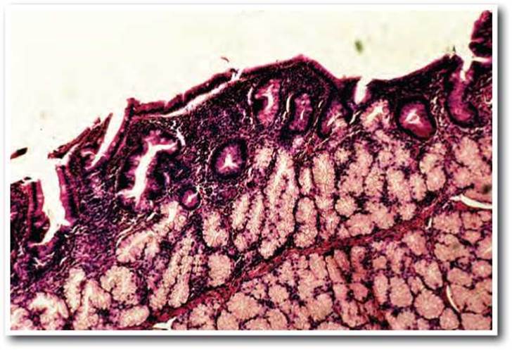
Fig. 3.13. Chronic duodenal ulcer complicated by acute gastrointestinal bleeding: thinning of the duodenal mucosa, hyperplasia of Brunner's glands. Hematoxylin-eosin stain x 100.
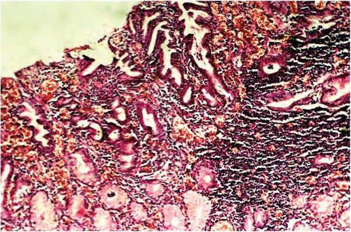
Fig. 3.14. Chronic gastric ulcer complicated by acute gastrointestinal bleeding: atrophy of the surface epithelium, reduced number of gastric glands, diffuse infiltration and hemorrhages in the stroma of the lamina propria of the gastric mucosa. Hematoxylin-eosin stain x 100.
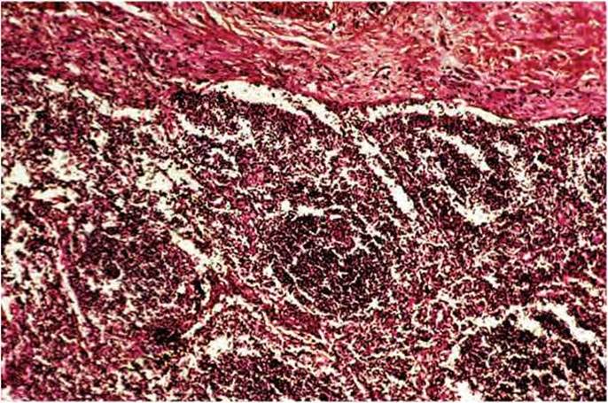
Fig. 3.15. Chronic gastric ulcer complicated by acute gastrointestinal bleeding: massive lymphoid aggregates extending to the muscularis mucosae of the stomach. Hematoxylin-eosin stain x 100.
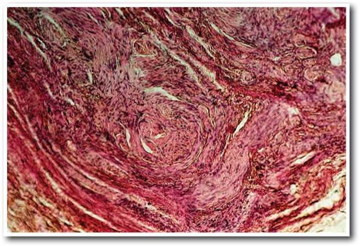
Fig. 3.16. Chronic gastric ulcer complicated by acute gastrointestinal bleeding: proliferation of newly formed nerve fibers in the scar zone of the gastric ulcer. Hematoxylin-eosin stain x 100.
The other type of atrophy included cases where the gastric pits were deepened and tortuous. The mucosa at the edges of the ulcer had the appearance of papillary structures covered with tall columnar epithelium. In place of chief and parietal cells, mucous neck cells appeared, locally completely replacing the glandular epithelium. In the neck Regions of the pits, lymphoid follicles were formed, sometimes with large reactive centers due to the proliferation of macrophages and large lymphocytes, sometimes with large reactive centers due to the proliferation of macrophages and large lymphocytes, and sometimes, instead of follicles, there were massive cellular infiltrates consisting mainly of lymphocytes and penetrating into the muscular layer (Fig. 3.15). Also, in 46% of cases, a combination of both types of atrophy was observed.
Such remodeling of the gastric mucosa showed a certain pattern depending on the localization of the ulcer. Thus, foveolar hyperplasia with atrophy of the secretory glands prevailed in antral ulcers. In ulcers of the gastric body, combined types and the first type of atrophy were observed. In all cases, the structural remodeling was accompanied by significant development of lymphoid tissue in place of the secretory glands and pits. Only in one case did we find goblet cells among the columnar epithelium of the stomach.
In the intramural nerve fibers, the following were found: a) regenerative proliferation, b) inflammatory changes in the form of perineuritis, c) degenerative and dystrophic changes. Destruction of nerve fibers was manifested by their granular disintegration, vacuolation, fragmentation, and lysis of fibrils. The regenerative reaction was in the form of ring-like and corkscrew-like growths, and proliferation of fibrils, which, layering upon each other, formed lateral club-like outgrowths. The number of newly formed nerve fibers and their bundles near the ulcer defect was in some cases so large that they occupied most of the muscular layer (Fig. 3.16) and sometimes penetrated into the mucosa.
These neuromas clearly exhibit signs of irritation and destruction: fibers are intensely impregnated, neuroplasmic accumulations form, and varicose swellings appear. Peripherally, these neuromas were surrounded by lymphoplasmacytic infiltrates and replaced by connective tissue. Involvement of nerve trunks in the pathological process was accompanied by their deformation and perineurial fibrosis.
In the deep sections of the ulcers within the serous coat, clusters of nerve fibers, large arteries, and veins were found in the form of nests. The nerve fibers here were large and proliferated in the manner of "amputation neuromas". Thus, lesions of the intramural Nervous Tissue in gastric and duodenal ulcer disease complicated by ARGIB manifested as the formation of neuromatous proliferations with degenerative changes and perineuritis, which were more intense and generalized in cases of gastric ulcer.
Thus, chronic gastric and duodenal ulcers complicated by recurrent gastrointestinal bleeding were characterized by:
1. A weakly expressed zone of exudation and a deeper zone of fibrinoid necrosis.
2. Absence of the vertical capillary zone in 68% of cases and predominantly horizontal vessel orientation in 83% of cases.
3. Low activity of regenerative processes at the ulcer margins in 94% of cases.
4. In cases of gastric ulcers, the presence of chronic active atrophic periulcerous gastritis with a large number of lymphoid aggregates was characteristic in 86% of patients. Duodenal ulcers were characterized predominantly by lymphoid infiltration of the mucosa with the development of chronic atrophic duodenitis and hyperplasia of Brunner's glands.
5. On the part of the vascular bed, gastric and duodenal ulcers complicated by recurrent bleeding were characterized by:
a) impaired vascular permeability in the form of numerous hemorrhages in the mucosal, submucosal layers, and in the zones of fibrinoid necrosis;
б) arterial intimal hyperplasia, fibrosis of the vascular muscular layer with lumen obliteration;
в) paretically dilated veins and venules with the formation of erythrocyte-fibrin thrombi.
6. Lesions of the intramural nervous tissue manifested as neuromatous proliferations with degenerative changes and perineuritis, which were more intense and generalized in cases of gastric ulcer.
Last update: 11/08/2026
Editorial and Educational Adaptation: This material has been compiled based on the primary/original source text. The project team performed an editorial review, corrected technical inaccuracies, structured sections, and adapted the content for an educational format.
What was processed:
- elimination of formatting defects (OCR errors, structural breaks, corrupted characters);
- editorial organization of content;
- standardization of terminology in accordance with academic sources;
- verification of factual statements against the original source text.
All mentions of the author, publication year, and origin of the primary text have been preserved in accordance with the source.