Tuberculosis - I. T. Pyatnochka 2005
Extrapulmonary tuberculosis
Tuberculosis of the cardiovascular system
There are no Organs or Tissues in The Human Body that cannot be affected by tuberculosis; however, the incidence and Diagnostics of these lesions vary considerably.
In developed countries, Extrapulmonary tuberculosis accounts for 1/4 to 1/2 of all newly diagnosed tuberculosis cases. In contrast, its share in Ukraine is less than 1/10, indicating that a significant number of patients with extrapulmonary tuberculosis remain underdiagnosed, primarily those with cardiovascular tuberculosis. All PARTS OF THE Circulatory system can be involved in the specific process—most commonly the Pericardium, and much less frequently the endocardium, myocardium, aorta, and Blood Vessels, which is due to differences in their local resistance to Mycobacterium tuberculosis.
Tuberculous pericarditis is the most frequent form of cardiac tuberculosis (Fig. 30). In recent years, the number of cases of tuberculous pericarditis has increased, although it does not exceed 1% of tuberculosis patients. Pathogenetically, pericardial tuberculosis is a secondary disease that develops As a result of the spread of mycobacteria, predominantly by contact from intrathoracic Lymph Nodes, the Mediastinum, and the Lungs. In addition, Mycobacterium tuberculosis can invade the pericardium via lymphatic or hematogenous routes, primarily in acute miliary Pulmonary Tuberculosis. With this form of tuberculosis, specific tubercles in the pericardium are detected in 5.8% of cases. In the Cytology/cytology/16.html">Early stages of pericarditis, fibrin is deposited on the epicardium and the inner surface of the parietal layer of the pericardial sac, and exudate accumulates, sometimes reaching 2 liters or more.
Class="center">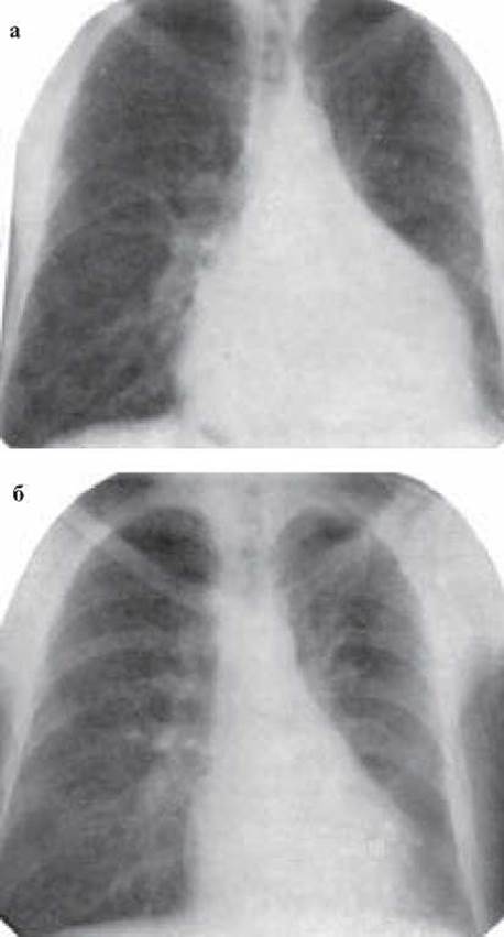
Fig. 30. Tuberculous pericarditis:
a) before Treatment; b) after 3 months of treatment
Productive and exudative forms of tuberculous pericarditis are distinguished. According to the character of the exudate, serous, serofibrinous, hemorrhagic, and purulent forms are identified. Adhesive or constrictive pericarditis occupies a separate place. Tuberculous tubercles are found beneath the fibrin on the epicardium and the inner surface of the pericardial sac. Sometimes the exudate in the pericardial sac resolves spontaneously, and the fibrin transforms into scar tissue with partial or complete fusion of the pericardium and epicardium, accompanied by calcium salt deposition ("armored heart").
The onset of tuberculous pericarditis is usually gradual, as the accumulation of exudate occurs slowly and is relatively well tolerated by the patient. The clinical manifestations of tuberculous pericarditis are nonspecific. However, when analyzed comprehensively, they often point to the correct Diagnosis. Middle-aged men are primarily affected. The clinical picture is dominated by symptoms of tuberculous intoxication: low-grade or high fever, excessive sweating, anorexia, weight loss, weakness, and rapid fatigue; this is followed by a dry, irritating cough, dyspnea, chest pain, and ankle edema. These last four symptoms serve as significant indicators, as they occur much more frequently in tuberculous pericarditis than in tuberculosis without pericardial involvement. The patient's general condition deteriorates sharply in the presence of a large amount of exudate due to mechanical compression of The Heart, which is accompanied by elevated systemic venous pressure, hepatomegaly, edema, and ascites.
In the early stage of tuberculous pericarditis, Cardiac Activity is accelerated (tachycardia) and mostly regular, heart sounds are muffled, and a "pericardial friction rub" can be auscultated. Subsequently, when the effusion is large, Percussion reveals a significant enlargement of cardiac dullness, and Auscultation reveals muffled heart sounds. At the same time, with a large volume of effusion, percussion dullness and bronchial breathing are detected near the lower angle of the left scapula (Ewart's sign), caused by pulmonary compression. Radiographically, the picture shows cardiomegaly, which is visualized in 98% of patients with effusion. Overall, the radiological findings depend on The amount of exudate; when the effusion is loculated, the shadow resembles a pericardial cyst. Computed tomography is also of great importance in diagnosing tuberculous pericarditis.
A dangerous complication of pericardial effusion is cardiac tamponade, as the accumulated fluid significantly impedes blood inflow into the ventricles. As a result, Cardiac Output decreases and systemic venous congestion develops, clinically manifested by a drop in blood pressure, elevated venous pressure, distension of the jugular Veins during inspiration (Kussmaul's sign), and pulsus paradoxus. In this condition, systolic blood pressure drops by more than 10 mmHg during inspiration.
Echocardiography (ultrasound) is a sensitive method (100%) for detecting even minimal amounts of fluid in the pericardial sac. Angiocardiography is also effective in diagnosing pericardial effusion, but the Procedure is time-consuming, often inconvenient, and sometimes painful.
Magnetic Resonance imaging allows for direct visualization of the pericardium and the detection of even minor thickening.
Other important diagnostic Methods of great significance in tuberculous pericarditis include electrocardiography, the Mantoux test, pericardial biopsy, and biochemical, cytological, and bacteriological Analysis of the pericardial effusion.
In the first days of the disease, the Electrocardiogram shows Displacement of the RS-T segment, a decrease in the amplitude of the QRS complex and the T wave in standard and chest leads. A large amount of exudate manifests as alteration of the atrial and ventricular complexes, Cardiac Arrhythmias, atrial fibrillation and flutter, premature contractions, and atrioventricular conduction disturbances.
Hemogram changes are usually moderately expressed. Tuberculin Skin tests (Mantoux) are positive and occasionally hyperergic.
Pericardial puncture, performed for diagnostic and therapeutic purposes, often helps determine the Etiology of the disease. The exudate in pericardial tuberculosis is predominantly lymphocytic, with a high protein content and low glucose level. Upon triple culture examination of the pericardial fluid, colonies of Mycobacterium tuberculosis grow in 50% of patients. Biopsy of the epicardium or pericardium followed by histological (cytological) examination of the biopsy specimen significantly increases diagnostic efficacy.
Overall, diagnosing tuberculous pericarditis is challenging. Signs of systemic venous congestion, a low-grade fever in the absence of other heart diseases, and a large amount of exudate with mild clinical manifestations are of substantial value in suspecting tuberculous etiology. The diagnosis becomes probable when Mycobacterium tuberculosis is found in the exudate or specific granuloma elements are present in the biopsy. Incidentally, pericarditis in patients with pulmonary or Other forms of tuberculosis must be considered specific, even with negative tuberculin reactions. In complex and controversial cases, a trial of antituberculosis therapy is justified.
The prognosis for the natural course of untreated tuberculous pericarditis is unfavorable, resulting in a 17–50% mortality rate. Death from tuberculous pericarditis results from circulatory failure, compression of The Heart and great vessels, narrowing of the coronary vessels, diffuse myocardial damage, and pathological Changes in the heart Valves.
Modern prolonged antimycobacterial therapy promotes recovery with a favorable prognosis in the vast majority of patients with tuberculous pericarditis. However, in 14–40% of patients, the disease still ends fatally despite current antimycobacterial treatment. In general, treatment is currently administered according to WHO-recommended regimens. The first two months (intensive phase): isoniazid 0.3–0.45 g; rifampicin 0.6 g; pyrazinamide 2.0 g; and ethambutol 1.2 g daily. Streptomycin is undesirable due to its coronarospastic properties. The subsequent 4–6 months (continuation phase): isoniazid 0.3–0.45 g and rifampicin 0.6 g daily, or isoniazid 0.6 g and rifampicin 0.6 g three times a week.
The best therapeutic results are achieved with the simultaneous use of antimycobacterial drugs and glucocorticosteroids, which promote the rapid resolution of the inflammatory reaction in the pericardium and minimize The Development of adhesions.
Surgical intervention—pericardiectomy—is indicated in the following cases: when there is no reduction in heart size after three months of antimycobacterial therapy; when heart size decreases but venous pressure rises or fails to normalize within 6 months of treatment; and when Heart Failure progressively worsens unexpectedly. In cases of cardiac compression due to adhesions or an "armored heart," pericardiectomy or cardiac decortication is performed.
Endocardial tuberculosis is an exceptionally rare disease. The infection may spread to the endocardium via hematogenous, lymphogenous, or contact routes from the myocardium or pericardium. Acute tuberculous endocarditis is severe. Its clinical picture resembles generalized tuberculosis in general. Uncomplicated nodular endocardial tuberculosis is frequently asymptomatic. The diagnosis is usually established during surgery or at autopsy. The prognosis is unfavorable.
Tuberculous myocarditis is more frequently observed in men. The Pathogenesis of the disease is similar to that of endocardial tuberculosis. Pathanatomically, macro-nodular, miliary, and the rarest form—diffuse—are distinguished. Clinically, the macro-nodular form of tuberculous myocarditis has a predominantly latent course; the miliary form largely mimics nonspecific myocarditis or Sepsis. Diffuse tuberculous myocarditis resembles the two previous forms in its clinical course. Diagnosing tuberculous myocarditis is difficult. The unexpected development of congestive heart failure, angina pectoris, heart murmurs, or rhythm and conduction disturbances provides grounds for suspecting specific myocardial involvement. The treatment of endocardial and myocardial tuberculosis is similar to that of tuberculous pericarditis.
Tuberculosis of the aorta and large visceral vessels is observed with equal frequency in young men and women, and is quite rare despite pronounced bacteremia and direct contact with caseous intrathoracic lymph nodes. This is due to the strong local Immunity of the aorta and blood vessels against tuberculosis infection. The Clinical presentation of aortic tuberculosis depends on the Location OF THE process (thoracic or abdominal), its form (miliary, polypoid, tuberculous aneurysm, mesoarteritis), and The Nature of the primary tuberculous process. Half of the patients with aortic tuberculosis present with aneurysmal dilation of its thoracic or abdominal segment, and the cause of death in such cases is rupture of the aneurysm.
When active tuberculosis in various organs is accompanied by an aortic aneurysm, particularly in the thoracic segment, tuberculous aortitis should be suspected. Clinically, aortic aneurysm is the most frequently diagnosed condition. The primary diagnostic method is aortography, which allows for the timely detection of aortic tuberculosis, the aneurysm, and its location. Furthermore, the affected aortic wall can rapidly undergo necrosis and rupture. The Treatment of tuberculosis of the aorta and blood vessels consists of prolonged administration of antimycobacterial drugs, whereas surgery is indicated for aneurysms (aneurysmectomy, resection, artificial grafting) to prevent rupture and massive Hemorrhage.
Last update: 10/08/2026
Editorial and Educational Adaptation: This material has been compiled based on the primary/original source text. The project team performed an editorial review, corrected technical inaccuracies, structured sections, and adapted the content for an educational format.
What was processed:
- elimination of formatting defects (OCR errors, structural breaks, corrupted characters);
- editorial organization of content;
- standardization of terminology in accordance with academic sources;
- verification of factual statements against the original source text.
All mentions of the author, publication year, and origin of the primary text have been preserved in accordance with the source.