Review of Medical Physiology - William F. Ganong 2002
Blood Circulation
Regional Blood Flow
Cerebral Circulation — Anatomical Features
Vessels
Arterial Blood is supplied to the Brain primarily through four Arteries: two internal carotid arteries and two vertebral arteries. The vertebral arteries join to form the Basilar artery, which, together with the carotids, forms the circle of Willis beneath the Hypothalamus. This circle gives rise to six major arteries that supply blood to the Cerebral Cortex. In some animals, the vertebral arteries are large and the internal carotids are small, whereas in humans, a relatively small fraction of the total arterial blood flow is delivered via the vertebral arteries. When substances are injected into one of the carotid arteries, a corresponding unilateral distribution to the cerebral hemisphere is observed. Normally, no crossover is detected, presumably due to the equal pressures on both sides. Even when pressures differ, anastomoses do not permit significant blood flow. Occlusion of one carotid artery, particularly in elderly individuals, frequently results in severe symptoms of cerebral ischemia. In humans and certain other species, pre-capillary anastomoses exist between cerebral arterioles and other vessels; however, Blood flow through these vessels is generally insufficient to maintain perfusion and prevent infarction resulting from cerebral artery occlusion. Venous drainage from the human brain occurs via deep Veins and Dural Venous Sinuses, primarily into the Internal jugular vein, although a small amount of venous blood passes through the ophthalmic and facial venous plexuses, via emissary veins into the scalp, and down the paravertebral Venous system into the spinal canal. In other species, the internal jugular vein is small, and venous blood from the brain mixes with blood draining from other structures.
Class="center">Table 32-1. Resting Blood Flow and O2 Consumption in Various Organs (Adult male weighing 63 kg, mean arterial pressure 90 mm Hg, and O2 consumption 250 mL/min)1
Organ/Region |
Mass, kg |
Blood Flow |
Arteriovenous O2 Difference, mL/dL |
O2 Consumption |
Resistance, R-units1 |
Percentage of Total, % |
||||
mL/min |
mL/100 g/min |
mL/min |
mL/100 g/min |
Absolute |
per kg |
Oxygen Consumption |
||||
2.6 |
1500 |
5 7.7 |
34 |
51 |
2.0 |
3.6 |
9.4 |
27.8 |
20.42 |
|
0.3 |
1260 |
420.0 |
14 |
18 |
6.0 |
4.3 |
1.3 |
23.3 |
7.2 |
|
Brain |
1.4 |
750 |
54.0 |
62 |
46 |
3.3 |
7.2 |
10.1 |
13.9 |
18.4 |
3.6 |
462 |
12.6 |
25 |
12 |
0.3 |
11.7 |
42.1 |
8.6 |
4.8 |
|
31.0 |
840 |
2.7 |
60 |
50 |
0.2 |
6.4 |
198.4 |
15.6 |
20.0 |
|
Heart muscle |
0.3 |
250 |
84.0 |
114 |
29 |
9.7 |
21.4 |
6.4 |
4.7 |
11.6 |
Rest of body |
23.8 |
336 |
1.4 |
129 |
44 |
0.2 |
16.1 |
383.2 |
6.2 |
17.6 |
Whole body |
63.0 |
5400 |
8.6 |
46 |
250 |
0.4 |
1.0 |
63.0 |
100.0 |
100.0 |
1 Reproduced with permission from Bard P [editor]: Medical Physiology. 11th ed. Mosby, 1961.
2 R-units are pressure (mm Hg) divided by blood flow (mL/s).
Cerebral Blood Vessels possess distinctive Anatomical Features. In the choroid plexus, gaps are present between the endothelial Cells of the Capillary Wall, whereas the choroidal epithelial cells separating them from the CEREBROSPINAL FLUID (CSF) are joined by tight junctions. Capillaries in brain tissue resemble the nonfenestrated capillaries of skeletal muscle (see Chapter 30), but tight junctions between endothelial cells restrict the passage of substances. In addition, the endothelial Cytoplasm contains a relatively small number of vesicles, reflecting low levels of Vesicular Transport. Brain capillaries are surrounded by astrocyte end-feet (Fig. 32-1). These end-feet are closely apposed to the capillary basement membrane, although they do not completely cover the capillary wall. Gaps of about 20 nm are present between the end-feet. The end-feet contribute to the tight junctions in capillaries (see Chapter 2). Astrocyte protoplasm is also found around synapses, where it isolates brain synapses from one another.
Innervation
Three nerve systems innervate cerebral blood vessels. The Cell bodies of postganglionic sympathetic Neurons are located in the superior cervical ganglia, and their terminals contain norepinephrine and neuropeptide Y. Cholinergic neurons, which likely originate in ganglia, also innervate cerebral vessels, and postganglionic vascular cholinergic neurons contain acetylcholine, VIP, and PHM-27 (see Chapter 26). These nerves terminate primarily on large arteries. Sensory nerves are found on distal arteries; their cell bodies are located in the trigeminal ganglion and contain substance P, neurokinin A, and CGRP. Substance P, CGRP, VIP, and PHM-27 induce vasodilation, whereas neuropeptide Y causes vasoconstriction. Traction on or manipulation of cerebral blood vessels causes pain.
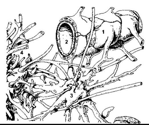
Fig. 32-1. Membrane of the end-FOOT (1) of a fibrous astrocyte surrounding a cerebral capillary (2). On the left, astrocyte end-feet (3) also encompass a portion of the dendrites of neighboring neurons (4) (reproduced with permission from Krstic RV: Die Gewebe des Menschen und der Säugetiere. Springer, 1978).
Last update: 10/08/2026
Editorial and Educational Adaptation: This material has been compiled based on the primary/original source text. The project team performed an editorial review, corrected technical inaccuracies, structured sections, and adapted the content for an educational format.
What was processed:
- elimination of formatting defects (OCR errors, structural breaks, corrupted characters);
- editorial organization of content;
- standardization of terminology in accordance with academic sources;
- verification of factual statements against the original source text.
All mentions of the author, publication year, and origin of the primary text have been preserved in accordance with the source.