Human Anatomy - H. I. Koliadenko 2009
Main periods and stages of human ontogenesis
Prenatal development of the organism
Human ontogeny is divided into two main developmental periods: prenatal (intrauterine) and postnatal (extrauterine). In turn, the prenatal period is subdivided into the Embryonic and Fetal periods. The Embryonic period is characterized by The Development of the embryo.
The embryo is formed As a result of Fertilization—the fusion of a male gamete, or spermatozoon, with a female gamete, the ovum (Figs. 6 and 7). A single ovum (very rarely two or more) from the female Organism participates in the act of fertilization, alongside up to 100 million or more spermatozoa from the male. Fertilization takes place in the fallopian tubes, where each spermatozoon has the potential to give rise to a new organism, though only a single, most viable one penetrates the ovum.
Following the penetration of the sperm HEAD into the ovum, the nuclei of the male and Female Germ Cells fuse, resulting in a qualitatively novel single-celled Structure known as a zygote. Immediately thereafter, The process of Cleavage begins—the division of the fertilized egg into two daughter cells (blastomeres). This marks the beginning of a new organism's life. Ten hours after the First Division, a Second Division occurs, producing four blastomeres. Cell Division proceeds continuously, and their number increases in geometric progression. By the end of the first week of development, they reach a count of one hundred. The embryo represents a small vesicle—the blastula—with an internal cavity; cell division at this stage continues, accompanied by the accumulation of cellular material.
Class="center">
Fig. 6. STRUCTURE OF THE male gamete:
(a — general view; b — sperm head):
1 — nucleus; 2 — mitochondrial spiral; 3 — axial filament; 4 — tail; 5 — body; b — neck 7 — head
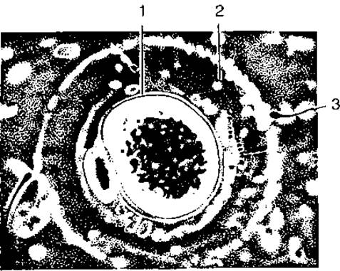
Fig. 7. Female ovum during fertilization:
1 — ovum; 2 — follicular cells; 3 — spermatozoon
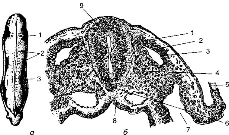
Fig. 8. Development of somites:
a — human embryo at approximately 3.5 weeks of development:
1 — otic pit; 2 — somites; 3 — line of incision of the amniotic membrane; b — early differentiation of somites.
Cross-section of the embryo:
1 — myotome; 2 — dermatome; 3 — somite cavity; 4 — sclerotome; 5 — amnion; b — notochord; 7 — coelom; 8 — gut; 9 — neural tube
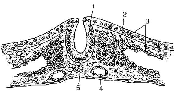
Fig. 9. Cross-section of a human embryo (schematic) at the stage of neural tube formation:
1 — neural tube; 2 — ectoderm; 3 — mesoderm; 4 — endoderm; 5 — notochord
At the beginning of the second week of prenatal development, the embryo implants into the uterine mucosa. A close connection is established between the maternal organism and the embryo, whose Nutrition is mediated through the maternal Circulatory system. The quantitative accumulation of cellular material by the embryo leads to a qualitative leap, initiating The formation of cellular plates. At this stage of development, the embryo is termed a gastrula. A portion of the blastula wall invaginates into the embryonic vesicle. The cells rearrange to form three germ layers: the ectoderm, the endoderm, and the mesoderm. Each of the three germ layers possesses distinct morphological and functional properties, a process known as the differentiation of cell layers.
The ectoderm is the outer layer of the embryo; during development, it gives rise to the Epithelial Tissue of The Cytology/cytology/66.html">Skin and its Derivatives.
The endoderm is the inner layer of the embryo, lining the primitive gut—the future digestive tract; it gives rise to the intestinal epithelium, digestive glands, and the epithelium of the Respiratory system. All types of connective, muscular, cartilaginous, and osseous tissues, as well as Blood and Hematopoietic Organs, develop from the mesoderm.
Embryonic development is characterized by the formation of so-called provisional organs that perform a temporary, auxiliary role. They originate from extraembryonic cellular material, which also differentiates into ectoderm, endoderm, and mesoderm. Extraembryonic cellular material develops intensively during the initial period of Embryogenesis. It forms the amniotic vesicle—the primary external environment of the organism—and the yolk sac, which performs a nutritional function.
The first period of development is the blastula stage—the accumulation of cellular material; the second is the gastrula stage—the initial period of histogenesis (histios — tissue, genesis — development).
Beginning in the third week (Fig. 8) of prenatal development, the axial organs begin to form: the notochord, the neural tube, and the mesodermal somites (Fig. 9). The notochord serves as the primary Axial Skeleton in the form of a cellular cord (Fig. 10). Later, it degenerates and is replaced by THE Vertebral Column. The neural tube and lateral ganglionic plates develop from ectodermal cells, forming the foundation of the future Nervous system. The dorsal part of the mesoderm consists of somites—the primary body segments. Mesenchymal cells, which constitute embryonic Connective Tissue, differentiate from the mesoderm.
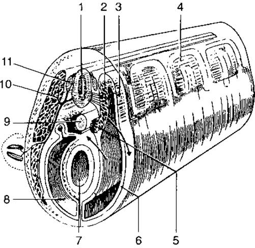
Fig. 10. Diagram of the trunk structure in a vertebrate embryo:
1 — neural tube; 2 — myotome; 3 — dermatome; 4 — somites of the dorsal mesoderm; 5 — sclerotome; 6 — parietal layer of the ventral mesoderm; 7 — gut; 8 — secondary body cavity (coelom); 9 — notochord; 10 — spinal nerve; 11 — spinal ganglion
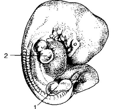
Fig. 11. Human embryo at 4.5–5 weeks of development:
1 — lower limb bud; 2 — upper limb bud
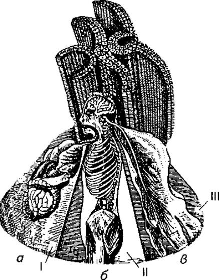
Fig. 12. Derivatives of the three germ layers (schematic):
a — endoderm; b — mesoderm; c — ectoderm.
The three germ layers give rise to three Organ Systems:
I — Internal Organs and glands; II — bones, Muscles, Blood Vessels; III — nerves, integument (skin)
Somites differentiate into sclerotomes, dermatomes, and myotomes. Bone and Cartilage Tissues develop from the sclerotomes; dermatomes give rise to the connective tissue layer of the skin; skeletal muscles are formed from the myotomes.
The ventral unsegmented part of the mesoderm forms thin plates of mesothelium that line the thoracic and abdominal Body Cavities.
By the end of the first month of prenatal development, the primordia of all tissue types are already formed: Epithelial Tissues (arising from the ectoderm and endoderm), connective tissue, muscular tissue, and Nervous Tissue.
Starting from the second month of prenatal development, almost all Organs of the systems and apparatuses of the developing embryo are formed. This period is known as Organogenesis (Fig. 11). The end of the second month is characterized by the Formation of the external Features of the head, trunk, and limbs.
Beginning in the third month of development, intensive growth of all organs and individual body PARTS OF THE embryo is observed (Fig. 12).
Last update: 08/08/2026
Editorial and Educational Adaptation: This material has been compiled based on the primary/original source text. The project team performed an editorial review, corrected technical inaccuracies, structured sections, and adapted the content for an educational format.
What was processed:
- elimination of formatting defects (OCR errors, structural breaks, corrupted characters);
- editorial organization of content;
- standardization of terminology in accordance with academic sources;
- verification of factual statements against the original source text.
All mentions of the author, publication year, and origin of the primary text have been preserved in accordance with the source.