Human Anatomy - Kotsan I. Ya. 2009
Vestibulocochlear organ (organ of hearing and balance)
Statokinetic (vestibular) analyzer
The statokinetic analyzer is a complex morphofunctional system responsible for the reception, transmission, analysis, and integration of vestibular stimuli.
Thanks to the vestibular apparatus, we receive information about the position and movement of the HEAD, and consequently, of the body in space.
Encompassing the vestibular apparatus, cortical and subcortical centers, as well as a system of connecting pathways, the vestibular analyzer—along with other analyzers (visual, motor, cutaneous)—participates in the orienting Reactions of the body relative to space. It serves to maintain static and dynamic equilibrium (ensuring balance during various movements) and allows the head to be held in its natural position without the involvement of Vision.
The peripheral division of the statokinetic analyzer is represented by sensory Cells located on the wall of the membranous labyrinth within cristae (in the ampullae of the semicircular ducts) and maculae (in the utricle and saccule). The receptor of this analyzer consists of specialized epithelial Hair cells, upon the hairs of which lies a gelatinous otolithic membrane containing calcium carbonate crystals, known as otoliths. During Head movements, the otoliths shift, pulling on or pressing against the hairs, which triggers a corresponding stimulus.
The receptor hair Cells of the utricle and saccule signal changes in body position relative to the center of gravity. The receptor hair cells of the saccule also perceive vibrational oscillations.
The receptor hair cells of the ampullae of the semicircular ducts signal body movements in a specific plane. The outer surface of these cells is adjacent to a cap-like gelatinous body into which the receptor Cell hairs are immersed. During active and passive Displacement of the body in space, inertial displacement of the endolymph occurs—primarily within the membranous semicircular duct lying in the plane of body movement—which causes a shift of the gelatinous body and deformation of the receptor cell hairs. In response to The stimulation of the cristae receptor cells (in the ampullae of the semicircular ducts), a reaction is triggered in the corresponding Muscle groups that level the BODY POSITION AND coordinate the Movements of the extraocular Muscles.
The transmission of nerve impulses from the receptor hair cells of the cristae (in the ampullae of the semicircular ducts) and maculae (in the utricle and saccule) to the cortical centers of the cerebral hemispheres is carried out via the vestibular (statokinetic) pathway (Fig. 275).
The first-order Neurons of this pathway are represented by bipolar cells whose cell bodies lie in the vestibular ganglion located at the bottom of the internal acoustic meatus. The peripheral processes—dendrites of these bipolar cells—contact the receptor hair cells. The central processes—axons of the bipolar cells—in the form of the vestibular nerve, together with the cochlear nerve, enter the cranial cavity through the internal acoustic opening, and then at the cerebellopontine angle enter the Brainstem tissue, where they divide into ascending and descending fibers. The ascending fibers terminate on the cells of the superior vestibular Nucleus (Bechterew's nucleus), while the descending fibers end in the lateral (Deiters's), medial (Schwalbe's), and inferior (Roller's) vestibular nuclei, which lie in the lateral angle of the Rhomboid fossa (vestibular area). The cells of the vestibular nuclei serve as the second-order neurons of the statokinetic pathway. A small number of bipolar neuron fibers proceed directly to the Cerebellum, bypassing the vestibular nuclei of the brainstem.
The axons of the vestibular nucleus cells (second-order neurons) form a series of tracts that lead to the cerebellum, the nuclei of the eye muscle nerves, the nuclei of autonomic centers, the Cerebral Cortex, and the Spinal Cord.
Class="center">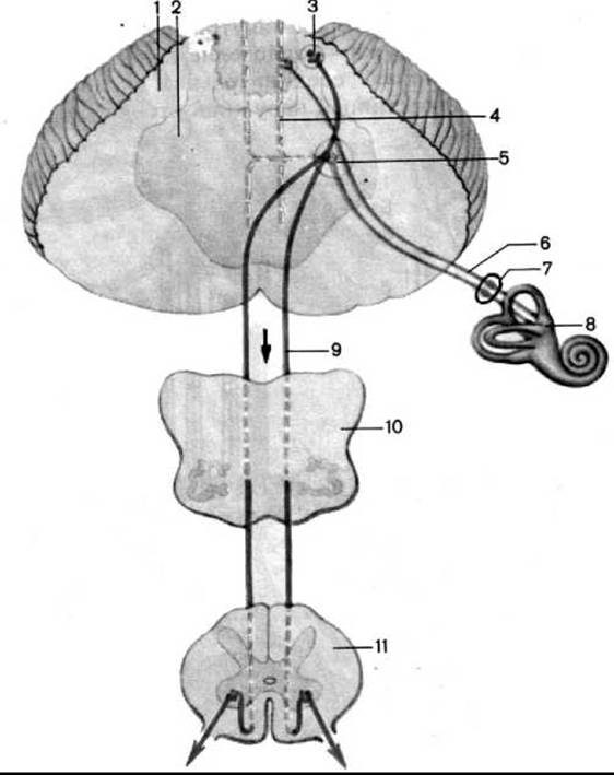
Fig. 275. Pathway of the static (vestibular) analyzer (after M. R. Sapin)
1 — cerebellum; 2 — Pons; 3 — fastigial nucleus; 4 — medial longitudinal fasciculus; 5 — vestibular nuclei; 6 — vestibular part of the Vestibulocochlear nerve; 7 — vestibular ganglion; 8 — Inner ear; 9 — vestibulospinal tract; 10 — section of the Medulla Oblongata; 11 — section of the spinal cord
Thus, the axons of the cells whose bodies lie in the superior, medial, and lateral vestibular nuclei pass through the inferior cerebellar peduncles, forming the vestibulocerebellar tract, to the fastigial nucleus or the cortex of the cerebellar vermis.
Furthermore, a portion of the axons from the lateral vestibular nucleus forms the vestibulospinal tract, which descends into the spinal cord and terminates segmentally on the motor cells of the anterior horns, transmitting vestibular impulses to the Muscles of the neck, trunk, and limbs, thereby ensuring the maintenance of body balance.
A portion of the neurons' axons from the lateral vestibular nucleus extends to the medial longitudinal fasciculus of the same and opposite sides, establishing a connection between the Organ of Balance—via the lateral nucleus—and the cranial nerve nuclei (III, IV, VI pairs) that innervate the extraocular muscles. This makes it possible to maintain the direction of gaze regardless of changes in head position. Maintaining body balance largely depends on the coordinated movements of the eyeballs and the head.
The axons of the vestibular nuclei cells form connections with the neurons of the brainstem Reticular Formation and with the nuclei of the Midbrain.
The Emergence of autonomic reactions (slowed pulse, lowered Blood pressure, nausea, vomiting, etc.) in response to stimulation of the vestibular apparatus can be explained by the connections of the vestibular nuclei—via the reticular formation—with the nuclei of the vagus and glossopharyngeal nerves.
Conscious perception of body (head) position is ensured by a crossed pathway running from the vestibular nuclei to the thalamus, where The Cell bodies of the third-order neurons reside, and further to the cerebral cortex. It is believed that the cortical portion of the statokinetic analyzer is scattered across the cortex of the superior temporal and postcentral gyri, as well as the superior parietal lobule of the cerebral hemispheres.
Damage to the vestibular nuclei, nerve, and labyrinth is accompanied by three main symptoms: vertigo (the patient feels as though the floor is swaying beneath them), nystagmus (characterized by rhythmic twitching of the eyeballs), and impaired balance and motor coordination.
Last update: 08/08/2026
Editorial and Educational Adaptation: This material has been compiled based on the primary/original source text. The project team performed an editorial review, corrected technical inaccuracies, structured sections, and adapted the content for an educational format.
What was processed:
- elimination of formatting defects (OCR errors, structural breaks, corrupted characters);
- editorial organization of content;
- standardization of terminology in accordance with academic sources;
- verification of factual statements against the original source text.
All mentions of the author, publication year, and origin of the primary text have been preserved in accordance with the source.