Human Anatomy and Physiology - I. V. Gaivoronsky 2011
Anatomy and Physiology of the Digestive System
Stomach
Structure. The Stomach, ventriculus (Greek: gaster), is a hollow muscular organ located in the Abdominal cavity, primarily in the left hypochondrium. Its lumen is significantly wider than that of other hollow Organs in the Digestive System. The shape of the stomach is individual and depends on body type. Furthermore, in the same individual, it varies depending on the degree of distension. The capacity of an adult stomach ranges from 1.5 to 4 liters.
The stomach has two surfaces: anterior and posterior, which merge at the margins. The upper margin is termed the lesser curvature, and the lower margin is the greater curvature. Several PARTS OF THE stomach are distinguished (see Fig. 7.10). The region bordering the Esophagus is called the cardiac part. To the left of it lies a dome-shaped upward protrusion known as the Cytology/practical/108.html">Fundus of the stomach. Bordering the cardiac part and the fundus is the largest section — the body of the stomach. The pyloric part transitions into the duodenum. Located at this junction is a sphincter that regulates the passage of food into the Small Intestine — the pyloric sphincter.
The stomach wall consists of three layers: the mucous membrane (mucosa), the muscular layer, and the serous membrane (serosa). The mucosa forms numerous folds and is lined with a single-layered columnar epithelium. It contains a vast number of glands (up to 35 million). Glands are classified into cardiac, body (gastric), and pyloric types. They comprise various Cell types: chief Cells secrete pepsinogen; parietal (oxyntic) cells produce Hydrochloric acid; and mucous cells (mucous neck cells and pit cells) secrete mucus (predominant in the cardiac and pyloric glands).
In the stomach lumen, the secretions of all glands mix to form gastric juice. Its daily volume reaches 1.5–2.0 liters. This amount of juice makes it possible to liquefy and digest ingested food, transforming it into a semi-liquid mass (chyme).
The muscular layer of the stomach consists of three layers of Smooth Muscle tissue oriented in different directions. The outer layer is longitudinal, the middle is circular, and oblique fibers lie adjacent to the mucosa.
The serous membrane (Peritoneum) covers the stomach externally on all sides, thereby allowing it to alter its shape and volume.
Composition of gastric juice. The acidity (pH) of gastric juice at the peak of Digestion is 0.8–1.5; at rest, it is about 6. Consequently, during digestion, it represents a strongly acidic environment. Gastric juice contains Water (99–99.5%), as well as organic and inorganic substances.
Organic substances are represented primarily by various Enzymes and mucin. Mucin is produced by mucous cells and facilitates better coating of food bolus particles, protecting the mucosa from the aggressive factors of gastric juice.
The principal enzyme of gastric juice is Pepsin. It is produced by chief cells as an inactive proenzyme, pepsinogen. Under METABOLISM/18.html">The Influence of hydrochloric acid in the gastric juice and air located in the fundus region, a specific Amino Acid Sequence is cleaved from pepsinogen, converting it into an active enzyme capable of catalyzing Protein Hydrolysis (breakdown). Pepsin activity is observed exclusively in a strongly acidic environment (pH 1–2). Pepsin breaks the bonds between two adjacent Amino Acids (peptide bonds). As a result, the protein molecule is cleaved into several smaller molecules of lower molecular weight (Polypeptides). Nevertheless, they are not yet capable of passing through the gastrointestinal (GI) epithelium and being absorbed into the Blood. Their further digestion takes place in the small intestine. It is worth noting that 1 g of pepsin can hydrolyze 50 kg of egg albumin and curdle 100,000 liters of milk within 2 hours.
In addition to the primary enzyme, pepsin, gastric juice contains Other Enzymes. For instance, gastrinxin and rennin (chymosin) also belong to protein-digesting enzymes. The former is active at a moderate acidity of gastric juice (pH 3.2–3.5); the latter operates in a mildly acidic environment close to neutral (pH 5–6). Gastric lipase breaks down fats, but its activity is negligible. Rennin and gastric lipase are most active in infants. They facilitate the Hydrolysis of Proteins and fats in breast milk, supported by the near-neutral environment of infant gastric juice (pH around 6).
Inorganic substances in gastric juice include: HCl, SO42-, Na+, K+, HCO3-, and Ca2+ ions. The primary inorganic substance of the juice is hydrochloric acid. It is secreted by the parietal Cells of the gastric mucosa and performs A number of Functions essential for normal digestion. Hydrochloric acid creates the acidic environment required to convert pepsinogen into pepsin and ensures the proper functioning of this enzyme. This specific level of acidity causes the Denaturation (loss of structure) of dietary proteins, facilitating the work of enzymes. The bactericidal properties of gastric juice are also due to the presence of hydrochloric acid. Few microorganisms can withstand the concentration of hydrogen ions generated in the stomach lumen through The activity of parietal cells.
The gastric glands synthesize a special substance known as the intrinsic factor of Castle. It is essential for the absorption of vitamin B12: the intrinsic factor binds with the vitamin, and the resulting complex passes from the GI lumen into the epithelial cells of the small intestine and subsequently into the blood. In the stomach, iron is treated with hydrochloric acid and converted into easily absorbable forms, which plays a major role in the synthesis of erythrocyte Hemoglobin. A decrease in the acid-forming function of the stomach and a reduction in intrinsic factor production (in gastritis with decreased secretory function) quite frequently lead to anemia.
MOTOR FUNCTION OF the stomach. Through the contractions of the muscular layer, food in the stomach is mixed, treated with gastric juice, and moved into the small intestine. Tonic and peristaltic contractions are distinguished. Tonic contractions adjust the stomach to the volume of ingested food, whereas peristaltic contractions are necessary for mixing and evacuating the contents. The latter process occurs gradually. Chyme passes into the duodenum in portions, as the hydrochloric acid contained in the food mass is neutralized by Liver and pancreatic secretions, as well as intestinal juice. Only then does the pyloric sphincter open for the next portion. Reverse muscular movements occur upon the intake of spoiled food or when it contains large amounts of aggressive substances that irritate the mucosa. This triggers the vomiting reflex. Food remains in the human stomach anywhere from 1.5–2 to 10 hours, depending on its Chemical Composition and consistency.
Furthermore, so-called hunger contractions occur, which are observed in an empty stomach at regular intervals. It is believed that they contribute to the sensation of hunger.
It should be particularly emphasized that a physiological antral sphincter is located between the body and the pyloric part, separating these regions. It is formed by the tonic contraction of the circular layer of the muscular coat. Due to this demarcation, the Primary processes of food DIGESTION IN THE stomach take place above the pyloric region (the cardiac part, fundus, and body of the stomach form the so-called digestive pouch). From the digestive pouch, digested food enters the pyloric region in small portions, often referred to as the evacuation channel. Here, the incoming food mixes with mucus, leading to a significant reduction in the acidic reaction of the chyme. Afterward, the food moves into the small intestine. Thus, the following processes occur in the stomach:
1) accumulation of food;
2) mechanical Processing of food masses (mixing);
3) Protein Denaturation under the influence of hydrochloric acid;
4) Protein Digestion under the influence of pepsin;
5) continuation of carbohydrate breakdown within the food bolus by salivary amylase (this enzyme is inactivated upon contact with gastric juice);
6) bactericidal Treatment of food by hydrochloric acid;
7) formation of chyme (semi-liquid food mass);
8) conversion of iron into easily absorbable forms and synthesis of Castle's intrinsic factor — an anti-anemic function;
9) propulsion of chyme into the small intestine.
The works of I. P. Pavlov on The Study of gastric digestion.
The most valuable research on digestion was conducted by the brilliant Russian scientist I. P. Pavlov, who was awarded the Nobel Prize in 1904 for his work in this field.
In his experiments on dogs, he transected the esophagus and brought both newly formed ends to the surface. This made it possible to perform the " sham feeding" experiment, in which food, after being processed in the Oral Cavity, did not enter the stomach but instead fell out through the opening in the esophagus. At the same time, the secretion of gastric juice was studied via a fistula. Nevertheless, food could still be introduced into the stomach through the lower opening in the neck.
To study the digestive process in the stomach, I. P. Pavlov isolated a small portion of the organ in dogs while preserving its innervation and blood supply. He separated this portion to create a so-called "small stomach," which had no direct connection with the main gastric cavity (Fig. 7.11).
The lumen of the small pouch was brought out to the exterior (creating a small stomach fistula). This made it possible to obtain pure gastric juice while food was present in the stomach. Through these experiments, I. P. Pavlov established the fundamental laws governing the REGULATION OF GASTRIC juice secretion. During sham feeding, gastric juice was secreted in the small pouch with virtually the same intensity as during normal feeding. Consequently, stimulation of receptors in the oral cavity reflexively triggered the secretion of gastric juice. This phenomenon was termed the unconditioned secretory reflex, the center of which is located in the Medulla Oblongata.
Gastric juice is also secreted at the mere sight or smell of food, even when there is no direct contact with the receptors of the oral cavity. This phenomenon was named the conditioned secretory reflex. Pavlov referred to the juice secreted under these conditions as "appetite juice." The COMPOSITION OF THE gastric juice was virtually identical to that observed during sham feeding.
Class="center">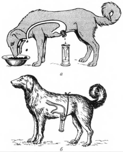
Fig. 7.11. Fistula Methods FOR STUDYING gastric secretion (after I. P. Pavlov):
a — sham feeding experiment; b — dog with an isolated small stomach
As a result of his groundbreaking work, I. P. Pavlov identified three Main phases of gastric juice secretion:
1) the cephalic phase, characterized by the secretion of "appetite gastric juice" in response to the sight, smell, or presence of food in the oral cavity; the Qualitative and quantitative composition of the gastric juice during this phase does not depend on the type or amount of food;
2) the gastric phase, during which juice is secreted while food is being digested in the stomach; the qualitative and quantitative composition of the juice in this phase directly depends on the type and amount of food;
3) the intestinal phase, mediated by the influence of intestinal receptors on the gastric glands. Stimulation of the gastric glands occurs as a result of chyme entering the duodenum that has not been sufficiently processed physically and chemically, which allows for necessary adjustments to be made to gastric secretion.
The regulation of gastric activity is governed by neural and humoral mechanisms. The parasympathetic Nervous system enhances gastric gland secretion and the motor activity of the muscular coat, whereas the sympathetic system exerts the opposite effect.
Humoral regulation involves Changes in the volume of secreted juice under the influence of various chemical substances. Glucose and amino acids absorbed into the bloodstream decrease secretion. Substances that increase gastric juice secretion include gastrin and histamine, which are produced by the cells of the gastric mucosa. Conversely, substances such as secretin and cholecystokinin inhibit secretion. The quantity and quality of the juice also depend on The Nature of the ingested food; for instance, a protein-rich diet increases the output of pepsin and hydrochloric acid.
Last update: 08/08/2026
Editorial and Educational Adaptation: This material has been compiled based on the primary/original source text. The project team performed an editorial review, corrected technical inaccuracies, structured sections, and adapted the content for an educational format.
What was processed:
- elimination of formatting defects (OCR errors, structural breaks, corrupted characters);
- editorial organization of content;
- standardization of terminology in accordance with academic sources;
- verification of factual statements against the original source text.
All mentions of the author, publication year, and origin of the primary text have been preserved in accordance with the source.