Human Anatomy and Physiology - I. V. Gayvoronsky 2011
Anatomy and Physiology of the Digestive System
Esophagus
Structure AND Functions. The Esophagus (oesophagus) is a hollow muscular tube, 25 — 30 cm in length. It begins at the Pharynx at the level of the VII cervical vertebra and terminates at the level of the XI thoracic vertebra, where it transitions into The Stomach. The largest portion of the esophagus is located within the thoracic cavity. Small segments, measuring 1.0—1.5 cm each, are situated in the cervical and Abdominal Regions. Consequently, the esophagus is divided into cervical, thoracic, and abdominal parts. The esophagus runs posterior to the Trachea and is related to the Thoracic Aorta (Fig. 7.10). Laterally, it is flanked by the vagus nerves, which intertwine to form nerve plexuses.
Class="center">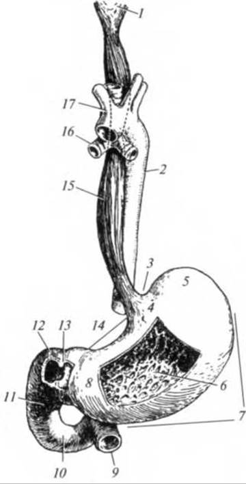
Fig. 7.10. Pharynx, esophagus, stomach, and duodenum:
1 — pharynx; 2 — thoracic aorta; 3 — cardiac notch; 4 — cardiac part of stomach; 5 — fundus of stomach; 6 — gastric folds; 7 — greater curvature; 8 — pyloric part; 9 — jejunum; 10 — horizontal part of duodenum; 11 — inferior part of duodenum; 12 — superior part of duodenum; 13 — pyloric sphincter; 14 — lesser curvature; 15 — esophagus; 16 — main bronchus; 17 — aortic arch
The esophagus features three permanent (anatomical) constrictions:
1) the pharyngeal constriction, located at the junction of the Pharynx and Esophagus;
2) the bronchial constriction, situated where the esophagus comes into contact with the left main bronchus;
3) the diaphragmatic constriction, at the level of the esophageal hiatus of the Diaphragm.
In addition, there are non-permanent (physiological) constrictions of the esophagus, which can be identified via X-ray Examination only in living individuals:
✵ the aortic constriction, located where the aortic arch abuts the esophagus;
✵ the cardiac constriction, at the junction of the esophagus and stomach.
The wall of the esophagus consists of three layers: the mucosa, the muscular layer, and the adventitia. Non-keratinized stratified squamous epithelium lines the inner surface of the mucosa, which features numerous longitudinal folds. As a result, the lumen of the organ appears star-shaped in cross-section. These folds allow the esophagus to expand as a bolus passes through. The muscular layer of the upper esophagus is composed of Striated Muscle tissue. In the middle third, it also contains smooth muscle Cells, while the lower third is entirely composed of Cytology/cytology/32.html">Smooth muscle tissue. The muscular layer is arranged into two strata: an outer longitudinal layer and an inner circular layer. In the cervical and thoracic regions, the esophagus is covered by an adventitia, whereas in the abdominal region, it is covered by a serosa.
The primary function of the esophagus is to convey food from the pharynx to the stomach. The food bolus is propelled by the force of gravity and peristaltic contractions of the organ's musculature. Liquid food passes through the esophagus in 1 — 2 s, without active contractions of the muscular layer. Denser food takes about 3 — 10 s to travel down, aided actively by the esophageal Muscles.
Swallowing (deglutition). This is a complex reflex action that transfers a food bolus from the Oral Cavity into the stomach. The swallowing center is located in the Medulla Oblongata and is functionally linked to the Neurons of the respiratory and vasomotor centers, which are also situated in this region of The Nervous system. Consequently, during swallowing, Respiration is automatically inhibited, and cardiovascular activity is modulated.
After being processed in the oral cavity, food is formed into a bolus. Chewing movements push the bolus toward the ROOT of the Tongue, which contains numerous sensory nerve endings. Nerve impulses from these receptors travel to the swallowing center in the medulla oblongata. From there, motor impulses are transmitted via Cranial Nerves to the Muscles responsible for swallowing. The tongue is tipped backward, propelling the bolus into the pharynx. The soft palate (velum palatinum) elevates, completely separating the nasopharynx from the oropharynx, thereby preventing the food bolus from entering the Nasal cavity. Simultaneously, the Pharynx and Larynx are elevated. The epiglottis folds down to seal the laryngeal inlet tightly, preventing food from entering the respiratory tract. Notably, speaking while eating can cause the food bolus to enter the Airways, leading to death by choking (asphyxia).
Strong contractions of the pharyngeal muscles push the bolus through the oro- and laryngopharynx into the esophagus. Peristaltic contractions of the esophagus facilitate the movement of food into the stomach. The musculature relaxes directly at the site of the bolus and just below it, while the proximal segments contract to push it downward. This movement takes the form of a peristaltic wave. Between the stomach and the esophagus, in the region of the cardiac constriction, There is a specialized valve — the cardiac sphincter — which allows food to enter the stomach while preventing the reflux of gastric contents back into the esophagus.
Last update: 08/08/2026
Editorial and Educational Adaptation: This material has been compiled based on the primary/original source text. The project team performed an editorial review, corrected technical inaccuracies, structured sections, and adapted the content for an educational format.
What was processed:
- elimination of formatting defects (OCR errors, structural breaks, corrupted characters);
- editorial organization of content;
- standardization of terminology in accordance with academic sources;
- verification of factual statements against the original source text.
All mentions of the author, publication year, and origin of the primary text have been preserved in accordance with the source.