Human Anatomy and Physiology - N. I. Fedyukovich 2003
Internal Organs
Urogenital System
Male Genital Organs
The Male reproductive Organs include the Testes with their epididymides, Ductus deferens and ejaculatory ducts, Seminal Vesicles, prostate and Bulbourethral Glands, Scrotum, and Penis (Fig. 84).
Internal Male Genitalia. The testes, or testicles (Testis), are paired male Gonads whose primary Functions are The production of male Gametes—spermatozoa—and the secretion of Male Sex Hormones into the bloodstream.
The testes are oval-shaped, measuring approximately 4.5 × 3 cm, with a mass of 20–30 g. They reside within the scrotum, the left testis typically hanging lower than the right. The testes are separated by the scrotal septum and enclosed within several tunics. Each testis is suspended by the Spermatic Cord, which contains the ductus deferens, Muscles AND FASCIAE, Blood and Lymphatic vessels, and nerves.
Each testis presents convex lateral and medial surfaces, an anterior and a posterior margin, as well as superior and inferior poles. The Epididymis, comprising a HEAD, body, and tail, is applied to the posterior margin of the testis.
Class="center">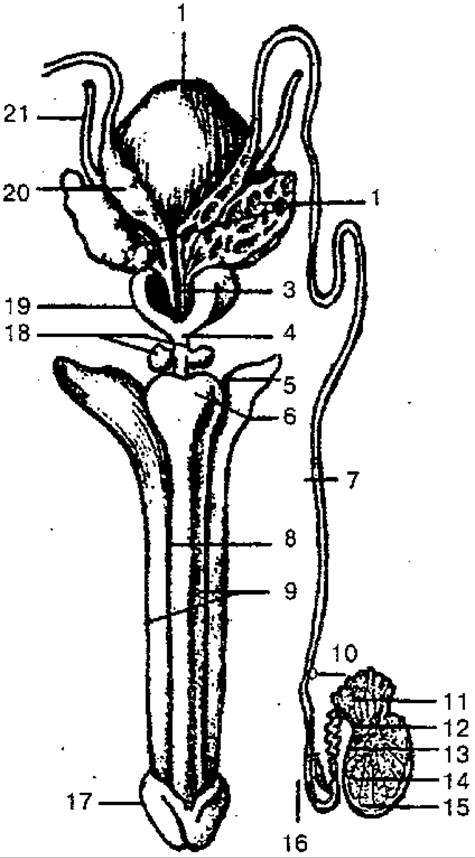
Fig. 84. Internal and external male reproductive organs (diagram):
1 — Urinary Bladder; 2 — Seminal Vesicle; 3 — ejaculatory duct; 4 — membranous part of the Urethra; 5 — crus of the penis; 6 — bulb of the penis; 7 — ductus deferens (vas deferens); 8 — corpus spongiosum; 9 — corpus cavernosum; 10 — epididymis; 11 — efferent ductules; 12 — rete testis; 13 — straight seminiferous tubules; 14 — convoluted seminiferous tubules; 15 — tunica albuginea; 16 — lower part of the ductus deferens; 17 — glans penis; 18 — Bulbourethral gland; 19 — Prostate Gland; 20 — ampulla of the ductus deferens; 21 — Ureter
The Peritoneum envelops the testis from all sides, creating a closed serous cavity. Externally, the testis is covered by a dense fibrous capsule known as the tunica albuginea, beneath which lies the testicular parenchyma. From the inner surface of the posterior margin of the tunica albuginea, a Connective Tissue extension—the Mediastinum testis—projects into the organ's interior. Delicate connective tissue septa radiate from the mediastinum, dividing the gland into numerous (250 to 300) pyramidal lobules, with their apices directed toward the mediastinum and their bases toward the tunica albuginea. Within each lobule lie two to three convoluted seminiferous tubules, 60–90 mm in length, surrounded by loose connective tissue and a rich network of Blood Vessels. The interior of the seminiferous tubules is lined with stratified spermatogenic epithelium, where Male Germ Cells (spermatozoa) are produced. These cells become part of the semen, the fluid component of which is derived from the secretions of the seminal vesicles and the prostate. The seminiferous tubules converge to form straight seminiferous tubules, which empty into the rete testis. From the rete testis, 12–15 efferent ductules arise, piercing the tunica albuginea to empty into the duct of the epididymis.
The ductus deferens (vas deferens) is a paired muscular tube approximately 50 cm long, with an outer diameter of 3 mm and a lumen diameter of about 0.5 mm. Based on its topographical features, it is divided into four parts: testicular, corresponding to the length of the testis; funicular, within the spermatic cord; inguinal, passing through the Inguinal Canal; and pelvic, extending from the deep inguinal ring to the prostate gland.
After passing through the inguinal canal, the ductus deferens arches downward along the lateral wall of the lesser pelvis to the Base of the urinary bladder. Near the prostate, its terminal portion widens to form an ampulla. Distally, the ampulla gradually narrows into a slender canal that joins the excretory duct of the seminal vesicle to form the ejaculatory duct, which opens via a small orifice into the prostatic urethra.
The seminal vesicle (vesicula (glandula) seminalis) is a paired secretory organ measuring 10–12 cm in length and 0.6–0.7 cm in thickness. These vesicles are located in the pelvic cavity, lateral and posterior to the base of the urinary bladder. Each seminal vesicle consists of a base (the broad end), a body (the central section), and a narrow inferior end that transitions into the excretory duct. The wall of the seminal vesicle comprises mucosal, muscular, and adventitial layers and contains numerous tortuous chambers filled with a proteinaceous fluid that contributes to the composition of semen.
The prostate gland (prostata) is an unpaired fibromuscular and glandular organ weighing 20–25 g that secretes a fluid contributing to seminal plasma. It is situated beneath the urinary bladder on the floor of the lesser pelvis. In shape, it resembles a chestnut slightly flattened in the anteroposterior direction.
The prostate presents a base, which abuts the base of the bladder, an anterior, posterior, and inferolateral surfaces, and an apex. The anterior surface faces the Pubic Symphysis, the posterior surface faces the rectum, the inferolateral surfaces are directed toward the levator ani Muscle, and the apex rests upon the urogenital Diaphragm.
The prostate is divided into right and left lobes and an isthmus; externally, it is enclosed by a capsule from which septa extend into the interior of the organ. It is composed of glandular and Cytology/cytology/32.html">Smooth Muscle tissue. The glandular tissue forms the parenchyma and consists of specialized alveolar-tubular lobules. The glandular ducts drain into prostatic ductules, which open via minute orifices into the lumen of the male urethra. Smooth muscle tissue occupies the anterior portion of the prostate and, blending with muscle fibers from the base of the bladder, forms the internal (involuntary) urethral sphincter.
The bulbourethral gland (Cowper's gland) is a paired Structure located posterior to the membranous urethra within the deep transverse perineal muscle. The gland has an alveolar-tubular architecture, a firm consistency, an oval shape, and a diameter of 0.3–0.8 cm. The ducts of the bulbourethral glands empty into the urethra. These glands secrete a viscous mucus that protects the urethral mucosa from irritation by urine.
External Male Genitalia. These are represented by the penis and the scrotum.
The penis is an organ designed for urination and the expulsion of semen. It consists of an anterior free portion—the body (shaft), which terminates in the glans—and a posterior portion anchored to the pubic bones. The glans penis features a broad margin known as the corona of the glans and a narrower constriction called the neck of the glans. The body of the penis is covered by thin, highly mobile Skin, marked along its inferior surface by a median raphe. In the anterior region of the body, the skin forms a fold known as the prepuce (foreskin), which covers the glans and then reflects onto its surface. On the underside of the organ, the prepuce is anchored to the glans by the frenulum. At the apex of the glans opens the external urethral orifice, which appears as a vertical slit.
The body of the penis is composed of two corpora cavernosa and one unpaired corpus spongiosum. Posteriorly, the corpus spongiosum expands into the bulb of the penis, while anteriorly it forms the glans penis. The urethra traverses the interior of the corpus spongiosum, expanding within the glans to form the intrabulbar fossa (navicular fossa). The corpora cavernosa are cylindrical structures whose divergent posterior ends form the crura of the penis, which attach to the inferior rami of the pubic bones.
The corpora cavernosa and corpus spongiosum are composed of specialized erectile tissue containing numerous vascular spaces (cavernae) that fill with blood to achieve rigidity; upon blood outflow, they flaccid. These bodies are encased in the tunica albuginea, which is further surrounded by deep and superficial fascias of the penis. The penis is anchored by two suspensory fascias: the superficial and deep fundiform ligaments. The former extends from the superficial Fascia of the abdomen to the corresponding fascia of the penis, while the latter originates from the pubic symphysis and attaches to the tunica albuginea of the corpora cavernosa.
The scrotum is a musculocutaneous sac housing the testes, epididymides, and the lower sections of the spermatic cords. The scrotal wall consists of seven distinct layers (tunics): skin, dartos fascia, external spermatic fascia, cremasteric fascia, cremaster muscle, internal spermatic fascia, and the tunica vaginalis (possessing parietal and visceral layers). These scrotal layers correspond to the strata of the anterior abdominal wall, as they develop concomitantly with the descent of the testis from the Abdominal cavity into the scrotum. The scrotal cavity is divided into two halves by a midline septum, each compartment lodging a single testis. The scrotal skin is thin, readily forms rugae, is darker in pigmentation than most other areas of the body, and bears Hair. Externally, the line of attachment of the internal septum corresponds to the scrotal raphe, which runs in a sagittal direction.
Spermatogenesis is the biological process of male gamete formation. It serves as the primary and definitive indicator of the onset of Puberty in males and continues virtually throughout life. Spermatogenesis comprises three distinct phases and takes place within the seminiferous tubules of the male gonads—the testes.
The First stage involves extensive mitotic division of spermatogonial stem cells; the second is Meiosis; and the third is spermiogenesis. Initially, spermatogonia are formed along the outer basement membrane of the seminiferous tubules. These subsequently develop into primary spermatocytes. Through meiotic division, the primary spermatocytes yield two identical daughter cells known as secondary spermatocytes. During the second meiotic division, the secondary spermatocytes produce four immature germ cells called spermatids. Finally, these four resulting spermatids gradually differentiate into mature, motile spermatozoa.
Last update: 08/08/2026
Editorial and Educational Adaptation: This material has been compiled based on the primary/original source text. The project team performed an editorial review, corrected technical inaccuracies, structured sections, and adapted the content for an educational format.
What was processed:
- elimination of formatting defects (OCR errors, structural breaks, corrupted characters);
- editorial organization of content;
- standardization of terminology in accordance with academic sources;
- verification of factual statements against the original source text.
All mentions of the author, publication year, and origin of the primary text have been preserved in accordance with the source.