Human Anatomy and Physiology - N. I. Fedyukovich 2003
Muscular System
Muscles and Fasciae of the Lower Limb
Depending on their Location and function, the Muscles of the Lower Limb are divided into the Muscles of the Pelvic Girdle and those of the Free part of the lower limb—the thigh, leg, and FOOT (Fig. 74).
Muscles of the pelvis. They are divided into two groups—internal and external. They originate from the pelvic bones and the spine, span the hip joint, and insert into the upper part of the Femur.
Internal Group of pelvic muscles. The iliopsoas Muscle (m. iliopsoas) consists of the psoas major and the iliacus; it originates from the XII thoracic and all lumbar vertebrae, and the iliac fossa; it inserts into the lesser trochanter of the femur. It flexes and rotates the thigh, and tilts the lumbar spine and trunk forward.
The psoas minor muscle (m. psoas minor) is inconstant (absent in 40% of cases), originating from the XII thoracic and I lumbar vertebrae and inserting into the iliopubic eminence and the iliac fascia. It tenses the iliac fascia, increasing support for the iliopsoas muscle.
The obturator internus muscle (m. obturatorius internus) originates from the inner surface of the obturator membrane, the obturator foramen, the pelvic surface of the ilium, and the obturator fascia; it inserts into the greater trochanter. It laterally rotates the thigh.
Class="center">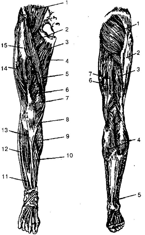
Fig. 74. Muscles of the lower limb:
A — anterior view: 1 — iliopsoas muscle; 2 — pectineus muscle; 3 — adductor longus muscle; 4 — gracilis muscle; 5 — sartorius muscle; 6 — vastus medialis muscle; 7 — quadriceps femoris tendon; 8 — patellar ligament; 9 — gastrocnemius muscle; 10 — soleus muscle; 11 — extensor digitorum longus; 12 — peroneus longus muscle; 13 — tibialis anterior muscle; 14 — vastus lateralis muscle; 15 — rectus femoris muscle; B — posterior view: 1 — gluteus maximus muscle; 2 — iliotibial tract (part of the fascia lata); 3 — biceps femoris muscle; 4 — gastrocnemius muscle; 5 — calcaneal (Achilles) tendon; 6 — semimembranosus muscle; 7 — semitendinosus muscle
The superior and inferior gemellus muscles (m. gemellus superior et inferior) originate from the ischium and the ischial tuberosity; they insert into the greater trochanter. They laterally rotate the thigh.
The piriformis muscle (m. piriformis) originates from the pelvic surface of the sacrum, passes through the sciatic foramen, and inserts into the greater trochanter of the femur. It laterally rotates and slightly abducts the thigh.
External Group of pelvic muscles. The muscles of this group are divided into three layers: superficial, middle, and deep. The superficial layer contains the gluteus maximus and the tensor fasciae latae; the middle layer contains the gluteus medius, the superior and inferior gemellus muscles, and the quadratus femoris; and the deep layer contains the gluteus minimus and the obturator externus.
The gluteus maximus muscle (m. gluteus maximus) originates from the iliac crest, the dorsal surface of the sacrum, the coccyx, and the aponeurosis of the erector spinae; it inserts into the gluteal tuberosity of the femur. It extends, laterally rotates, and abducts the thigh, and stabilizes the pelvis and trunk.
The gluteus medius muscle (m. gluteus medius) originates from the ilium and the fascia lata, and inserts into the greater trochanter of the femur. It abducts and rotates the thigh, and, together with the gluteus minimus, helps stabilize the pelvis and trunk in an upright position when the lower limb is fixed.
The gluteus minimus muscle (m. gluteus minimus) originates from the ilium and inserts into the greater trochanter of the femur. It abducts and rotates the thigh medially and laterally, and extends the trunk.
The tensor fasciae latae muscle (m. tensor fasciae latae) originates from the anterior superior iliac spine, runs downward, and merges into the iliotibial tract of the fascia lata. Upon contraction, it tenses the fascia and helps stabilize the knee joint in extension.
The quadratus femoris muscle (m. quadratus femoris) runs from the ischial tuberosity and inserts into the intertrochanteric crest. It laterally rotates the thigh.
The obturator externus muscle (m. obturatorius externus) originates from the outer surface of the pubis, the ramus of the ischium, and the obturator membrane; it inserts into the trochanteric fossa of the femur and the Joint Capsule. It laterally rotates the thigh.
Muscles of the free part of the lower limb. The muscles of the thigh surround the femur and form three groups: anterior (hip flexors), medial (hip adductors), and posterior (hip extensors).
Anterior Group of thigh muscles.
The quadriceps femoris muscle (m. quadriceps femoris) has four heads: the rectus femoris, vastus lateralis, vastus medialis, and vastus intermedius.
The rectus femoris originates from the anterior inferior iliac spine and from the ilium above the acetabulum; the vastus lateralis arises by muscle bundles from the greater trochanter, intertrochanteric line, gluteal tuberosity of the femur, and lateral intermuscular septum; the vastus medialis originates from the intertrochanteric line, medial lip of the linea aspera, and medial intermuscular septum; the vastus intermedius arises by muscle fiber bundles from the anterior and lateral surfaces of the femoral shaft, inserting into the Patella along with the other vasti muscles. Uniting into a common tendon, the quadriceps femoris is a powerful extensor of the leg at the knee joint, while the rectus femoris also flexes the thigh.
The sartorius muscle (m. sartorius) is the longest muscle in The Human Body. It runs from the anterior superior iliac spine and inserts into the Tibia. It flexes, rotates, and abducts the thigh, and flexes the leg.
Medial Group of thigh muscles. This group consists of the following muscles: gracilis, pectineus, and adductor (longus, brevis, and magnus). The primary function of this muscle group is adduction of the thigh, hence their name, the adductors.
The gracilis muscle (m. gracilis) is a long, flat muscle located on the Medial surface of the thigh; it arises from the lower half of the Pubic Symphysis and the pubic bone, and inserts into the tuberosity of the tibia. Upon contraction, it adducts the thigh, flexes the leg, and rotates it medially.
The pectineus muscle (m. pectineus) is a short, flat muscle; it originates from the pectineal line and superior ramus of the pubis, and inserts between the posterior surface of the lesser trochanter and the linea aspera of the femur. It adducts and flexes the thigh.
The adductor longus muscle (m. adductor longus) is triangular in shape, originating from the superior pubic ramus and inserting into the middle third of the medial lip of the linea aspera of the femur. It adducts, flexes, and laterally rotates the thigh.
The adductor brevis muscle (m. adductor brevis) is a thick, triangular muscle that originates from the body and inferior pubic ramus. It inserts via short tendinous bundles into the linea aspera on the shaft of the femur.
The adductor magnus muscle is a large, powerful muscle of the adductor group. It originates from the ischial tuberosity and the rami of the ischium and pubis, inserting into the medial lip of the linea aspera of the femur. It adducts and flexes the thigh.
The posterior compartment of the thigh muscles. The biceps femoris muscle (m. biceps femoris) originates by its long HEAD from the ischial tuberosity and the sacrotuberous ligament, and by its short head from the lateral lip of the linea aspera, the upper part of the lateral epicondyle, and the lateral intermuscular septum of the thigh; it courses downward and transitions into a tendon that inserts into the head of the Fibula. It extends the thigh, flexes the leg, and laterally rotates it.
The semitendinosus muscle (m. semitendinosus) originates from the ischial tuberosity and inserts into the medial surface of the upper part of the tibia. Upon contraction, it extends the thigh and flexes the leg; it medially rotates the leg when the knee is flexed.
The semimembranosus muscle (m. semimembranosus) originates from the ischial tuberosity and inserts via three tendinous slips into the posterolateral surface of the medial condyle of the tibia. These tendinous slips form the so-called pes anserinus. Some fibers transition into the oblique popliteal ligament and the popliteal fascia. It extends the thigh, flexes the leg, medially rotates it, and draws back the capsule of the knee joint.
Muscles of the Leg. They form the anterior, lateral, and posterior compartments. The interosseous membrane and the BONES OF THE leg separate the anterior muscle compartment from the posterior one.
The anterior compartment of the leg muscles. The tibialis anterior muscle (m. tibialis anterior) originates from the lateral condyle of the tibia and inserts into the medial cuneiform bone and the Base of the I metatarsal bone. It dorsiflexes the foot, elevates its medial margin, and helps maintain the foot in an upright position.
The extensor digitorum longus muscle (m. extensor digitorum longus) originates from the lateral condyle of the tibia and the head of the fibula; on the dorsum of the foot, it divides into four tendons that insert into the distal Phalanges of digits II—V and the base of the V metatarsal bone. It extends digits II—V and dorsiflexes the foot, elevates its lateral margin, and maintains the leg in an upright position.
The extensor hallucis longus muscle (m. extensor hallucis longus) originates from the lower part of the fibula and the interosseous membrane of the leg, inserting into the distal and partially proximal phalanx of the great toe. It extends the great toe, dorsiflexes the foot, and elevates its medial margin.
The posterior compartment of the leg muscles. This muscle group of the leg is divided into two layers: superficial and deep. The superficial layer includes the triceps surae and plantaris muscles, while the deep layer consists of the popliteus muscle, three long muscles (the flexor digitorum longus, located medially; the flexor hallucis longus, located laterally), and the tibialis posterior muscle, which is positioned intermediately.
1. The Superficial layer of the posterior compartment of the leg muscles. The triceps surae muscle (m. triceps surae) consists of the gastrocnemius and soleus muscles, which share a common tendon. The gastrocnemius muscle (m. gastrocnemius) originates by two heads (lateral and medial) from the lateral and medial condyles of the femur. At the midpoint of the leg, the heads of the gastrocnemius muscle unite, forming a thick (Achilles) tendon together with the tendon of the soleus muscle; it inserts into the calcaneal tuberosity.
The soleus muscle (m. soleus) lies beneath the gastrocnemius, originating from the posterior surface of the tibia and the tendinous arch, and transitions into the common tendon that inserts into the calcaneal tuberosity of the calcaneus. Upon contraction, the triceps surae muscle flexes the leg and plantarflexes the foot; it stabilizes the leg, preventing it from tilting forward.
The plantaris muscle (m. plantaris) is inconstant, having a small belly and a long, slender tendon. It originates from the lateral condyle of the femur and the oblique popliteal ligament, inserting into the calcaneal tuberosity. It tenses the capsule of the knee joint and assists in flexing the leg and foot.
2. The Deep Layer of the posterior compartment of the leg muscles. The popliteus muscle (m. popliteus) runs from the lateral condyle of the femur and the capsule of the knee joint, inserting into the posterior surface of the tibia. It flexes the leg, medially rotates it, and draws back the capsule of the knee joint.
The flexor digitorum longus muscle (m. flexor digitorum longus) originates from the middle part of the tibia, the fascia, and the posterior intermuscular septum of the leg; it inserts into the distal phalanges of digits II—V. It flexes the phalanges of digits II—V and plantarflexes the foot, laterally rotates it, and Supports the arch of the foot.
The flexor hallucis longus muscle (m. flexor hallucis longus) originates from the lower two-thirds of the shaft of the fibula and the interosseous membrane of the leg, inserting into the base of the distal phalanx of the great toe. It flexes the great toe and assists in plantarflexion, supination, and adduction of the foot; it supports the arch of the foot.
The tibialis posterior muscle (m. tibialis posterior) originates from the posterior surfaces of the tibia and fibula and the interosseous membrane of the leg; it inserts into the tuberosity of the navicular bone, the three cuneiform bones, and the base of the IV metatarsal bone. It plantarflexes, adducts, and laterally rotates the foot.
The lateral compartment of the leg muscles. The fibularis longus muscle (m. peroneus longus) originates from the head and upper two-thirds of the lateral surface of the fibula, the condyle of the tibia, the deep Fascia of the leg, and the intermuscular septa of the leg; it inserts into the bases of the I and II Metatarsal Bones and the medial cuneiform bone. It plantarflexes the foot, elevates its lateral margin, and supports the arch of the foot.
The fibularis brevis muscle (m. peroneus brevis) originates from the lower two-thirds of the lateral surface of the fibula and the intermuscular septum of the leg, inserting into the base of the V metatarsal bone. Upon contraction, it abducts and plantarflexes the foot, elevating its lateral margin.
Muscles of the foot. The muscles of the foot are divided into dorsal and plantar groups.
Muscles of the dorsum of the foot. The extensor digitorum brevis muscle (m. extensor digitorum brevis) originates from the calcaneus and inserts into the bases of the middle and distal phalanges of digits II—IV. It extends digits II—IV.
The extensor hallucis brevis muscle (m. extensor hallucis brevis) originates from the calcaneus and inserts into the proximal phalanx of the great toe. It extends the great toe.
Muscles of the SOLE OF THE foot. The plantar muscles are divided into three groups: medial, lateral, and central.
1. The medial group of plantar muscles. The abductor hallucis muscle (m. abductor hallucis) originates from the calcaneal tuberosity, the flexor retinaculum, and the plantar aponeurosis; it inserts into the base of the proximal phalanx of the great toe. It flexes and abducts the great toe.
The flexor hallucis brevis muscle (m. flexor hallucis brevis) arises from the plantar surface of the cuboid bone, the cuneiform bones, and the plantar ligaments, and inserts into the proximal phalanx and the sesamoid bone. It flexes the great toe.
The adductor hallucis muscle (m. adductor hallucis) has oblique and transverse heads, arising from the cuneiform and cuboid bones, the II—IV metatarsal bones, and the III—V metatarsophalangeal joints. Uniting, they insert via a tendon into the lateral sesamoid bone and the proximal phalanx of the great toe. It flexes and adducts the great toe.
2. Lateral group of plantar muscles. The abductor digiti minimi muscle (m. abductor digiti minimi) arises from the plantar surface of the calcaneus, the V metatarsal bone, and the plantar aponeurosis; it inserts into the proximal phalanx of the little toe. It flexes and abducts the little toe.
The flexor digiti minimi brevis muscle (m. flexor digiti minimi brevis) arises from the V metatarsal bone and the long plantar ligament; it inserts into the proximal phalanx of the little toe. It flexes the little toe.
The opponens digiti minimi muscle (m. opponens digiti minimi), which is inconstant, arises from the long plantar ligament and inserts into the V metatarsal bone. It helps support the arch of the foot.
3. Intermediate group of plantar muscles. The flexor digitorum brevis muscle (m. flexor digitorum brevis) arises from the anterior part of the calcaneal tuberosity and the plantar aponeurosis, and inserts by four tendons into the bases of the middle phalanges of the II—V toes. It flexes the middle phalanges of the II—V toes and supports the arch of the foot.
The quadratus plantae muscle (m. quadratus plantae) arises by two heads from the inferior and medial margins of the plantar surface of the calcaneus and inserts into the lateral margin of the tendons of the flexor digitorum longus. It assists in flexing the toes.
The lumbrical muscles (mm. lumbricales) are four spindle-shaped muscles that arise from the tendons of the flexor digitorum longus, with the first muscle arising by a single head, and the remaining three by two heads. They insert into the proximal phalanges and the tendons of the extensor digitorum longus of the II—V toes. They flex the proximal and extend the middle and distal phalanges, moving them toward the great toe.
The interossei muscles (mm. interossei) are the deepest of the short muscles of the foot, lying between the metatarsal bones. They are divided into plantar interossei and dorsal interossei muscles.
The plantar interossei muscles (mm. interossei plantares) are located on the plantar side in the interosseous spaces of the II—V metatarsal bones. Each muscle arises from the medial side of the III—V metatarsal bones. They insert into the bases of the proximal phalanges of the III—V toes, partially extending into the dorsal aponeurosis. They adduct the III—V toes toward the II toe and flex the proximal phalanges of these toes.
The dorsal interossei muscles (mm. interossei dorsales) are located in the interosseous spaces between the metatarsal bones. Each muscle arises from the adjacent surfaces of neighboring metatarsal bones. They insert into the bases of the proximal phalanges of the II—IV toes, partially extending into the dorsal aponeurosis. The first dorsal interosseous muscle abducts the II toe from the midline of the foot, while the others abduct the II—IV toes laterally; they also flex the proximal phalanges of the II—IV toes.
Fasciae of the lower limb. Since several muscles of the lower limb arise from the spine and pelvic bones, the fasciae covering them are closely continuous with the fasciae lining the walls of the abdominal and pelvic cavities.
The psoas fascia is a part of the intra-abdominal fascia; it covers the psoas major muscle anteriorly, attaches to the vertebral bodies and the upper part of the sacrum, and also blends with the fascia of the quadratus lumborum muscle. Inferiorly, the psoas fascia continues as the iliac fascia.
The iliac fascia arises from the lateral surfaces of the lumbar vertebrae and covers the iliopsoas muscle. Laterally, it is firmly attached to the inguinal ligament and continues into the transverse ligament, while medially, spanning from the inguinal ligament to the pubic bone, it forms the iliopectineal arch, which separates the vascular and muscular lacunae.
The gluteal fascia is located on the outer surface of the pelvis. It covers the gluteus maximus muscle superficially and then continues into the fascia lata. The deep layer of the gluteal fascia separates the gluteus maximus from the gluteus medius and the tensor fasciae latae muscle.
The fascia lata covers the thigh muscles like a dense sheath and consists of two layers: deep and superficial. The deep layer covers the pectineus muscle and part of the iliopsoas muscle, and is known as the iliopectineal fascia. The superficial layer is somewhat thinner below the inguinal ligament and features a small opening called the saphenous opening, through which the great saphenous vein passes to drain into the femoral vein. The saphenous opening is covered by the cribriform fascia, which has numerous perforations for Vessels and nerves. The saphenous opening serves as the external opening of the femoral canal, through which femoral hernias can protrude. Under normal conditions, there is no actual femoral canal, and this short space is filled with Connective Tissue. Its anterior wall is formed by the inguinal ligament and the superior horn of the falciform margin of the fascia lata; its posterior wall by the iliopectineal fascia; and its lateral wall by the femoral vein.
Fascia of the leg. Inferiorly, the fascia lata continues as the crural fascia, which covers the muscles and gives off intermuscular septa that separate the anterior, posterior, and lateral muscle groups of the leg. The fascia also forms a septum that divides the superficial and deep layers of the flexor muscles. In the lower third of the leg, There is a fairly broad band (the superior extensor retinaculum), and in the region of the malleoli, the inferior extensor retinaculum. On the lateral surface of the leg, the fascia forms the superior and inferior fibular retinacula. Near the medial malleolus, the fascia forms the flexor retinaculum.
The fascia of the foot is a continuation of the crural fascia and is divided into dorsal and plantar parts. The dorsal fascia covers the muscles of the dorsum of the foot, and its deep layer separates the interossei muscles from the extensor tendons. On the sole, the fascia thickens to form the plantar aponeurosis, from which septa extend deep between the muscle groups, along with tendon slips to the five toes.
In the distal part of the leg and foot, there are synovial sheaths of the tendons of the leg muscles. The anterior group consists of three sheaths—medial, intermediate, and lateral—through which the tendons of the anterior leg muscles pass. On the lateral surface, behind the malleoli, lies the common synovial Sheath of the fibular muscles. On the plantar side, the synovial sheaths of the digital tendons of the foot are formed.
Topography of the lower limb. In the region of the greater sciatic foramen, there are the suprapiriform and infrapiriform foramina, through which major vessels and nerves exit the pelvic cavity. In the region of the obturator foramen, the obturator canal is located, through which the obturator vessels and nerve exit the pelvic cavity to reach the adductor muscles of the thigh.
On the anterior surface of the thigh, below the inguinal ligament, lies the femoral triangle, bounded medially by the adductor longus muscle and laterally by the sartorius muscle.
Within the femoral triangle, the iliopectineal groove is clearly visible, continuing distally into the femoral groove.
The anterior region of the thigh communicates with the popliteal fossa via the adductor (femoral-popliteal) canal, which has three openings (inlet, inferior, and anterior). The femoral artery, femoral vein, and saphenous nerve pass through this canal.
In the middle third of the leg, the inferior musculofibular canal branches off laterally from the cruropopliteal canal, containing the fibular artery and Veins. The superior musculofibular canal is an independent Structure in the leg, through which the superficial fibular nerve passes.
The popliteal fossa is diamond-shaped and covered by fascia. It contains Lymph Nodes, Arteries, veins, and nerves, which then continue into the cruropopliteal canal. The popliteal fossa is bounded by the biceps femoris, semimembranosus, and gastrocnemius muscles.
Last update: 08/08/2026
Editorial and Educational Adaptation: This material has been compiled based on the primary/original source text. The project team performed an editorial review, corrected technical inaccuracies, structured sections, and adapted the content for an educational format.
What was processed:
- elimination of formatting defects (OCR errors, structural breaks, corrupted characters);
- editorial organization of content;
- standardization of terminology in accordance with academic sources;
- verification of factual statements against the original source text.
All mentions of the author, publication year, and origin of the primary text have been preserved in accordance with the source.