MEDICAL BIOLOGY, HUMAN ANATOMY, PHYSIOLOGY AND PATHOLOGY - Y.I. Fedoniuk 2010
ANATOMY, PHYSIOLOGY, PATHOLOGY
SECTION 1. ORGAN. ORGAN SYSTEM. ORGANISM
INJURY
3. Necrosis
Necrosis, or local death, is the demise of individual Cells, tissue areas, part of an organ, or an entire organ in a living Organism. In this process, METABOLISM completely and irreversibly ceases, resulting in the loss of all Functions. This process goes through several stages:
1) paranecrosis – changes may still be reversible;
2) necrobiosis – irreversible changes;
3) Cell death;
4) autolysis – The breakdown of dead substrate under The Influence of hydrolytic Enzymes.
The causes of necrosis are diverse. Tissue destruction can be triggered by mechanical (trauma) and thermal (Burns, frostbite) factors, ionizing radiation, chemical substances (acids, alkalis), and impaired neural and vascular trophic supply to Tissues. Furthermore, necrosis can result from The impact of toxins in infectious (diphtheria, tuberculosis) and non-infectious diseases, as well as pathological conditions (uremia).
The primary manifestations of necrosis are irreversible Changes in the cell nuclei and Cytoplasm. During necrobiosis, cells lose Water, and their nuclei shrink and condense, a process known as karyopyknosis. Nucleic Acids emerge from The Nucleus into the cytoplasm in the form of discrete clumps, and the nucleus breaks apart (karyorrhexis). Finally, the nuclear material dissolves, leading to karyolysis. Necrotic cells lack nuclei, which serves as one of the hallmark features of necrosis.
<Identical dynamics of necrotic changes are observed in the cytoplasm, where plasmarrhexis and plasmorrhysis develop. Ultimately, the entire cell dissolves, resulting in cytolysis. In Necrosis of the interstitial tissue, its ground substance swells and liquefies, while fibrous structures become denser or likewise undergo dissolution. The unstructured, homogeneous necrotic masses formed As a result of tissue destruction—consisting of protein and fat granules, remnants of nuclear substance—are referred to as necrotic detritus. Necrotic tissues differ from surrounding healthy tissues in consistency, color, and odor.
Based on their external characteristics, the following types are distinguished:
- dry (coagulative) necrosis;
- moist (colliquative) necrosis;
- gangrene;
- sequestrum;
- infarction.
In dry necrosis, the dead areas are dry, condensed, and clay-yellow or grayish-yellow in color. Microscopic examination reveals karyopyknosis, karyorrhexis, and plasmarrhexis. Dry necrosis is characteristic of tuberculosis, Syphilis, lymphogranulomatosis, and rheumatic diseases.
In moist necrosis, the dead areas dissolve, transforming into a fine-grained emulsion or a liquid opaque mass. Examples of moist necrosis include gray softening of the Brain (ischemic stroke) and fetal tissue necrosis during antenatal death (maceration).
In gangrene, dead tissues that remain in contact with the external environment acquire a grayish-brown or even black color (depending on alterations in Blood pigments).
Moist gangrene is a consequence of microorganisms penetrating dead tissues. Tissue breakdown is caused by common pyogenic flora (staphylococci, streptococci) as well as anaerobic microorganisms.
Class="center">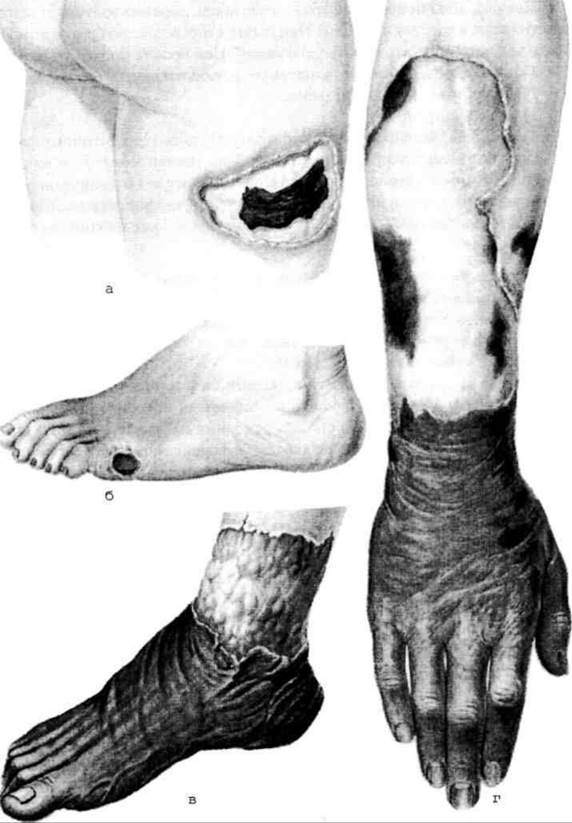
Fig. 1.2. Gangrene: a – moist Gangrene of the thigh Skin; b – moist gangrene of the FOOT skin; c – dry gangrene of the foot; d – dry gangrene of the forearm and hand.
Moist gangrene (Fig. 1.2 a, b) typically develops in fluid-rich tissues. It is preceded by Circulatory Disorders (venous congestion) and impaired Lymph flow (edema, lymphostasis). This type of gangrene complicates inflammatory processes in the Lungs and intestines associated with mesenteric artery obstruction (thrombosis, embolism), as well as in children suffering from measles (noma).
A specific form is anaerobic gangrene—a distinct infectious disease caused by a specific group of microorganisms. It most frequently occurs in gunshot and other wounds accompanied by massive Muscle destruction and bone fragmentation.
Dry gangrene, or mummification, occurs when superficial necrotic tissues lose their moisture to the environment, dry out, and become dense. In this process, blood diffuses into the surrounding dead tissue, where blood pigments decompose and, in the presence of hydrogen sulfide, are converted into iron sulfide. This is why necrotic tissues turn black (Fig. 1.2 c, d). Examples of dry gangrene include mummification of the umbilical cord stump in newborns and marantic (senile) gangrene. Most commonly, dry gangrene develops in the extremities as a result of ischemic coagulative necrosis.
A variant of gangrene is a bedsore (pressure ulcer), which is the necrosis of body areas (skin, soft tissues) resulting from prolonged compression.
Bedsores appear in the sacral region, the spinous processes of the spine, and the greater trochanter of the Femur in severely ill patients suffering from cardiovascular, infectious, or neurological disorders.
Their Pathogenesis is of a trophic-neurotic nature due to the compression of blood Vessels and nerves.
A sequestrum is an area of necrotic
tissue that is neither enzymatically liquefied nor replaced by Connective Tissue, remaining free among living tissues.
It most commonly occurs in Osteomyelitis, where a sequestered cavity filled with pus forms, containing a bone fragment (sequestrum). Sequestration can also occur in soft tissues (lungs, Kidneys), although these sequestra are rapidly liquefied.
Last update: 08/08/2026
Editorial and Educational Adaptation: This material has been compiled based on the primary/original source text. The project team performed an editorial review, corrected technical inaccuracies, structured sections, and adapted the content for an educational format.
What was processed:
- elimination of formatting defects (OCR errors, structural breaks, corrupted characters);
- editorial organization of content;
- standardization of terminology in accordance with academic sources;
- verification of factual statements against the original source text.
All mentions of the author, publication year, and origin of the primary text have been preserved in accordance with the source.