Plant Physiology - Musienko M. M. 2001
Physiology of Secretion in Plant Organisms
External secretory structures: trichomes, glandular hairs, nectaries, osmophores, hydathodes
Most secretory Cells differ from other cells by their electron-dense Cytoplasm, high content of Cell/35.html">Mitochondria, Golgi apparatus, numerous vesicles, and small vacuoles. They possess a large Nucleus and are interconnected by numerous plasmodesmata. Interestingly, in plants, hairs and glands with identical anatomical structures often secrete different substances, whereas similar secretions can be produced by various secretory structures.
Trichomes, hairs, salt glands. Most superficial secretory structures are of epidermal origin, although derivatives not only of the epidermis but also of deeper-lying cells can be found.
Trichomes are biserial hairs possessing a unicellular or multicellular HEAD on a slender stalk. They develop from epidermal cells As a result of uneven growth and subsequent division.
More complex secretory structures are called glands, which are most frequently associated with The excretion of Mineral Substances. Stinging hairs of nettles (Urtica) have a special mechanism for releasing their secretion. Each Hair resembles a capillary impregnated with calcium carbonate at the base and silicon at the top. The lower, expanded end of the hair is embedded in the epidermal cells. The upper part terminates in a head that breaks off upon contact. The sharp edges of the fracture pierce the Skin, and the Contents of the hair (nettle stinging juice is quite complex, containing histamine and acetylcholine) enter the wound, causing a stinging effect. Glandular hairs secrete their substance into the space between The Cell wall and the cuticle; in this process, the cuticle stretches strongly and bursts. Sticky hairs, which produce mucilage and a mixture of Terpenes, are found on the buds of young leaves. During excretion, the cuticle ruptures without stretching.
A rather diverse group of secretory structures is involved in excreting mineral substances. These include salt glands on the leaves and stems of halophytes, which excrete an excess of various salt ions; a group of salt hairs consisting of a stalk and a head that is shed once a specific salt concentration is reached within it; and finally, a group of secretory structures in carnivorous plants that secrete ions, Water, and proteinaceous secretions (Fig. 146).
Class="center">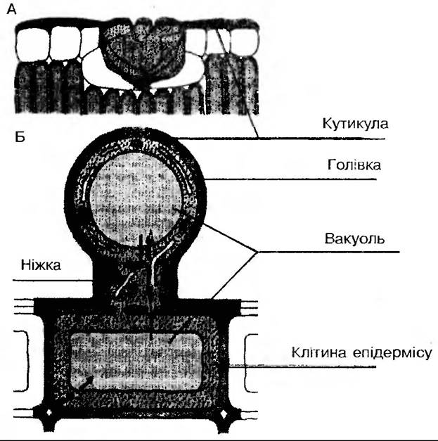
Fig. 146. Salt-secreting cells: A — salt gland; B — salt hair (arrows indicate Active ion transport)
Salt-excreting glands consist of Two Types of cells: concentrating (accumulating) cells and secretory (excretory) cells that are not connected to the plant's Vascular System. Accumulating cells resemble epidermal and parenchymal cells in Structure and are closely connected to the adjacent mesophyll via plasmodesmata. The Cell wall structure of these cells is specific. Externally, the glands are covered by a cuticle, while the lateral and basal walls are impregnated with Lignin and suberin. Therefore, ion transport occurs primarily via the symplast. Numerous protuberances are found on the cell wall, and accordingly, the Plasmalemma is also folded, which increases its overall surface area.
Mechanism of salt excretion. In plants growing on saline soils, salts move through the xylem and the apoplast. In the region of transfer cells, ion pumps pump them into the symplast, and they are transported via the symplast all the way to their external excretion. It is believed that there is active electronic transport of chloride ions in the glands, coupled with the passive transport of sodium or organic ions. It is possible that chloride ions are expelled from the secretory cell through the operation of a chloride pump, a function performed by a chlorine-activated ATPase. This may serve as an important part of the homeostatic mechanism of physiological adaptation to salinity in halophytes.
Nectaries and the physiology of nectar secretion. Nectaries are glandular structures that secrete a sugary fluid known as nectar. Depending on their Location, G. Caspary divides them into floral nectaries (located inside the flower), extrafloral nectaries (located outside the flower on leaves, petioles, and stems), and septal nectaries (found in the depressions, or septa, of the Ovary in monocots).
In dicotyledonous flowers, nectar may be secreted by the basal PARTS OF THE stamens, and sometimes nectaries appear as a ring or disk at the Base of the ovary. Diverse in Morphology (papillae on the petals of lime and mallow; convex nectaries in barberry; disk-shaped in lamiids; flat in legumes, etc.), nectaries consist of typical Tissues—epidermis, parenchymal and secretory cells, and vascular elements. The simplest ones consist of secretory cells and intercellular cavities adjacent to Stomata and phloem elements. Vascular bundles approach the nectaries; in small nectaries, the solution is transported from the vessels to the secretory tissue by parenchymal cells, whereas in large ones (such as squash), vascular bundles penetrate the secretory tissue. Nectar is secreted first into the subculticular cavity and then to the outside through pores or a rupture of the cuticle. The difference in The Nature of The vascular system (the presence and ratio of phloem and xylem) is related to the type of nectar secreted. The sugars in nectar are produced by the phloem. Depending on the type of nectary and weather conditions, the concentration of sugars (sucrose, glucose, fructose) varies from 7 to 100%.
The compositional constancy of nectar has been noted. For instance, out of 893 plant species sampled from a single region, variations in nectar composition were detected in only 61 species, which accounts for 7%. Nectar contains low concentrations of K+, Na+, Ca2+, Mg2+, PO43-, Trace Elements, organic acids, Amino Acids, Proteins, and Vitamins. Characteristically, the set of amino acids in nectar is also constant for each plant species, although their quantitative composition varies widely.
Coumarins, phenols, Alkaloids, and antibiotic substances have been detected in nectar. It is hypothesized that floral nectaries secrete substances necessary for the normalization of pollination processes and The Development of the ovary, fruits, and seeds.
The chemicalization of agricultural production leads to the appearance of toxic compounds in nectar, which can cause not only bee poisoning but also allergic diseases in humans. Flowering plants should not be treated with pesticides.
Flowers of central shoots secrete the most nectar, and There is a direct correlation between its quantity and flower size. Endogenous rhythms in nectar secretion exist that do not depend on changes in environmental factors, but their physiology remains insufficiently studied. A positive correlation has been established between leaf surface area and The amount of sugars secreted in nectar, which is related to the processes of plant photosynthetic productivity. Optimal levels of mineral Nutrition (low doses), Temperature, and water supply (60% of full field capacity) correlate positively with nectar secretion.
Active nectaries are characterized by intensive Respiration, indicating a link between nectar production and energy processes as well as membrane transport systems. Enzyme systems of nectaries (hexose and fructokinases, Phosphatases, cytochrome oxidase, polyphenol oxidase, peroxidase) play a crucial role in sugar transformations.
In plants that accumulate starch, α-amylase and starch phosphorylase break down starch in buds before flower opening as a preparatory stage of the secretory process.
Back in the 1940s, D. A. Sabinin hypothesized that The activity of nectaries is based on The phenomenon of physiological polarity of the secretory tissue cells, which enables the vectorial transport of the secretion and maintains its concentration gradient. Following this gradient, sugars move osmotically from the lower to the upper cells.
MODERN CONCEPTS OF the transport mechanism suggest the possibility of Active Transport. Since phloem sap is the initial product for nectar, its exit from the phloem (phloem unloading) must be the reverse of the mechanism by which assimilates are loaded into the phloem within the leaf blade. As is well known, assimilates are loaded against a concentration gradient with the participation of sugar and H+ cotransport. Sucrose enters the phloem terminals in symport with hydrogen ions driven by the pH gradient across the plasmalemma of phloem terminals. Transmembrane transfer of H+ occurs along the concentration gradient, while that of sucrose occurs against the gradient. This process is carried out by carrier proteins in the plasmalemma whose affinity for sucrose increases upon protonation, with H+ being pumped out by a hydrogen pump coupled with potassium ion uptake.
Further transport of the nectar precursor hypothetically can occur via both the apoplast and the symplast.
Along with flower coloration and scents, nectar has become one of the primary elements of entomophily—an adaptation that attracts insects for cross-pollination. Floral nectaries are capable of synthesizing substances such as Steroid Hormones, which are necessary for reproductive processes in insects. All of this has proven to be a powerful driver in the co-evolution of plants and insects throughout their phylogenetic development.
Osmophores. In many plants, the floral aroma emanates from specialized glands called osmophores (from Greek osmo — odor, phoros — bearing). Various floral tissues can differentiate into osmophores in the form of wings or hairs, particularly in representatives of Araceae and Orchidaceae. The volatile secretion is released for a short duration. Sometimes, osmophore tissues can be compact and possess a vascular system.
Hydathodes. These are structures that ensure the release of water from the internal parts of the leaf to the surface during guttation. Salts, sugars, and Organic compounds may be dissolved in this water; they are conventionally classified among secretory structures, although most of them lack tissues comparable to the glandular tissues of true secretory Organs. The excreted water originates from the tracheids of the vascular bundle. However, there are plants in which hydathodes may be quite complex and potentially associated with Secretory Tissues.
Last update: 07/08/2026
Editorial and Educational Adaptation: This material has been compiled based on the primary/original source text. The project team performed an editorial review, corrected technical inaccuracies, structured sections, and adapted the content for an educational format.
What was processed:
- elimination of formatting defects (OCR errors, structural breaks, corrupted characters);
- editorial organization of content;
- standardization of terminology in accordance with academic sources;
- verification of factual statements against the original source text.
All mentions of the author, publication year, and origin of the primary text have been preserved in accordance with the source.